. 2025 Feb 28;14(2):949-965.
doi: 10.21037/tcr-24-1532. Epub 2025 Feb 26.

A novel necroptosis-related miRNA signature for predicting the prognosis of esophageal cancer and immune infiltration analysis

Affiliations

A novel necroptosis-related miRNA signature for predicting the prognosis of esophageal cancer and immune infiltration analysis

Miao Zhang et al. Transl Cancer Res. .

Abstract

Background: The prognostic value of necroptosis-related microRNAs (miRNAs), which are important in tumorigenesis and development, remains unclear. Therefore, we aimed to screen prognostic necroptosis-related miRNAs in esophageal cancer (EC).

Methods: Nine necroptosis-related miRNA expression profiles and associated clinical data of EC patients were obtained from The Cancer Genome Atlas (TCGA) database. The relationships between necroptosis-related miRNAs and overall survival (OS) were determined via Cox regression model analysis. Target genes of the miRNAs were investigated in TargetScan, miRDB, and miRTarBase. The biological functions of these genes were evaluated by Gene Ontology (GO) and Kyoto Encyclopedia of Genes and Genomes (KEGG) analyses. For the most significant correlation between miR-425-5p expression and the survival of EC patients, the effect of miR-425-5p on necroptosis was explored in EC cells. The relationship between targeted gene expression and immune infiltration was also analyzed and validated.

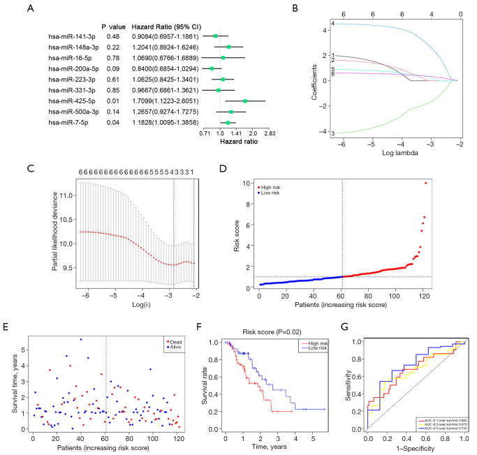
Results: Hsa-miR-425-5p, hsa-miR-500a-3p, hsa-miR-7-5p and hsa-miR-200a-5p were selected for the construction of a prognostic signature based on their correlation with the survival of EC patients. EC patients were divided into high- and low-risk groups according to the median value of the risk score. Patients in the high-risk group tended to have higher death rates than those in the low-risk group (P<0.05). The risk score was an independent prognostic indicator for the OS of EC patients [hazard ratio (HR) >1, P<0.05]. The prognostic model had good predictive efficiency. The genes targeted by necroptosis-related miRNAs were significantly enriched in apoptosis etc. The inhibition of miR-425-5p promoted necroptosis in EC cells by targeting branched chain amino acid transaminase 1 (BCAT1). The expression level of BCAT1 was significantly correlated with immune infiltration.

Conclusions: A necroptosis-related four-miRNA model was constructed successfully to predict the potential value of the four miRNAs in the prognosis of EC, which can be conducive to promoting the therapeutic effect on EC.

Keywords: Esophageal cancer (EC); miRNA signature; necroptosis; prognosis.


Conflict of interest statement

Conflicts of Interest: All authors have completed the ICMJE uniform disclosure form (available at https://tcr.amegroups.com/article/view/10.21037/tcr-24-1532/coif). The authors have no conflicts of interest to declare.

Figures

Figure 1
Figure 1
Flow chart of the study. miRNA, microRNA; EC, esophageal cancer; TCGA, The Cancer Genome Atlas; LASSO, least absolute shrinkage and selection operator.
Figure 2
Figure 2
Screening of the nine necroptosis-related miRNAs in the TCGA database. (A) Heatmap of the expression levels and distributions of the nine necroptosis-related miRNAs in normal and esophageal cancer tissues (green: low expression; orange: high expression; N: normal; T: tumor). (B) The correlation network of the nine miRNAs (red line: positive correlation; blue line: negative correlation. The depth of the colour indicates the intensity of the correlation). TCGA, The Cancer Genome Atlas; miRNA, microRNA.
Figure 3
Figure 3
Construction of the necroptosis-related miRNA model in the TCGA training cohort. (A) Forest plots of the univariate Cox regression analysis between the miRNAs and overall survival. (B,C) LASSO regression model of the miRNAs. (D) Distribution of the risk scores in the training cohort. (E) Analysis of the survival status in the training cohort. (F) Kaplan-Meier analysis of the overall survival of esophageal cancer patients in the high- and low-risk groups. (G) AUC of time-dependent ROC curves to evaluate the predictive efficiency. CI, confidence interval; TCGA, The Cancer Genome Atlas; miRNA, microRNA; LASSO, least absolute shrinkage and selection operator; AUC, area under the curve; ROC, receiver operating characteristic.
Figure 4
Figure 4
Validation of the risk model in the TCGA testing cohort. (A) Analysis of risk scores in the validation cohort. (B) Distribution of survival status in the validation cohort. (C) Kaplan-Meier survival curves in the high- and low-risk groups. (D) AUCs of time-dependent ROC curves for the 1-, 2-, and 3-year OS in the validation cohort. AUC, area under the curve; TCGA, The Cancer Genome Atlas; ROC, receiver operating characteristic; OS, overall survival.
Figure 5
Figure 5
Independent prognostic value of the risk model. (A,C) Univariate (A) and multivariate (C) Cox analyses in the training cohort. (B,D) Univariate (B) and multivariate (D) Cox analyses in the validation cohort. (E) Heatmap of the four necroptosis-related miRNAs and clinical features in the high- and low-risk groups. CI, confidence interval; miRNAs, microRNAs.
Figure 6
Figure 6
The target genes of the four necroptosis-related miRNAs. (A) Venn diagram of the miRNA targets predicted by miRDB, miRTarBase and TargetScan. (B) Network of four miRNAs and their target genes. miRNAs, microRNAs.
Figure 7
Figure 7
Functional enrichment analysis of the target genes in the TCGA cohort. (A) GO analysis of the miRNA-target genes into three functional groups, including BP, CC and MF. (B) The enriched items of the miRNA-target genes identified via KEGG analysis. GO, Gene Ontology; BP, biological process; CC, cellular component; MF, molecular function; KEGG, Kyoto Encyclopedia of Genes and Genomes; TCGA, The Cancer Genome Atlas; miRNA, microRNA.
Figure 8
Figure 8
Effects of miR-425-5p on necroptosis and downstream target genes in EC cell lines in vitro. (A) Flow cytometry of EC109 and EC9706 cell lines treated with NC inhibitor or miR-425-5p inhibitor (necroptosis: left upper). (B) The expression levels of four necroptotic markers were detected by real-time PCR. (C) The expression levels of four target genes of miR-425-5p were assessed by real-time PCR. *, P<0.05; **, P<0.01; ***, P<0.001; ns, not significant. NC, negative control; FADD, fas associated via death domain; RIPK1, receptor interacting serine/threonine-protein kinase 1; RIPK3, receptor interacting serine/threonine-protein kinase 3; MLKL, mixed lineage kinase domain-like protein; PAK2, P21 (RAC1) activated kinase 2; LCOR, ligand dependent nuclear receptor corepressor; CRLS1, cardiolipin synthase 1; BCAT1, branched chain amino acid transaminase 1; EC, esophageal cancer; PCR, polymerase chain reaction.
Figure 9
Figure 9
BCAT1 expression and immune infiltration analysis in EC. (A) The biological pathway between the low- and high-BCAT1 expression group by GSVA (red: activated pathways; blue: inhibited pathways). (B) Relationship between BCAT1 expression and immune cell infiltration. (C) Analysis of immune checkpoint expression in BCAT1 low- and high-expression group. (D) Representative images and quantification of immunohistochemistry staining of BCAT1 and Ly6G in esophageal cancer tissue microarray (scale bar: 100 µm). (E,F) The expression levels of BCAT1 and CD274 in EC9706 cells after EGR240 treatment were assayed by real-time PCR (E) and Western blotting (F). *, P<0.05; **, P<0.01; ***, P<0.001; ns, not significant. BCAT1, branched-chain amino acid transaminase 1; EC, esophageal cancer; PCR, polymerase chain reaction; GSVA, gene set variation analysis; BCAT1_exp, BCAT1 expression; KEGG, Kyoto Encyclopedia of Genes and Genomes; ESCA, esophageal cancer; Ly6G, lymphocyte antigen 6G; PD-L1 (CD274), programmed cell death 1 ligand 1; GAPDH, glyceraldehyde-3-phosphate dehydrogenase.

Similar articles

Cited by

References

    1. Bray F, Laversanne M, Sung H, et al. Global cancer statistics 2022: GLOBOCAN estimates of incidence and mortality worldwide for 36 cancers in 185 countries. CA Cancer J Clin 2024;74:229-63. 10.3322/caac.21834 - DOI - PubMed
    1. Harada K, Yamamoto S, Kato K. Pembrolizumab for the treatment of advanced esophageal cancer. Future Oncol 2022;18:2311-9. 10.2217/fon-2022-0108 - DOI - PubMed
    1. Huang FL, Yu SJ. Esophageal cancer: Risk factors, genetic association, and treatment. Asian J Surg 2018;41:210-5. 10.1016/j.asjsur.2016.10.005 - DOI - PubMed
    1. Uhlenhopp DJ, Then EO, Sunkara T, et al. Epidemiology of esophageal cancer: update in global trends, etiology and risk factors. Clin J Gastroenterol 2020;13:1010-21. 10.1007/s12328-020-01237-x - DOI - PubMed
    1. Degterev A, Huang Z, Boyce M, et al. Chemical inhibitor of nonapoptotic cell death with therapeutic potential for ischemic brain injury. Nat Chem Biol 2005;1:112-9. 10.1038/nchembio711 - DOI - PubMed

LinkOut - more resources