. 2025 Mar 19;74(5):153.
doi: 10.1007/s00262-025-03995-4.

A novel peptide targeting CCR7 inhibits tumor cell lymph node metastasis

Affiliations

A novel peptide targeting CCR7 inhibits tumor cell lymph node metastasis

Yixuan Sun et al. Cancer Immunol Immunother. .

Abstract

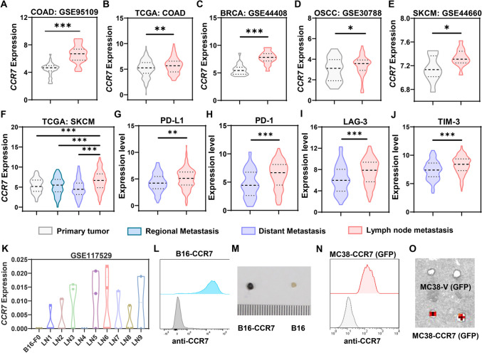
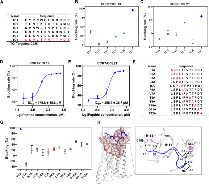
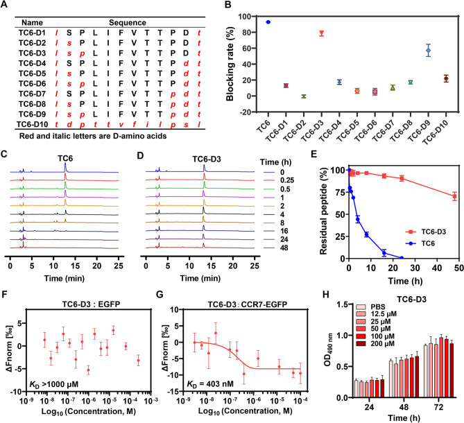
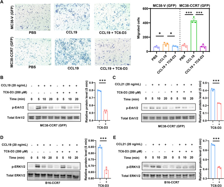
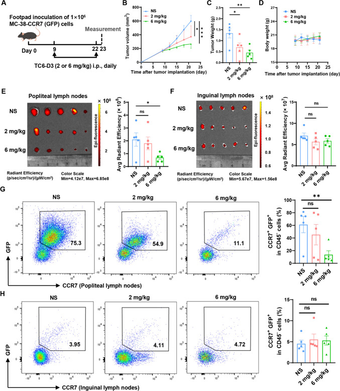
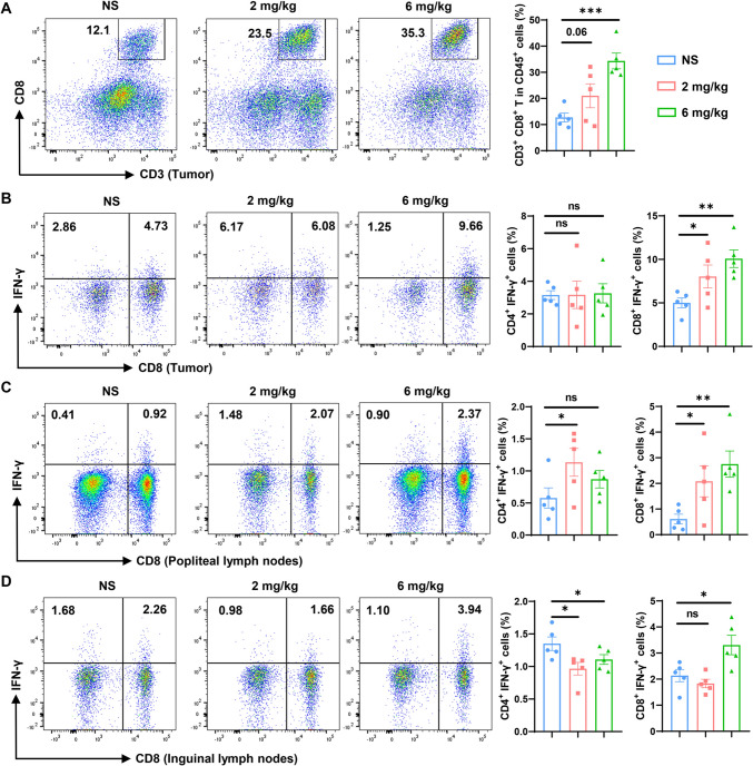
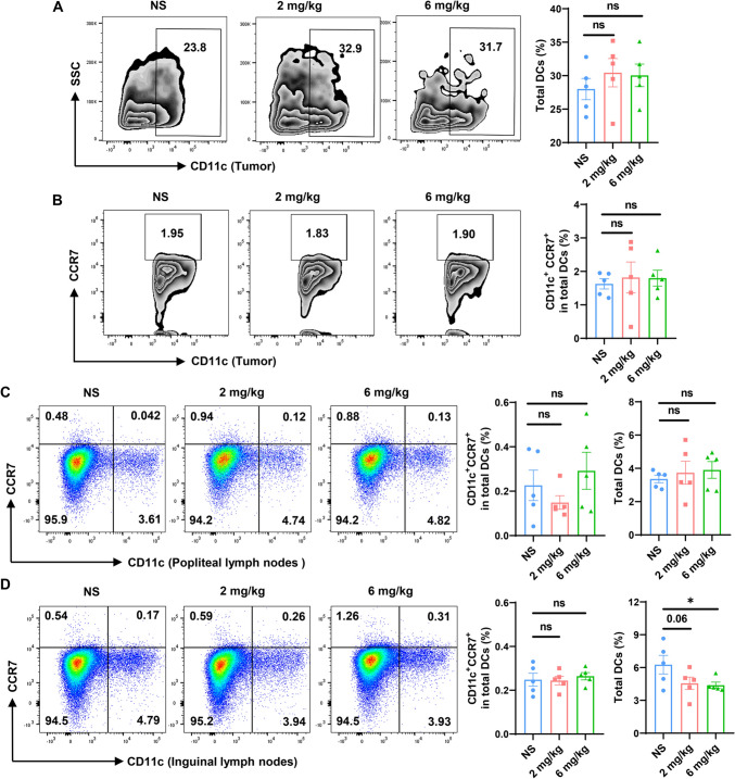
Lymph nodes are the most common metastasis sites for tumor cells, which are intimately linked to patient prognosis. It has been reported that cancer cells can upregulate CC Chemokine Receptor 7 (CCR7) expression and hijack its normal functions, enabling them to migrate along the gradient of CCL19 and CCL21 toward the lymph nodes and colonies as the initial stage of distant metastasis. In tumor patients, the metastatic tumor in the lymph nodes exhibited higher expression of CCR7, as well as inhibitory immune checkpoints PD-1, LAG-3, and TIM-3 compared to the primary tumors with the analysis of TCGA and GEO databases. Also, in mouse tumor model, tumor cells with elevated CCR7 expression were more susceptible to develop popliteal lymph node metastasis. Subsequently, we successfully identified a CCR7 binding peptide TC6 by phage display biopanning, which specifically blocks the interaction of CCR7/CCL19 and CCR7/CCL21. Further, the D-amino acids were introduced to substitute the N- and C-terminus of TC6 peptide to obtain the proteolysis-resistant TC6-D3 peptide, which decreased tumor cell migration in vitro via ERK1/2 pathway and inhibited tumor growth and lymph nodes metastasis in vivo, as well as effectively restored T cells cytotoxicity in both primary tumors and lymph nodes. In conclusion, CCR7 promoted tumor cell metastasis to lymph node and inhibited the anti-tumor immune responses in lymph nodes. Specific blockade of the CCR7 pathway with TC6-D3 peptide can significantly reduce lymph node tumor burden, promoting CD8+ T cell infiltration in primary tumors, meanwhile, enhancing anti-tumor immune responses in lymph nodes.

Keywords: Blockade; CCR7; Immune responses; Lymph node metastasis; Peptide.


Conflict of interest statement

Declarations. Conflict of interest: The authors declare no competing interests.

Figures

Fig. 1
Fig. 1
The expression of CCR7 correlated with the LN metastasis. AE The expression of CCR7 in primary tumor and lymph node metastasis tumor tissue among multifarious cancer types analyzed by GEO datasets (GSE95019, GSE44408, GSE30788, and GSE44660) and TCGA datasets. F The expression of CCR7 in primary tumor, regional metastasis, distant metastasis, and lymph node metastasis tissue in skin cutaneous melanoma (SKCM) analyzed by TCGA dataset. GJ The level of PD-L1, PD-1, LAG-3, and TIM-3 in distant metastasis and lymph node metastasis tissue of SKCM. K The expression level of CCR7 in LN metastatic tumor lines developed by serial in vivo lymph node metastases selection in the GSE117529 dataset. L The expression of CCR7 in B16-CCR7 cell line tested by Flow cytometer. M The effect of B16-CCR7 and B16 cell lines metastasized to adjacent popliteal lymph node after being injected into mouse footpad for 22 days. The popliteal lymph nodes were surgically excised and photographed. N The expression of CCR7 in the MC38-CCR7 (GFP) cell line tested by Flow cytometer. O The effect of MC38-CCR7 (GFP) and MC38-V (GFP) cell lines metastasized to adjacent popliteal lymph node after injected into mouse footpad for 22 days. The popliteal lymph nodes were surgically excised and detected with IVIS imaging. Statistical significance was determined by unpaired Student’s t test. *P < 0.05, **P < 0.01, ***P < 0.001
Fig. 2
Fig. 2
Screening of CCR7 peptide by phage display library. A The sequences of candidate peptides targeting CCR7 screened by phage peptide library. B, C The blocking efficacy of candidate peptides on interfering CCR7/CCL19 (B) and CCR7/CCL21 (C) at the concentration of 200 μM (n = 3). D, E Dose–response curves of TC6 on interfering CCR7/CCL19 (D) and CCR7/CCL21 (E) interaction (n = 3). F The sequences of alanine scanning peptides. G The blocking efficacy of alanine scanning peptides on interfering CCR7/CCL19 interaction at the concentration of 200 μM (n = 3). H Molecular docking was carried out in MOE software. The data are representative of at least three independent experiments and presented as mean ± SEM
Fig. 3
Fig. 3
Enhancing the stability of TC6 peptide by D-amino acid substitution. A The sequences of peptides substituted with D-amino acids from both N- and C- terminus of TC6. B The blocking efficacy of D-amino acid modified peptides on interfering CCR7-CCL19 interaction at the concentration of 200 μM (n = 3). CE RP-HPLC was utilized to detect the degradation of TC6 and TC6-D3 in 10% mouse serum (n = 3). F, G The binding affinity of TC6-D3 peptide to CHOK1-CCR7-EGFP (F) and CHOK1-EGFP (G) cells membrane fusion protein was tested by MST (n = 3). H The effect of TC6-D3 on the proliferation of MC3-CCR7 (GFP) tumor cell was determined by MTT assay (n = 3). The data are representative of at least three independent experiments and presented as mean ± SEM
Fig. 4
Fig. 4
TC6-D3-attenuated ERK1/2 phosphorylation. A Transwell assay was performed using MC38-V or MC38-CCR7 (GFP) cell lines with or without 200 μM TC6-D3 peptide when induced with 100 ng/mL chemokines CCL19 (n = 5). B, C Western blot analysis of ERK1/2 phosphorylation in MC38-CCR7 (GFP) cells with or without 200 μM TC6-D3 peptide in the condition of 20 ng/mL chemokines CCL19 (B) or CCL21 (C) (n = 3). D, E The same ERK1/2 phosphorylation in B16-CCR7 cells under the treatment of 20 ng/mL chemokines CCL19 (D) or CCL21 (E) (n = 3). The data are presented as mean ± SEM. Student’s t test, *P < 0.05, **P < 0.01, ***P < 0.001
Fig. 5
Fig. 5
TC6-D3 reduced the LN tumor burden in popliteal LN metastasis model. A Schematic of TC6-D3 peptide treatment on MC38-CCR7 (GFP) footpad tumor model. Mice were treated with TC6-D3 peptide intraperitoneally daily for 14 days. B Tumor growth curve of MC38-CCR7 (GFP) tumor-bearing mice treated with normal saline or TC6-D3 peptide (n = 5). C Statistical analysis of tumor weight between different groups at the experimental endpoint (n = 5). D TC6-D3 peptide did not affect the body weight after treatment for 14 days (n = 5). E, F Tumor cells metastasized to popliteal (E) and inguinal (F) lymph nodes were detected with IVIS imaging (n = 5). G, H The frequencies of CCR7+GFP+ tumor cells infiltrated in popliteal (G) and inguinal (H) lymph nodes were analyzed by flow cytometry (n = 5). Data are represented as means ± SEM. Student’s t test, *P < 0.05, **P < 0.01, ***P < 0.001, ns, not significant
Fig. 6
Fig. 6
TC6-D3 restored T cell-mediated anti-tumor immunity. A The frequencies of intratumoral CD8+ T cells in CD45+ lymphocytes were analyzed by flow cytometry (n = 5). B Cells from tumors were stimulated with 20 ng/mL PMA and 1 μM ionomycin containing 1 μM protein transport inhibitor for 4 h. IFN-γ-secreting CD8+ T cells (CD45+CD3+CD8+) and CD4+ T cells (CD45+CD3+CD8) were analyzed (n = 5). C, D IFN-γ-secreting CD8+ T cells (CD3+CD8+ T cells) and CD4+ T cells (CD3+CD8 T cells) in popliteal lymph nodes (C) and inguinal lymph nodes (D) were analyzed by flow cytometry (n = 5). Data are represented as means ± SEM. Student’s t test, *P < 0.05, **P < 0.01, ***P < 0.001, ns, not significant
Fig. 7
Fig. 7
TC6-D3 has no obvious effects on CCR7+ immunocytes. The frequencies of total DCs (CD45+CD11bCD11c+) and CCR7+ DCs (CD45+CD11bCD11c+CCR7+) subsets in tumors (A, B), popliteal lymph nodes (C), and inguinal lymph nodes (D) were detected by flow cytometer (n = 5). The data are presented as mean ± SEM, and the statistical significance was determined by unpaired Student’s t test, *P < 0.05, ns, not significant

Similar articles

Cited by

References

    1. Li Y, Byun AJ, Choe JK et al (2023) Micropapillary and solid histologic patterns in N1 and N2 lymph node metastases are independent factors of poor prognosis in patients with stages II to III lung adenocarcinoma. J Thorac Oncol 18:608–619. 10.1016/j.jtho.2023.01.002 - PMC - PubMed
    1. Al-Sukhni E, Attwood K, Gabriel EM, LeVea CM, Kanehira K, Nurkin SJ (2017) Lymphovascular and perineural invasion are associated with poor prognostic features and outcomes in colorectal cancer: a retrospective cohort study. Int J Surg 37:42–49. 10.1016/j.ijsu.2016.08.528 - PubMed
    1. Riedel A, Shorthouse D, Haas L, Hall BA, Shields J (2016) Tumor-induced stromal reprogramming drives lymph node transformation. Nat Immunol 17:1118–1127. 10.1038/ni.3492 - PMC - PubMed
    1. Reticker-Flynn NE, Zhang W, Belk JA et al (2022) Lymph node colonization induces tumor-immune tolerance to promote distant metastasis. Cell 185:1924–42.e23. 10.1016/j.cell.2022.04.019 - PMC - PubMed
    1. Ubellacker JM, Tasdogan A, Ramesh V et al (2020) Lymph protects metastasizing melanoma cells from ferroptosis. Nature 585:113–118. 10.1038/s41586-020-2623-z - PMC - PubMed

LinkOut - more resources