. 2025 Mar 19;25(1):88.
doi: 10.1007/s10238-025-01622-1.

MiR-101-3p targets the PI3K-AKT signaling pathway via Birc5 to inhibit invasion, proliferation, and epithelial-mesenchymal transition in hepatocellular carcinoma

Affiliations

MiR-101-3p targets the PI3K-AKT signaling pathway via Birc5 to inhibit invasion, proliferation, and epithelial-mesenchymal transition in hepatocellular carcinoma

Wenyuan Zhu et al. Clin Exp Med. .

Abstract

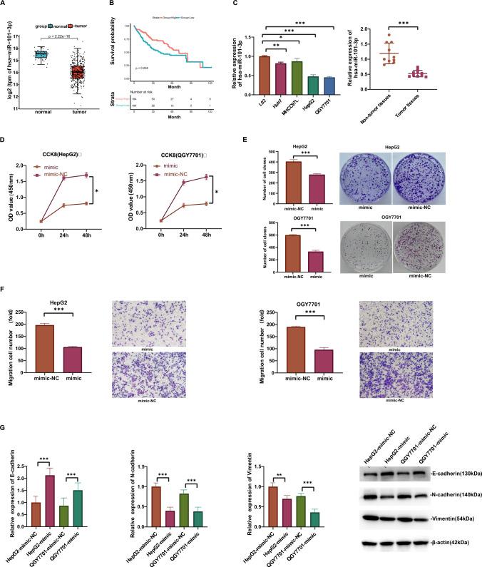
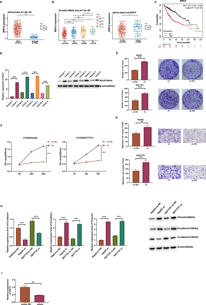
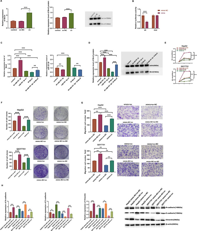
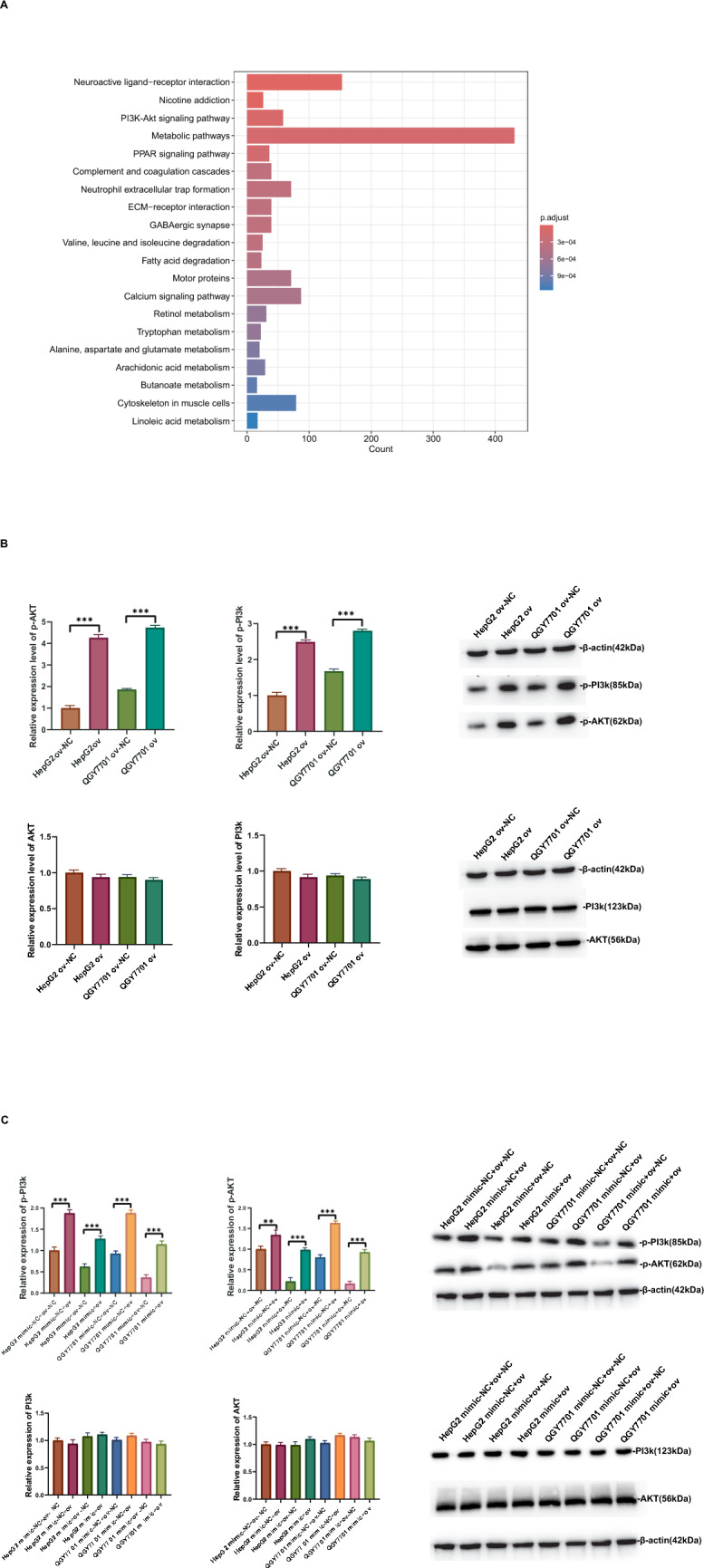
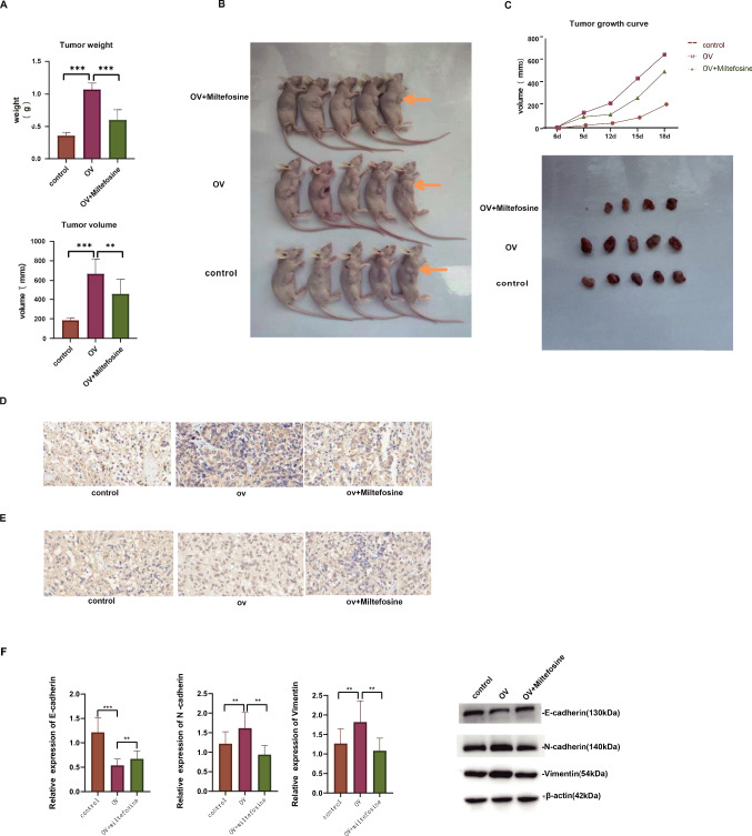
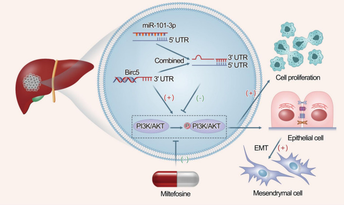
MicroRNAs (miRNAs) are small non-coding RNA molecules that regulate numerous genes in cells. Abnormal expression of miRNAs can lead to cancer. However, the roles and underlying mechanisms of miRNAs in hepatocellular carcinoma (HCC) are not fully understood. Using molecular biology techniques, we designed eukaryotic expression vectors with enhanced expression of miR-101-3p to transfect human hepatocellular carcinoma cell lines. Subsequent to this, cell cloning experiments, CCK8 assays, and Transwell migration experiments were executed to assess their impact on liver cancer cell proliferation and invasion. Dual-luciferase assays were employed to validate the molecular interaction between miR-101-3p and Birc5. Through rescue experiments aimed at manipulating the expression levels of Birc5, we scrutinized the influence of miR-101-3p on liver cancer cell proliferation and invasion. Furthermore, Western blot analysis was utilized to monitor alterations in the expression levels of E-cadherin, N-cadherin, and vimentin proteins within each cell group. In vivo investigations were conducted using nude mice implanted with hepatocellular carcinoma cells transfected with Birc5. Additionally, further exploration was carried out by combining this model with the PI3K/AKT pathway inhibitor miltefosine to elucidate its effects on tumor proliferation. In vitro functional analysis of miR-101-3p revealed that treatment of HCC cells with its corresponding mimic significantly inhibited cell proliferation, colony formation, invasion, and epithelial-mesenchymal transition. Additionally, miR-101-3p exerts its anti-tumor effects by targeting the shared gene Birc5. Experiments using nude mouse models demonstrate that Birc5 promotes tumor proliferation by phosphorylating the PI3K/AKT signaling pathway. Inhibiting the PI3K/AKT signaling pathway shows suppressive effects on liver cancer proliferation. MiR-101-3p plays crucial roles in inhibiting the proliferation, invasion and epithelial-mesenchymal transition of HCC cells by targeting Birc5 and downregulating the PI3K-AKT signaling pathway. These findings provide new insights for the molecular treatment of HCC.

Keywords: Birc5; Epithelial–mesenchymal transition; Invasion; Metastasis; MiR-101-3p.


Conflict of interest statement

Declarations. Conflict of interests: The authors of this manuscript have no conflict of interest. Ethical approval and consent to participate: The studies involving human participants were reviewed and approved by the Ethics Committee of Shandong Provincial Hospital (approval no. SWYX:NO.2022–477). All institutional and national guidelines for the care and use of laboratory animals were followed in this study. Consent for publication: Not applicable.

Figures

Fig. 1
Fig. 1
High miR-101-3p expression inhibits proliferation and invasion in HCC cells. A miR-101-3p expression levels were significantly higher in normal tissues than in HCC tissues. B Low miR-101-3p expression was associated with poor patient prognosis. C qRT-PCR was used to measure the expression levels of miR-101-3p in normal cell lines, HCC cell lines, HCC tissues, and normal tissues. The results demonstrate that miR-101-3p expression is lower in HCC cells and tissues compared to normal cells and tissues. D mimic-NC was the negative control group, and mimic was the experimental group with miR-101-3p overexpression. CCK-8 assay analyzing cell proliferation. E Colony formation assay was conducted to compare the proliferative capacity of HCC cells between the two groups. F Transwell assay analyzing invasion capacity in HCC cells. G WB analysis was performed to compare the expression levels of E-cadherin, N-cadherin, and Vimentin proteins in HCC cells between the two groups
Fig. 2
Fig. 2
Association between Birc5 expression and malignant behavior in HCC cells. A Assistant for Clinical Bioinformatics analysis revealed elevated Birc5 expression in HCC tissues. B Birc5 expression was significantly higher in HCC stages III and IV than in stages I and II. C Kaplan–Meier analysis of the association between high Birc5 expression and unfavorable prognosis in patients. D Protein immunoblotting experiment analyzing the Birc5 expression level in four pairs of cancer tissues and corresponding adjacent tissues, showing a significantly higher expression level in HCC tissues than in normal tissues. E ov-NC was the negative control group, and ov was the experimental group with Birc5 overexpression. Colony formation assay was performed in HepG2 and QGY7701 cells to compare the proliferative capacity of HCC cells between the Birc5 overexpression group and the negative control group. F Evaluation of HCC cell proliferation using the CCK-8 assay. G Transwell assay analyzing the migration and invasion capabilities of HCC cells in Birc5-overexpression and negative control groups. H Western blotting analysis of E-cadherin, N-cadherin, and vimentin expression levels in Birc5-overexpression and negative control groups. I qRT-PCR analyzing the Birc5 mRNA expression level in HCC cells in miR-101-3p overexpression and negative control groups
Fig. 3
Fig. 3
Elevated Birc5 expression reverses the effects of miR-101-3p, promoting proliferation and invasion in HCC cells. A In the control group, negative control group, and Birc5 overexpression group, qRT-PCR and WB were performed to compare the expression levels of Birc5 in HCC cells among the three groups. B Luciferase assay to detect the relative luciferase activity of mRNA reporter constructs containing wild-type or mutant Birc5 3′-UTR downstream of the luciferase gene after transfection with miR-101-3p mimic or NC. C miR-101-3p and Birc5-specific siRNA were transfected, and the mRNA expression levels of miR-101-3p and Birc5 in each group of cells were detected by qRT-PCR. D miR-101-3p and Birc5-specific siRNA were transfected, and the protein expression level of Birc5 in each group was detected by Western blotting analysis. E In HepG2 and QGY7701 HCC cells, based on the miR-101-3p overexpression group and the control group, the expression of Birc5 was upregulated and downregulated, respectively. CCK-8 assay was performed to compare the proliferation of cells in the four groups. F Colony formation assay was conducted in the four groups of HCC cells to compare their proliferation capabilities. G Transwell assay analyzing invasive ability in HCC cells. H Western blot analysis was performed to compare the protein expression levels of E-cadherin, N-cadherin, and Vimentin in the four groups of HCC cells. “Mimic” refers to miR-101-3p mimic; “NC” refers to negative control
Fig. 4
Fig. 4
miR-101-3p overexpression regulates Birc5 to inhibit the PI3K/AKT signaling pathway in HCC cells. A Bar plot of KEGG signaling pathway enrichment analysis. This figure shows the results of the signaling pathway enrichment analysis based on the KEGG database, which is used to evaluate the enrichment of genes related to liver cancer invasion, proliferation, and EMT in different signaling pathways. The vertical axis in the figure represents the adjusted p–value (p.adjust), reflecting the significance level of pathway enrichment. The smaller the p–value, the more significant the pathway enrichment. The horizontal axis represents the number of enriched genes (Count), reflecting the number of genes involved in each pathway. B Western blotting analysis of PI3K, AKT, p-PI3K and p-AKT expression levels  in the Birc5 overexpression group and the control group. C Based on the Birc5 overexpression group and the control group, the expression of miR-101-3p was upregulated and downregulated, respectively. Western blot was used to detect the protein expression levels of PI3K, AKT, p-PI3K, and p-AKT in the four groups of cells. “p- “ denotes phosphorylated forms of PI3K and AKT proteins
Fig. 5
Fig. 5
Blocking the PI3K/AKT signaling pathway inhibits HCC cell proliferation and EMT. A Tumor cells with and without the PI3K/AKT pathway inhibitor were subcutaneously implanted in male BALB/c nude mice. Tumor mass and volume were compared among the three groups over an 18-day observation period. B Macroscopic appearance of tumors in nude mice after 18 days. C Tumor volume changes in each mouse and the tumor growth curve. Immunohistochemical assessment of D Birc5 expression and E Ki67 expression. F Western blotting analysis of the effects of the PI3K/AKT signaling pathway inhibitor on EMT in HCC cells
Fig. 6
Fig. 6
Schematic representation of the function of miR-101-3p in HCC cells. In HCC cells with miR-101-3p overexpression, miR-101-3p binds to Birc5 and inhibits its expression, thereby suppressing phosphorylation of the PI3K/AKT signaling pathway and the proliferation and EMT of HCC cells

Similar articles

Cited by

References

    1. Ferlay J, Colombet M, Soerjomataram I, et al. Estimating the global cancer incidence and mortality in 2018: GLOBOCAN sources and methods. Int J Cancer. 2019;144(8):1941–53. - PubMed
    1. He J, Gu D, Wu X, et al. Major causes of death among men and women in China. N Engl J Med. 2005;353(11):1124–34. - PubMed
    1. Fedele M, Sgarra R, Battista S, Cerchia L, Manfioletti G. The epithelial-mesenchymal transition at the crossroads between metabolism and tumor progression. Int J Mol Sci. 2022;23(2):800. - PMC - PubMed
    1. Greenburg G, Hay ED. Epithelia suspended in collagen gels can lose polarity and express characteristics of migrating mesenchymal cells. J Cell Biol. 1982;95(1):333–9. - PMC - PubMed
    1. Onoue T, Uchida D, Begum NM, Tomizuka Y, Yoshida H, Sato M. Epithelial-mesenchymal transition induced by the stromal cell-derived factor-1/CXCR4 system in oral squamous cell carcinoma cells. Int J Oncol. 2006;29(5):1133–8. - PubMed

MeSH terms