

Beam hardening of K-edge contrast agents: a phantom study comparing clinical energy-integrating detector and photon-counting detector CT systems
- PMID: 40106074
- PMCID: PMC11923337
- DOI: 10.1186/s41747-024-00530-5
Beam hardening of K-edge contrast agents: a phantom study comparing clinical energy-integrating detector and photon-counting detector CT systems
Abstract
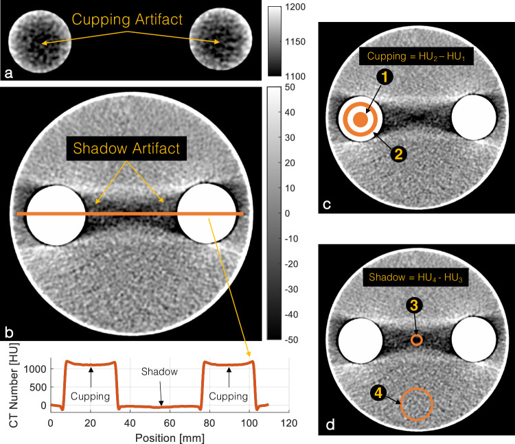
Background: Beam hardening (BH) artifacts negatively influence computed tomography (CT) measurements, especially when due to dense materials or materials with high effective atomic numbers. Photon-counting detectors (PCD) are more susceptible to BH due to equal weighting of photons regardless of their energies. The problem is further confounded by the use of contrast agents (CAs) with K-edge in the diagnostic CT energy range. We quantified the BH effect of different materials comparing energy-integrating detector (EID)-CT and PCD-CT.
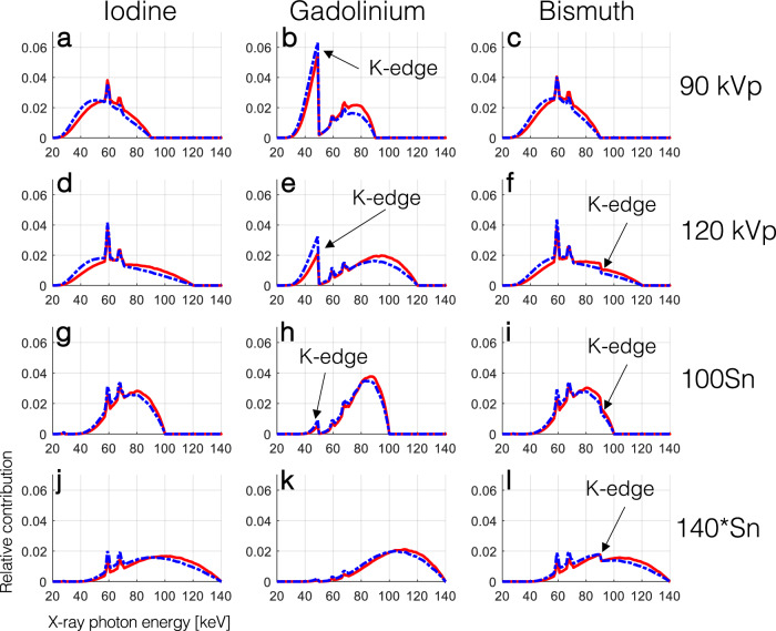
Methods: Pairs of test tubes were filled with dense CA (iodine-, gadolinium-, and bismuth-based) and placed inside a water phantom. The phantoms were scanned on EID- and PCD-CT systems, at all available tube voltages for the PCD scanner. Images were reconstructed with standard water BH correction but without any iodine/bone BH corrections. Virtual monoenergetic images (VMI) were calculated from PCD-CT data.
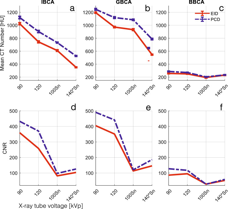
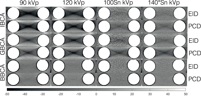
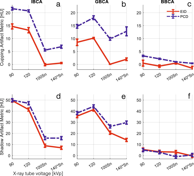
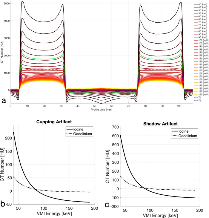
Results: PCD-CT had higher CT numbers in all x-ray spectra for all CAs (p < 0.001) and produced larger cupping artifacts in all test cases (p < 0.001). Bismuth-based CA artifacts were 3- to 5-fold smaller than those of iodine- or gadolinium-based CA. PCD-CT-based VMI completely removed iodine BH artifacts. Iodine BH artifacts decreased with increasing tube voltage. However, gadolinium-based BH artifacts had a different trend increasing at 120 kVp.
Conclusion: EID had fewer BH artifacts compared to PCD at x-ray tube voltages of 120 kVp and higher. The inherent spectral information of PCDs can be used to eliminate BH artifacts. Special care is needed to correct BH artifacts for gadolinium- and bismuth-based CAs.
Relevance statement: With the increasing availability of clinical photon-counting CT systems offering the possibility of dual contrast imaging capabilities, addressing and comprehending the BH artifacts attributed to old and novel CT CAs grows in research and ultimately clinical relevance.
Key points: EID-CT provides fewer BH artifacts compared to PCD-CT at x-ray tube voltages of 120 kVp and higher. K-edge CAs, such as those based on gadolinium, further confound BH artifacts. The inherent spectral information of photon counting detector CT can be used to effectively eliminate BH artifacts.
Keywords: Artifacts; Bismuth; Gadolinium; Iodine; Tomography (x-ray computed).
© 2025. The Author(s).
Conflict of interest statement
Declarations. Ethics approval and consent to participate: Not applicable. Consent for publication: Not applicable. Competing interests: AP, MvA, and TE are members of the scientific editorial board for European Radiology Experimental (sections: cardiovascular, artificial intelligence, augmented reality, computer science and radiomics, and section editor computed tomography). AV-S is a Deputy Editor for European Radiology Experimental. These authors have not participated in the selection nor review processes for this article. AP, UJS, TE, and AV-S have an active sponsored research agreement with Siemens Healthineers. Other authors declare they do not have any competing interests.
Figures
Similar articles
-
Energy-integrating-detector multi-energy CT: Implementation and a phantom study.Med Phys. 2021 Sep;48(9):4857-4871. doi: 10.1002/mp.14943. Epub 2021 Jul 29. Med Phys. 2021. PMID: 33988849 Free PMC article.
-
Performance evaluation of single- and dual-contrast spectral imaging on a photon-counting-detector CT.Med Phys. 2024 Nov;51(11):8034-8046. doi: 10.1002/mp.17367. Epub 2024 Sep 5. Med Phys. 2024. PMID: 39235343
-
Multi-energy CT imaging for large patients using dual-source photon-counting detector CT.Phys Med Biol. 2020 Aug 31;65(17):17NT01. doi: 10.1088/1361-6560/ab99e4. Phys Med Biol. 2020. PMID: 32503022 Free PMC article.
-
An introduction to photon-counting detector CT (PCD CT) for radiologists.Jpn J Radiol. 2023 Mar;41(3):266-282. doi: 10.1007/s11604-022-01350-6. Epub 2022 Oct 18. Jpn J Radiol. 2023. PMID: 36255601 Free PMC article. Review.
-
Photon-Counting Detector CT: Key Points Radiologists Should Know.Korean J Radiol. 2022 Sep;23(9):854-865. doi: 10.3348/kjr.2022.0377. Korean J Radiol. 2022. PMID: 36047540 Free PMC article. Review.
Cited by
-
Editorial: Photon counting CT technology in cardiovascular imaging.Front Cardiovasc Med. 2025 Jun 27;12:1641175. doi: 10.3389/fcvm.2025.1641175. eCollection 2025. Front Cardiovasc Med. 2025. PMID: 40656771 Free PMC article. No abstract available.
-
Photon-counting detector CT: a disrupting innovation in medical imaging.Eur Radiol Exp. 2025 Mar 25;9(1):38. doi: 10.1186/s41747-025-00571-4. Eur Radiol Exp. 2025. PMID: 40131583 Free PMC article.
-
Photon-counting CT in cancer radiotherapy: technological advances and clinical benefits.Phys Med Biol. 2025 May 16;70(10):10TR01. doi: 10.1088/1361-6560/add4ba. Phys Med Biol. 2025. PMID: 40328288 Free PMC article. Review.
References
-
- Barrett JF, Keat N (2004) Artifacts in CT: recognition and avoidance. Radiographics 24:1679–1691. 10.1148/rg.246045065 - PubMed
-
- Willemink MJ, Persson M, Pourmorteza A, Pelc NJ, Fleischmann D (2018) Photon-counting CT: technical principles and clinical prospects. Radiology 289:293–312. 10.1148/radiol.2018172656 - PubMed
-
- Sandfort V, Persson M, Pourmorteza A, Noel PB, Fleischmann D, Willemink MJ (2021) Spectral photon-counting CT in cardiovascular imaging. J Cardiovasc Comput Tomogr 15:218–225. 10.1016/j.jcct.2020.12.005 - PubMed
Publication types
MeSH terms
Substances
LinkOut - more resources
Full Text Sources
Medical