. 2025 Mar 21;11(12):eadq0558.
doi: 10.1126/sciadv.adq0558. Epub 2025 Mar 19.

Structural basis for the substrate recognition and transport mechanism of the human y+LAT1-4F2hc transporter complex

Affiliations

Structural basis for the substrate recognition and transport mechanism of the human y+LAT1-4F2hc transporter complex

Lu Dai et al. Sci Adv. .

Abstract

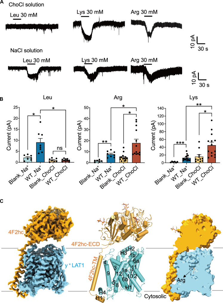
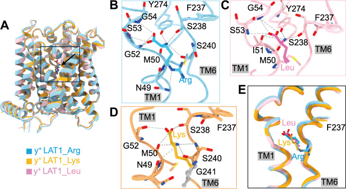
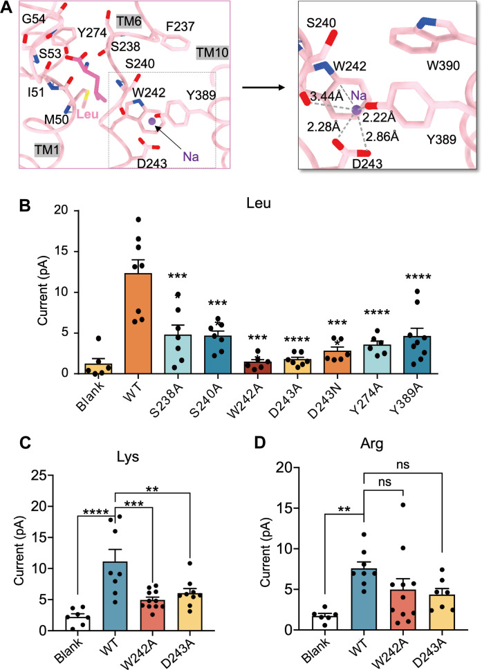
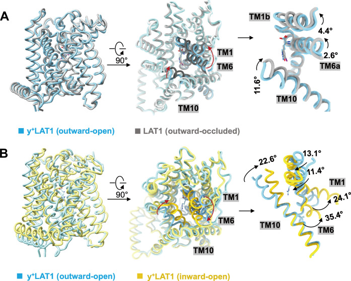
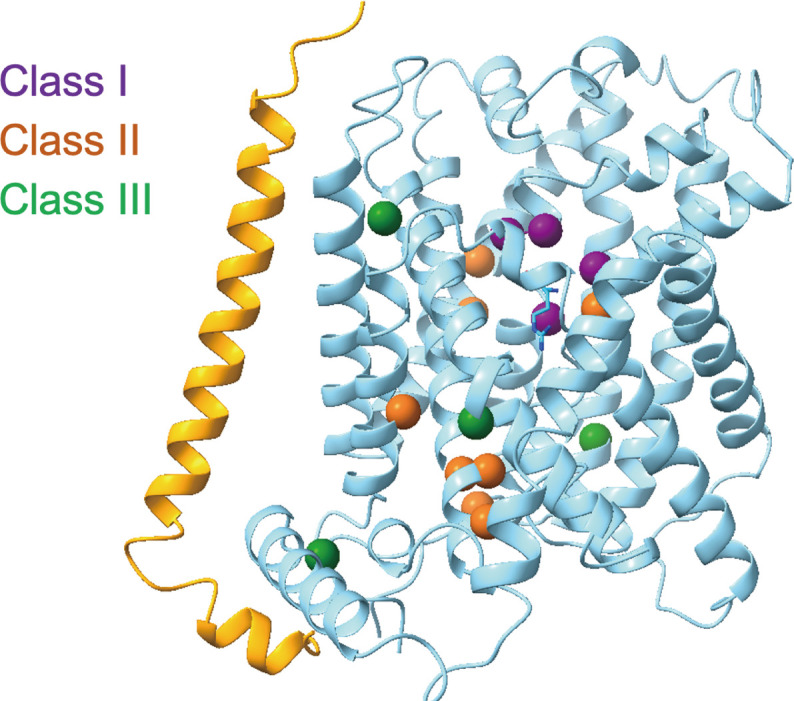
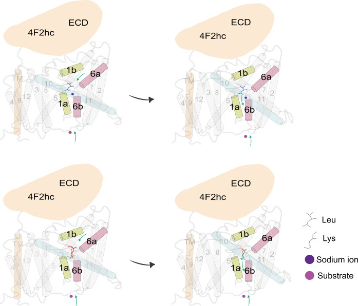
Heteromeric amino acid transporters (HATs), including y+LAT1-4F2hc complex, are responsible for transporting amino acids across membranes, and mutations in y+LAT1 cause lysinuric protein intolerance (LPI), a hereditary disorder characterized by defective cationic amino acid transport. The relationship between LPI and specific mutations in y+LAT1 has yet to be fully understood. In this study, we characterized the function of y+LAT1-4F2hc complex in mammalian cells and determined the cryo-EM structures of the human y+LAT1-4F2hc complex in two distinct conformations: the apo state in an inward-open conformation and the native substrate-bound state in an outward-open conformation. Structural analysis suggests that Asp243 in y+LAT1 plays a crucial role in coordination with sodium ion and substrate selectivity. Molecular dynamic (MD) simulations further revealed the different transport mechanism of cationic amino acids and neutral amino acids. These results provide important insights into the mechanisms of the substrate binding and working cycle of HATs.


Figures

Fig. 1.
Fig. 1.. Overall structures of y+LAT1-4F2hc complex.
(A and B) The absence of sodium ions in extracellular solution hampers the transport of Leu and enhances the transport of Lys and Arg. *P < 0.05, **P < 0.01, and ***P < 0.001, one-way analysis of variance (ANOVA) followed by Tukey’s test, data are presented as mean ± SEM. (C) Cartoon structure of the y+LAT1-4F2hc Arg-bound (blue) and y+LAT1 adopts an outward-open conformation with Arg in the center of the substrate binding pocket. ns, not significant.
Fig. 2.
Fig. 2.. Three-substrate binding analysis.
(A) The overall structure of y+LAT1 bound with three different substrates. The direction indicated by the black arrow in the box is the substrate binding site. (B to D) The interaction networks of Arg (blue), Leu (pink), and Lys (orange) with y+LAT1 are shown. (E) Structural comparison of the substrate binding sites of y+LAT1 with Arg, Leu, and Lys. Arg, Leu, and Lys are colored blue, pink, and orange, respectively. The gray dashed lines represent hydrogen bonds, and the gray amino acids are involved in the hydrophobic interactions.
Fig. 3.
Fig. 3.. Na+ binding site in the y+LAT1-4F2hc Leu-bound structure.
(A) The interaction interface of the Na+ binding site and the polar interactions are shown as gray dashes. Leu is displayed as bright pink, and the sodium ion is displayed as purple. (B) Key residue mutations inhibit the transport activity of y+LAT1, currents induced by 30 mM Leu. ****P < 0.0001, compared with WT-y+LAT1 and calculated by one-way ANOVA followed by Tukey’s test. Data present mean ± SEM. (C) W242A and D243A mutations inhibit the transport activity of y+LAT1. Currents were induced by 30 mM Lys perfusion. ***P < 0.001 and **P < 0.01 compared with WT-y+LAT1, calculated by one-way ANOVA followed by Tukey’s test. (D) W242A and D243A mutations have no effect on the transport activity of y+LAT1. Currents were induced by 30 mM Arg perfusion; not significant compared with WT-y+LAT1, calculated by one-way ANOVA followed by Tukey’s test. Data are presented as mean ± SEM.
Fig. 4.
Fig. 4.. MD simulations for the Na+ binding site.
(A) The interaction energy scan for a Na+ and indole was obtained using DLPNO-CCSD(T) methods with aug-cc-pVTZ basis set (green line) in comparison with that using C36m force field (purple line) by varying the distance at the direction perpendicular to the indole six-membered ring. The dashed pink line represents the NBFIX modification to C36m that restores the QM cation-π interactions between the sodium ion and indole. (B) The distance between Na+ and the minimum carboxyl oxygen of Asp243, the hydroxyl oxygen of Tyr389, backbone oxygen of Ser240, and the six-membered ring center of Trp242 during the simulation with NBFIX modification. (C) Snapshot of the simulation with NBFIX modification for the sodium binding pocket. The two water molecules coordinated with Na+ were also highlighted.
Fig. 5.
Fig. 5.. The conformational change of y+LAT1.
(A and B) Structural comparison among the y+LAT1-4F2hc bound with Arg (blue), the outward-occluded structure of LAT1 bound with 3,5-diiodo-L-tyrosine (gray), and the inward-open structure of y+LAT1 apo (yellow). The movements of TM1 and TM6 are shown in the middle and right, respectively.
Fig. 6.
Fig. 6.. LPl-related mutations mapping in the y+LAT1.
LPI-related mutations mapping in the complex, which can be classified into classes I, II, and III that are colored in purple, orange, and green, respectively.
Fig. 7.
Fig. 7.. Putative working model for the y+LAT1-4F2hc complex.
The model shows a transport mechanism of y+LAT1. y+LAT1 loads extracellular substrates, and then TM1b and TM6a rotate to close the outward gate. During the transition from the outward-open conformation to the outward-occluded conformation, the rotation of TM1b and TM6a triggers the close of the gating residue Phe237 to the occluded configuration. At the same time, the TM10 are also involved in an obvious shift. ECD, extracellular domain of 4F2hc.

References

    1. Bröer S., Palacin M., The role of amino acid transporters in inherited and acquired diseases. Biochem. J. 436, 193–211 (2011). - PubMed
    1. Taylor P. M., Role of amino acid transporters in amino acid sensing. Am. J. Clin. Nutr. 99, 223S–230S (2014). - PMC - PubMed
    1. Kandasamy P., Gyimesi G., Kanai Y., Hediger M. A., Amino acid transporters revisited: New views in health and disease. Trends Biochem. Sci. 43, 752–789 (2018). - PubMed
    1. Fotiadis D., Kanai Y., Palacin M., The SLC3 and SLC7 families of amino acid transporters. Mol. Aspects Med. 34, 139–158 (2013). - PubMed
    1. Mastroberardino L., Spindler B., Pfeiffer R., Skelly P. J., Loffing J., Shoemaker C. B., Verrey F., Amino-acid transport by heterodimers of 4F2hc/CD98 and members of a permease family. Nature 395, 288–291 (1998). - PubMed

MeSH terms

Substances