. 2025 Mar 21;11(12):eads2923.
doi: 10.1126/sciadv.ads2923. Epub 2025 Mar 19.

Transfer RNA acetylation regulates in vivo mammalian stress signaling

Affiliations

Transfer RNA acetylation regulates in vivo mammalian stress signaling

Supuni Thalalla Gamage et al. Sci Adv. .

Abstract

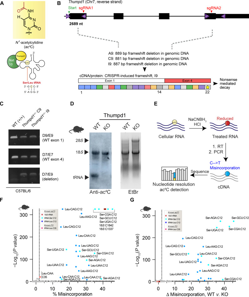
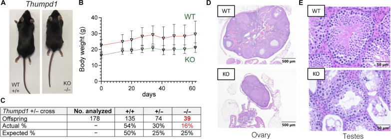
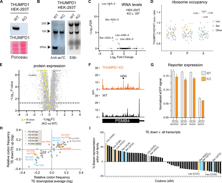
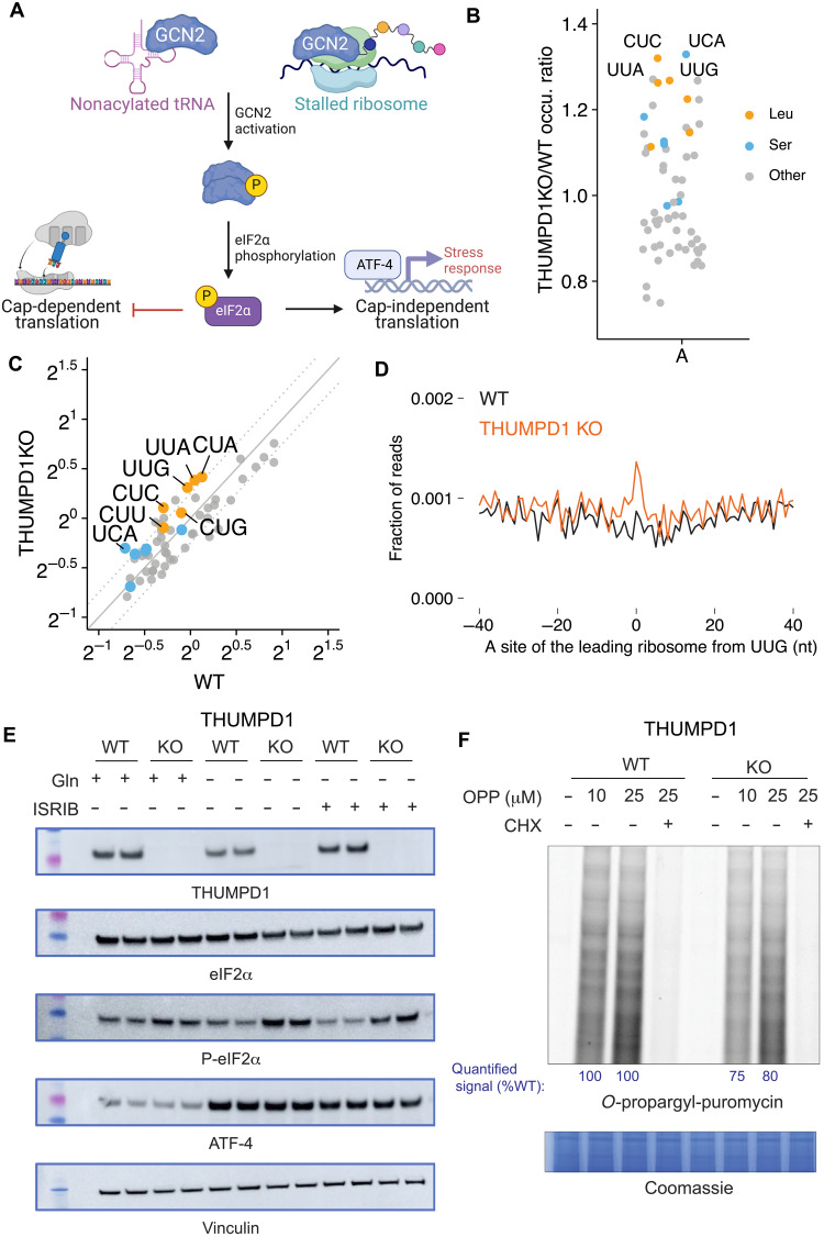
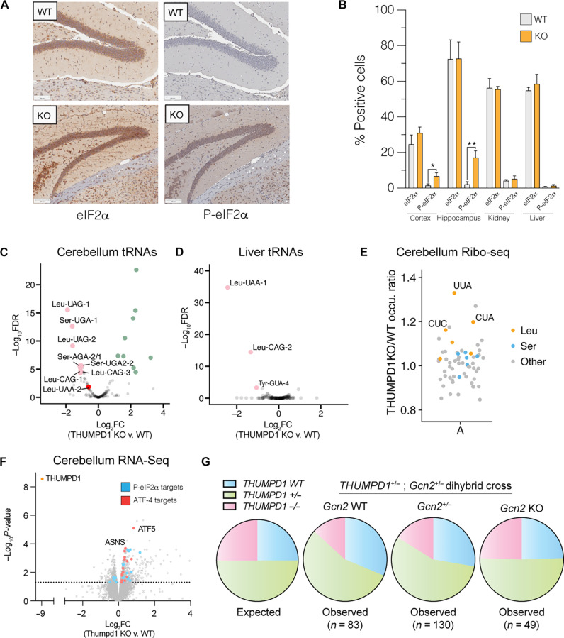
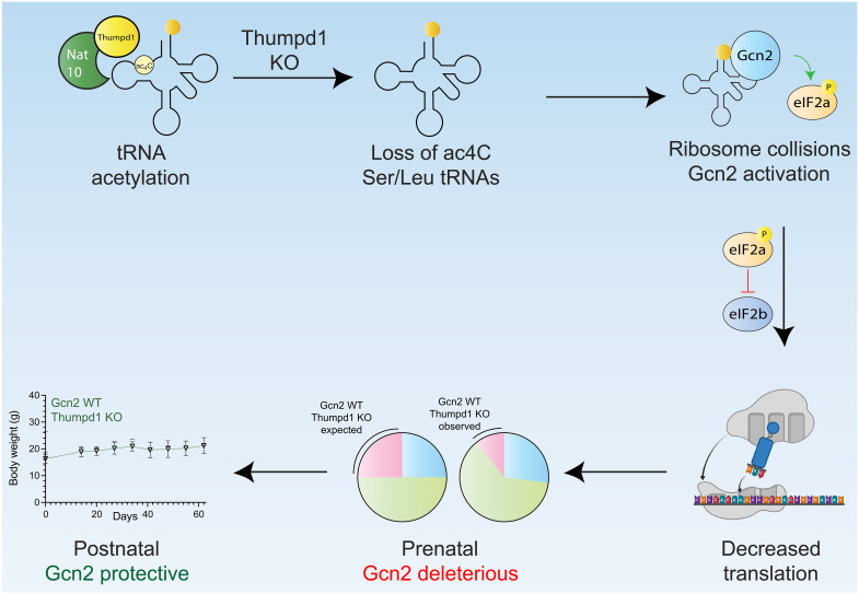
Transfer RNA (tRNA) modifications are crucial for protein synthesis, but their position-specific physiological roles remain poorly understood. Here, we investigate the impact of N4-acetylcytidine (ac4C), a highly conserved tRNA modification catalyzed by the essential acetyltransferase Nat10. By targeting Thumpd1, a nonessential adapter protein required for Nat10-catalyzed tRNA acetylation, we determine that loss of tRNA acetylation leads to reduced levels of tRNALeu, increased ribosome stalling, and activation of eIF2α phosphorylation. Thumpd1 knockout mice exhibit growth defects and sterility. Concurrent knockout of Thumpd1 and the stress-sensing kinase Gcn2 causes penetrant postnatal lethality in mice, indicating a critical genetic interaction. Our findings demonstrate that a modification restricted to a single position within type II cytosolic tRNAs can regulate ribosome-mediated stress signaling in mammalian organisms, with implications for our understanding of translational control and therapeutic interventions.


Figures

Fig. 1.
Fig. 1.. In vivo model for studying mammalian tRNA acetylation.
(A) Deposition of the RNA modification ac4C in eukaryotic tRNA requires Nat10 and Thumpd1. (B) Schematic of murine Thumpd1 locus and CRISPR-Cas9 genome editing strategy. Multiple murine lines (A9, C9, and I9) were obtained, and genotypes were characterized by next-generation sequencing. (C) PCR-based genotyping confirms Thumpd1 deletion. (D) Immuno-Northern blotting confirms loss of ac4C in tRNA upon Thumpd1 KO. EtBr, ethidium bromide. (E) Schematic for ac4C-seq. (F) Distribution of ac4C in unfractionated murine tRNA-enriched total RNA. Data represent mean values from three independent mouse liver samples (n = 3 biological replicates). (G) Applying ac4C-seq to unfractionated total RNA isolated from WT or Thumpd1 KO mice highlights C12 in the D-arm of tRNALeu/Ser as the dominant position of Thumpd1-regulated RNA acetylation. Data represent mean values from three independent mouse liver samples (n = 3 biological replicates per genotype).
Fig. 2.
Fig. 2.. In vivo phenotypes associated with loss of Thumpd1 and tRNA acetylation.
(A) Thumpd1−/− mice are runted. (B) Thumpd1−/− mice are viable for several weeks. (C) Offspring produced in Thumpd1+/− breeding studies. (D) Ovarian atrophy and (E) testicular seminiferous tubule degeneration in Thumpd1−/− mice. Immunohistochemistry results are representative of n = 4 biological replicates per genotype.
Fig. 3.
Fig. 3.. Molecular characterization of THUMPD1/tRNA acetylation in mammalian cells.
(A) Western blot confirms THUMPD1 KO. Data are representative of n = 3 biological replicates. (B) Immuno-Northern blotting confirms loss of tRNA ac4C upon THUMPD1 KO (HEK-293T). Data are representative of n = 2 biological replicates. (C) mim-tRNA-seq analysis of WT v. THUMPD1 KO cells indicates the effect on tRNALeu/Ser isodecoders. Data are derived from n = 2 biological replicates. (D) Ribosome profiling of WT v. THUMPD1 KO cells indicates ribosome pausing at codons (UUG, CUU, CUA, and UUA) decoded by tRNALeu in the ribosomal A-site. Codons encoding Leu, Ser, and other amino acids are shown in blue, orange, and gray, respectively. Data are derived from n = 4 biological replicates. (E) Proteomic profiling of WT v. THUMPD1 KO cells. Genes showing decreased TE (TE down) are highlighted in yellow. Values derived from n = 3 biological replicates. (F) Ribosome footprints in PSMB4 gene indicates that stalling at Leu codons in KO cells does not limit downstream ribosome occupancy. (G) eGFP reporters with 3x copies of Leu or Ser codons do not produce significantly less protein in THUMPD1 KO cells. Data are derived from n = 3 technical replicates. (H) Scatterplot of codon frequency in mRNAs differentially translated in Thumpd1 KO cells. Global average refers to average codon usage of all CCDS-defined consensus coding sequences. Values shifted up/right indicate codons more frequent in TE down transcripts. Leu codons: orange; Ser codons: blue. (I) Analysis of amino acid family–specific codon bias in TE down transcripts. Representation of individual codons (e.g., Leu-UUA) relative to amino acid family (e.g., all Leu) was calculated. Average values for TE down sequences and all CCDS-defined coding sequences were compared. Leu codons; orange; Ser codons: blue. U/A-rich Leu codons are labeled on the x axis in red.
Fig. 4.
Fig. 4.. THUMPD1-mediated tRNA acetylation regulates ribosome collisions.
(A) Defective tRNA function can activate the sensor kinase GCN2 and trigger the ISR. (B) Disome occupancies of WT v. THUMPD1 KO cells at the ribosomal A-site of the leading ribosome indicate increased disome formation at codons (UCA, CUC, UUG, and UUA) decoded by tRNALeu/Ser. Codons encoding Ser, Leu, and other amino acids are shown in orange, blue, and gray, respectively. Data are representative of n = 2 biological replicates. (C) Scatterplot of disome occupancies at 61 sense codons in THUMPD1 KO v. WT cells indicate increased ribosome collisions at multiple codons decoded by tRNALeu/Ser. (D) Meta-codon plot centered at UUG codons shows increased ribosome collisions in THUMPD1 KO (orange) compared to WT cells (black). (E) Analysis of eIF2a phosphorylation in THUMPD1 KO cells. Amino acid deprivation (−Gln) and ISRIB (1 μM) were used to induce translational stress. Biological replicates are loaded in adjacent lanes. (F) Analysis of global translation in THUMPD1 KO cells. OPP was used to label nascent transcripts, which were then ligated to a fluorophore-azide and visualized via SDS-PAGE. Densitometry analysis was calculated using the ImageJ software and is graphed in fig. S7I. CHX, cycloheximide.
Fig. 5.
Fig. 5.. Thumpd1 and Gcn2 interact in vivo.
(A) IHC staining of total eIF2a (left) and (Ser51) P-eIF2a (right) in brain tissue isolated from age-matched WT and Thumpd1 KO mice. Data are representative of n = 4 biological replicates. (B) Quantification of percent positive cells in the cerebral cortex, hippocampus, kidney cortex, and liver. (C and D) mim-tRNA-seq analysis of tRNA levels in the cerebellum (C) and liver (D) of WT and Thumpd1 KO mice shows reduced levels of tRNALeu/Ser isodecoders. Green points represent nonacetylated tRNA species that showed significant increases in abundance not marked by ac4C. Data are derived from n = 2 biological replicates for each tissue. (E) A-site ribosome occupancies of cerebella isolated from WT and THUMPD1 KO mice indicate increased ribosome stalling at codons (UUA, CUA, and CUC) decoded by tRNALeu. Codons encoding Leu, Ser, and other amino acids are shown in orange, blue, and gray, respectively. Data are representative of n = 2 biological replicates. (F) Gene expression profiling of cerebella isolated from WT and Thumpd1 KO mice. Literature annotated targets of Atf4 (red) and P-eIF2a (blue) exhibit a skew toward greater expression upon Thumpd1 KO. Values represent the average of n = 4 biological replicates. (G) Analysis of offspring produced by Thumpd1+/−, Gcn2+/− dihybrid cross. Pie charts are organized by Gcn2 genotype, with WT on the left, heterozygotes in middle, and KO on the right. The proportion of mice born on a given background is color coded as follows: blue: Thumpd1+/+; green: Thumpd1+/−; pink: Thumpd1−/−. Full numbers for dihybrid cross are provided in fig. S11A.
Fig. 6.
Fig. 6.. Model for Thumpd1-mediated tRNA ac4C in mammalian ribosome function.
Nat10/Thumpd1 is responsible for C12 modification of tRNALeu and tRNASer. Genetic mutations and potentially other stimuli can cause a loss of ac4C from Ser/Leu tRNAs. This reduces the levels of tRNALeu/Ser isodecoders and possibly slows translocation, resulting in increased A-site occupancy at Leu/Ser codons and ribosome collisions. This activates the sensor kinase Gcn2, which causes tissue-specific phosphorylation of eIF2α. Cellular studies suggest that differential activation of Gcn2 may reflect the ability of tissues to access compensatory mechanisms upon ribosome collisions, for example, down-regulating ribosomal proteins. The sub-Mendelian production of Thumpd1−/− offspring is rescued by concurrent KO of Gcn2, suggesting that Thumpd1-dependent Gcn2 activation is deleterious during prenatal development. However, dual KO animals perish shortly after birth, suggesting that, in the postnatal setting, Gcn2 plays a protective role.

Update of

Similar articles

Cited by

References

    1. McCown P. J., Ruszkowska A., Kunkler C. N., Breger K., Hulewicz J. P., Wang M. C., Springer N. A., Brown J. A., Naturally occurring modified ribonucleosides. Wiley Interdiscip. Rev. RNA. 11, e1595 (2020). - PMC - PubMed
    1. Delaunay S., Helm M., Frye M., RNA modifications in physiology and disease: Towards clinical applications. Nat. Rev. Genet. 25, 104–122 (2024). - PubMed
    1. Wiener D., Schwartz S., The epitranscriptome beyond m6A. Nat. Rev. Genet. 22, 119–131 (2021). - PubMed
    1. Gamage S. T., Manage S. A. H., Chu T. T., Meier J. L., Cytidine acetylation across the tree of life. Acc. Chem. Res. 57, 338–348 (2024). - PMC - PubMed
    1. Ito S., Horikawa S., Suzuki T., Kawauchi H., Tanaka Y., Suzuki T., Suzuki T., Human NAT10 is an ATP-dependent RNA acetyltransferase responsible for N4-acetylcytidine formation in 18 S ribosomal RNA (rRNA). J. Biol. Chem. 289, 35724–35730 (2014). - PMC - PubMed

LinkOut - more resources