. 2025 Mar 19;15(1):9494.
doi: 10.1038/s41598-025-90896-2.

Insights into GLP-1 and insulin secretion mechanisms in pasireotide-induced hyperglycemia highlight effectiveness of Gs-targeting diabetes treatment

Affiliations

Insights into GLP-1 and insulin secretion mechanisms in pasireotide-induced hyperglycemia highlight effectiveness of Gs-targeting diabetes treatment

Junichiro Sato et al. Sci Rep. .

Abstract

Pasireotide frequently causes severe hyperglycemia; however, its detailed mechanism remains unknown. There are no published guidelines regarding the optimal management of pasireotide-induced hyperglycemia based on its pathophysiology. Herein, we successfully switched a patient with acromegaly from a dipeptidyl peptidase-4 (DPP-4) inhibitor to a glucagon-like peptide-1 (GLP-1) analog due to pasireotide-induced deterioration of glycemic control, and we examined the underlying mechanism for glycemic control. An in vitro study was conducted using pancreatic β-cell line, MIN-6, stably expressing GLP-1R (GLP-1R-MIN-6 cells) and intestinal L-cell line, GLUTag. High glucose levels and Gs-coupled receptor stimulation synergistically triggered insulin and GLP-1 secretion. Gs-coupled receptor stimulation primarily triggered GLP-1 secretion, which was amplified by high glucose levels in GLUTag cells. Pasireotide drastically inhibited GLP-1 secretion induced by Gs-coupled receptor stimulation through SSTR5-Gi-dependent inhibition of cAMP levels, suggesting that the main pathway was completely blocked. Furthermore, administering GLP-1 partially overcame the inhibitory effect of pasireotide in GLP-1R-MIN-6 cells, leading to a partial recovery of insulin secretion. The drastic inhibition of GLP-1 secretion via shutdown of the main pathway is the primary cause of pasireotide-induced hyperglycemia. GLP-1 analogs, rather than DPP-4 inhibitors, can spare pasireotide-induced depletion of endogenous GLP-1 and restore insulin secretion.

Keywords: GLP-1; GLP-1 analog; GPCRs; Insulin; L-cells; Pasireotide.


Conflict of interest statement

Declarations. Competing interests: The authors declare no competing interests.

Figures

Fig. 1
Fig. 1
mRNA expression analysis of mouse SSTR (mSSTR) in GLP-1R-MIN-6 cells and in GLUTag cells by RNA-seq and RT-PCR. (a) mRNA analysis of mouse SSTR subtypes 1–5 (Sstr1-5) in MIN-6 and GLUTag cells was conducted using RNA-Seq. A read count analysis using StringTie was performed to determine the relative abundance of genes. (b) mRNA expression analysis of Sstr1-5 in GLP-1R-MIN-6 cells was performed using RT-PCR. (c) mRNA expression analysis of Sstr1-5 in GLUTag cells was also performed using RT-PCR.
Fig. 2
Fig. 2
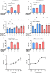
The main pathway for GLP-1 secretion is via cAMP signaling, and pasireotide inhibits GLP-1 secretion via SSTRs-Gi activation in GLUTag cells. (a, b) Effects of low glucose (0.1 mM) level or high glucose (25 mM) levels, DCA with low or high glucose levels, and L-glutamine (L-glu.) with low or high glucose levels on active GLP-1 secretion and cAMP accumulation in GLUTag cells. (c, d) Effects of low glucose level, triplet*, and DCA with or without PAS and/or PTX on active GLP-1 secretion and cAMP accumulation in GLUTag cells. PAS: pasirotide. PTX: pertussis toxin, an irreversible Gi inhibitor. The term “triplet*” refer to a high glucose (25 mM) level combined with 10 mM L-glutamine and 5 mM sodium butyrate, as indicated by the green square patterns. The blue and red square patterns indicate low glucose (0.1 mM) and high glucose (25 mM) levels, respectively. Values represent the mean ± SEM of triplicate experiments. Each set of results is representative of at least two additional experiments. The Y-axis shows the pM in the culture medium for active GLP-1 (a, c) and nM in the culture medium for cAMP (b, d) on the graphs. Data are expressed as means (SEM). The Tukey–Kramer multiple comparison test was conducted for statistical analysis. n.s., not significant; *P < 0.05; **P < 0.01; ***P < 0.001.
Fig. 3
Fig. 3
Pasireotide inhibits insulin secretion via SSTRs-Gi activation in GLP-1R-MIN-6 cells, and GLP-1 reverses the inhibitory action of pasiretoide on insulin secretion. (a, b) Effects of low glucose (2 mM) levels, high glucose (20 mM) levels, and GLP-1 with low or high glucose levels on insulin secretion and cAMP accumulation in GLP-1R-MIN-6 cells. (c, d) Effects of low glucose levels or high glucose levels with or without PAS and/or PTX on insulin secretion and cAMP accumulation in GLP-1R-MIN-6 cells. (e, f) The effects of GLP-1 on insulin secretion or cAMP accumulation in GLP-1R-MIN-6 cells incubated with high glucose levels and PAS. (g, h) Dose-dependent cAMP accumulation stimulated by GLP-1 and dulaglutide in GLP-1R-MIN-6 cells. The EC50 values were 31.0 nM in GLP-1 and 0.10 nM in dulaglutide. PAS: pasirotide. PTX: pertussis toxin, an irreversible Gi inhibitor. Values represent the mean ± SEM of n = 3 or n = 6 experiments. Each set of results is representative of at least two additional experiments. The Y-axis shows µg/L in the culture medium for insulin (a, c, e) and the nM in the culture medium for cAMP (b, d, f, g, h) on the graphs. Data are expressed as means (SEM). The Tukey–Kramer multiple comparison test was conducted for statistical analysis. n.s., not significant; *P < 0.05; **P < 0.01; ***P < 0.001.
Fig. 4
Fig. 4
PKA and EPAC signals via Gs activation partly increase GLP-1 secretion in GLUTag cells and insulin secretion in GLP-1R-MIN-6 cells.(a) Effects of DCA with or without H-89 and/or ESI-09 in the low-glucose (0.1 mM) state on active GLP-1 secretion in GLUTag cells. (b) Effects of 007-AM or 007-AM with ESI-09 in the low-glucose (2 mM) state on active GLP-1 secretion in GLUTag cells. (c) Effects of low (2 mM) or high glucose (20 mM) levels with or without GLP-1, H-89, and/or ESI-09 on insulin secretion in GLP-1R-MIN-6 cells. (d) Effects of 007-AM with low or high glucose levels, and the effect of ESI-09 on insulin secretion in GLP-1R-MIN-6 cells. H-89, is an inhibitor of protein kinase A. ESI-09, is an inhibitor of EPAC1 and EPAC2. 007-AM is a selective EPAC activator. The blue and red square patterns indicate low glucose (0.1 or 2 mM) and high glucose (20 mM) levels, respectively. Values represent the mean ± SEM of n = 3 or n = 6 determinations. Each set of results is representative of at least two additional experiments. The Y-axis shows pM in the culture medium for active GLP-1 (a, b) and µg/L in the culture medium for insulin (c, d) on the graphs. Data are expressed as means (SEM). The Tukey–Kramer multiple comparison test was conducted for statistical analysis. n.s., not significant; *P < 0.05; **P < 0.01; ***P < 0.001.
Fig. 5
Fig. 5
The detailed mechanism of pasireotide-induced hyperglycemia and its optimal management in relation to pathophysiology. Drastic inhibition of GLP-1 secretion through shutdown of the main pathway is the primary cause of pasireotide-induced hyperglycemia. GLP-1 analogs, rather than DPP-4 inhibitors, spare pasireotide-induced depletion of endogenous GLP-1 and restore insulin secretion.
Fig. 6
Fig. 6
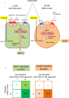
(a) Model of the molecular mechanisms involved in GLP-1 secretion from intestinal L-cells and insulin secretion from pancreatic β-cells. A schematic summary of the results. We demonstrated the commonality that GLP-1 from intestinal L-cells and insulin from pancreatic β-cells are synergistically regulated by Gs signaling and glucose. We also highlighted that the main pathways of GLP-1 and insulin secretion are different; Gs signaling in L-cells and glucose in β-cells. In intestinal L-cells, bile acids (acting on GPBAR1) and L-glutamine (acting on TAS1R3) stimulate GLP-1 secretion via protein kinase A and EPAC1/2 (the main pathway). A glucose-induced signal amplifies GLP-1 secretion (the amplification pathway). Suppression of adenylyl cyclase (AC) activity via SSTR2,5-Gi activation by pasireotide drastically inhibits GLP-1 secretion by shutting down the main pathway. In pancreatic β-cells, high glucose stimulates insulin secretion (the main pathway). GLP-1-amplified insulin secretion via protein kinase A and EPAC1/2 (the amplification pathway). Suppression of adenylyl cyclase (AC) activity via SSTRs-Gi activation by pasireotide inhibits insulin secretion, which can be overcome by GLP-1 analogs (b) Synergy between glucose and Gs-coupled GPCR agonist for GLP-1 and insulin secretion. We demonstrated that the main pathway can independently stimulate hormone secretion, even if the amplification pathway is suppressed (Gs activation is suppressed by PAS or in a low glucose state). In contrast, the amplification pathway cannot stimulate hormone secretion when the main pathway is significantly suppressed (Gs activation is suppressed by PAS or in a low glucose state). However, it can potentiate hormone secretion induced by the main pathway. PAS: pasireotide. Intestinal L-cells (left , drawn in green) . Gs-coupled GPCR agonists, such as bile acids or L-glutamine, can independently stimulate GLP-1 secretion in the low-glucose (0.1 mM) state (+, upper-right column). A high glucose (25 mM) level can stimulate GLP-1 secretion under “basal” Gs activation, that is, Gs-coupled receptor activation without agonists; however, it cannot stimulate GLP-1 secretion when Gs activation is significantly suppressed by pasireotide (−, lower-left column). However, a high glucose level can potentiate Gs-coupled GPCR-stimulated GLP-1 secretion (++, lower-right column). Pancreatic β-cells (right , drawn in orange) . A high glucose (20 mM) level can independently stimulate insulin secretion even when Gs activation is significantly suppressed by pasireotide (+, lower-left column). In contrast, Gs-coupled GPCR agonists such as GLP-1 cannot independently stimulate insulin secretion in the low-glucose (2 mM) state (−, upper-right column), but they can potentiate insulin secretion stimulated by high glucose levels (++, lower-right column).

References

    1. Iiri, T., Farfel, Z. & Bourne, H. R. G-protein diseases furnish a model for the turn-on switch. Nature394, 35–38. 10.1038/27831 (1998). - PubMed
    1. Lania, A. G., Mantovani, G. & Spada, A. Mechanisms of disease: mutations of G proteins and G-protein-coupled receptors in endocrine diseases. Nat. Clin. Pract. Endocrinol. Metab.2, 681–693. 10.1038/ncpendmet0324 (2006). - PubMed
    1. Makita, N. et al. Human G(salpha) mutant causes pseudohypoparathyroidism type Ia/neonatal diarrhea, a potential cell-specific role of the palmitoylation cycle. Proc. Natl. Acad. Sci. U S A. 104, 17424–17429. 10.1073/pnas.0708561104 (2007). - PMC - PubMed
    1. Vassart, G. & Costagliola, S. G protein-coupled receptors: mutations and endocrine diseases. Nat. Rev. Endocrinol.7, 362–372. 10.1038/nrendo.2011.20 (2011). - PubMed
    1. Schöneberg, T. & Liebscher, I. Mutations in G protein-coupled receptors: mechanisms, pathophysiology and potential therapeutic approaches. Pharmacol. Rev.73, 89–119. 10.1124/pharmrev.120.000011 (2021). - PubMed

MeSH terms