. 2025 Aug;104(9):983-992.
doi: 10.1177/00220345251323869. Epub 2025 Mar 19.

Potential of High-Intensity Focused Ultrasound in Enamel Remineralization

Affiliations

Potential of High-Intensity Focused Ultrasound in Enamel Remineralization

B Shrestha et al. J Dent Res. 2025 Aug.

Abstract

Remineralization is an essential interventional strategy for intercepting enamel white spot lesions (WSLs). Given the limitations of both natural and/or fluoride-mediated repair processes, there is a need to develop novel strategies for repairing enamel WSLs via a minimally invasive approach while restoring the unique ultrastructural integrity and functional properties. Inspired by the unique capability of high-intensity focused ultrasound (HIFU) in facilitating the crystallization process, we propose a novel strategy of employing HIFU for in vitro repair of WSLs through synergizing the crystallization process required for hydroxyapatite (HAP) formation from its precursor (calcium phosphate ion clusters; CPICs). Following CPIC formulation and characterization including the resultant amorphous calcium phosphate (ACP), the effect of HIFU on the ACP-to-HAP transition on the amorphous substrate was investigated using transmission electron microscopy and high-resolution transmission electron microscopy, selected area electron diffraction, and X-ray diffraction (XRD). The results showed profound amorphous-to-crystalline phase transition, within 5- to 30-min HIFU exposure, whereas the long axis of the resultant HAP corresponded with the (002) plane, and a lattice spacing of 0.34 nm indicated a preferred c-axis growth direction consistent with the orientation of natural enamel crystallites. For enamel repair, artificial WSLs were created on enamel specimens and then subjected to CPICs, followed by HIFU exposure for 2.5, 5, or 10 min. Scanning electron and atomic force microscopies revealed the decreased surface roughness and the gradual obliteration in the WSL porous structure with continuous linear coaxial arrangement of HAP crystallites filling the prismatic/interprismatic gaps closely resembling sound enamel specifically with 5-min HIFU exposure. Enamel WSL ultrastructural repair was further confirmed from XRD and Raman spectral analyses with the associated regaining of mineral density and nanomechanical properties as reflected from micro-computed tomography (CT) and nanoindentation results, respectively. Micro-CT further validated the subsurface remineralization of WSLs with HIFU exposure. Within the same exposure parameters, HIFU exhibited a potent antibiofilm effect against Streptococcus mutans. This study introduced a new approach for remineralizing enamel WSLs through the potent synergy between HIFU and CPICs.

Keywords: biomaterials; cariogenic biofilms; enamel repair; enamel white spot lesions; minimal invasive dentistry; ultrasonic energy.


Conflict of interest statement

Declaration of Conflicting InterestsThe authors declared no potential conflicts of interest with respect to the research, authorship, and/or publication of this article.

Figures

Figure 1.
Figure 1.
Schematic demonstration of the proposed concept of the potential of high-intensity focused ultrasound (HIFU) in synergizing the remineralization process of artificially simulated enamel carious lesion (white spot lesion; WSL) coupled with calcium phosphate ionic clusters (CPICs), along with its associated drug-free antibiofilm effect. (A) Mineral loss from the tooth enamel due to a reduction in pH associated with an acidic oral environment induced by cariogenic bacterial biofilms. (B) Scanning electron microscopy image showing the structure and orientation of hydroxyapatite (HAP) crystallites in sound tooth enamel. (C) Continuous exposure to an acidic environment resulting in mineral loss from HAP, affecting its enamel prismatic structure and leading to the formation of WSLs. (D) Schematic presentation summarizing the proposed dual action of HIFU in synergizing the remineralization of enamel WSL and eradicating the associated cariogenic biofilm. (E) The acoustic cavitation event generated by HIFU results in both chemical and physical effects (including the formation of highly reactive radical species, shockwaves, and microjets) enabling the accelerated formation of HAP crystallites in the presence of CPICs for repairing enamel WSLs (F). Schematic presentation (G) and confocal laser scanning microscopy images (H) showing the drug-free eradication of cariogenic biofilms attached to the enamel surface through HIFU’s mechanical effect.
Figure 2.
Figure 2.
Schematic diagram representing the experimental setup for investigating the synergizing effect of high-intensity focused ultrasound (HIFU) and calcium phosphate ion clusters (CPICs) on enamel remineralization along with characterizing the antibiofilm effect of HIFU on Streptococcus mutans. (A) Illustration of the methodology for creating an artificially simulated white spot lesion (WSL). (C) Application of synthesized CPICs on the artificially created enamel WSL, followed by subjecting the specimens to HIFU with 250 KHz operated at 30 W (continuous mode) to evaluate the synergistic effect of HIFU and CPICs for remineralizing WSL enamel (E). (B, D) Cultivation of S. mutans biofilm on the WSL enamel, followed by HIFU exposure for different time points (F). (G) HIFU components and attachments including a bowl-shaped piezoelectric ceramic transducer with a resonance frequency of 250 KHz, attached to a transparent polycarbonate coupling cone for acoustic delivery, along with HIFU transducer specifications (H).
Figure 3.
Figure 3.
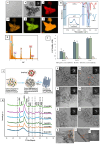
Transmission electron microscopy (TEM) images showing the morphology of the synthesized (A) calcium phosphate ion cluster (CPIC) nanoparticles and (B) amorphous calcium phosphate (ACP) resulting from the fusion of CPICs (scale bars 200 nm). Inset in (B): selected area electron diffraction (SAED) of the amorphous ACP. (C–G) TEM–energy dispersive spectroscopy mapping and elemental analysis of ACP nanoparticles confirming the presence of calcium, phosphorous, and oxygen, respectively. (H) The acquired Fourier transform infrared spectra of synthesized CPICs (red spectrum) and ACP nanoparticles (blue spectrum). Inset in (H): X-ray diffraction of ACP reconfirming the amorphous phase. (I) Acid phosphatase assay of oral fibroblasts against formulated CPIC nanoparticles at different time points (data are presented as mean and SD; dissimilar letters indicate statistical difference). (J) Schematic demonstration of mineral phase transformation leading to hydroxyapatite (HAP) formation facilitated by high-intensity focused ultrasound (HIFU) exposure. (K) X-ray diffraction patterns of the HIFU-exposed ACP specimens showing the major diffraction peaks observed with increasing exposure time identified as the (002), (211), (300), (310), (222), (213), and (004) planes confirming the formation of crystalline HAP. (L–Q) Corresponding TEM images: (L) no structural changes were observed with the amorphous ACP nanoparticles without HIFU exposure. (M) Following 2.5 min of HIFU exposure, chainlike aggregates begin to partially transform into a needlelike structure. (N–Q) After 5 to 30 min of HIFU exposure, more well-defined needlelike particles became more visible. The respective TEM-SAED (insets) showed an increase in the intensity of the diffraction rings with an increase in HIFU exposure time confirming the transformation of the amorphous precursor to the crystalline phase. (R, S) High-resolution TEM (HR-TEM) images and (T) the associated fast Fourier transform (FFT) of the formed HAP following 30 min of HIFU exposure showing that the long axis of the nanorod corresponds with the (002) plane, with lattice spacing of 0.34 nm. Scale bar 200 nm for TEM images (L–Q), 5 1/nm for TEM-SAED, and 50 and 20 nm for HR-TEM (R, S).
Figure 4.
Figure 4.
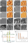
(A–G) Scanning electron microscopy (SEM) and (A1–F1) atomic force microscopy (AFM) images of the enamel specimens. (A, A1) Enamel with untreated white spot lesion (WSL; DE) displayed a highly porous surface and interprismatic gaps. (B, B1) Enamel WSL exposed to high-intensity focused ultrasound (HIFU) alone for 10 min demonstrated close morphological similarities to untreated WSL. (C, C1) The application of calcium phosphate ion clusters (CPICs) alone resulted in a smoother surface texture and formation of an amorphous calcium phosphate (ACP) coating layer masking the underlying prismatic and interprismatic structure. Enamel treated with CPICs/HIFU for 2.5 (D, D1), 5 (E, E1), and 10 min (F, F1) exposure time, respectively, revealed a gradual obliteration of the typical prismatic–interprismatic structure. (G) Sound enamel. (H–N) The corresponding cross-sectional SEM images of the enamel specimens. (H) Sound enamel showing a well-defined longitudinally oriented prismatic structure with closely packed rodlike hydroxyapatite (HAP) crystallites extending to the outermost surface layer. (I) Untreated WSL displayed a structureless, porous, disorganized surface layer with no defined orientation. More or less similar morphological features to the untreated WSL (I) were found following HIFU exposure alone (J), CPIC application alone (K), and CPIC/HIFU exposure for 2.5 min (L). A well-defined longitudinally oriented prismatic structure having a rodlike morphological appearance was observed with HIFU exposure for 5 min (M) and 10 min (N) following CPIC application. Specifically, CPICs followed by HIFU exposure for 5-min treatment (M) demonstrated a continuous organized linear coaxial arrangement of closely packed HAP crystallites. (O) X-ray diffraction (XRD) spectral analyses of enamel specimens. The blue spectrum represents untreated WSL (DE). Specimens treated with CPICs alone (orange spectrum) showed the formation of an amorphous ACP layer reflected from the broad hump in the XRD pattern at the 15 to 30.15° 2θ position (inset). However, following subsequent HIFU exposure for 5 min (green spectrum), the reappearance of a sharp peak within the same 2θ position inferred the conversion of amorphous ACP to crystalline HAP. (P) Mean roughness value (Sa) showing the gradual decrease in surface roughness with the increase in HIFU exposure time (error bars represent SD; dissimilar letters indicate statistically differences). SEM scale bars: 10 µm.
Figure 5.
Figure 5.
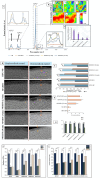
(A) Raman spectral analysis showing the intensity increase of the characteristic phosphate peaks (v1 PO43−, v2 PO43−, v4 PO43−) in the experimental groups treated with calcium phosphate ion cluster nanoparticles (NP) followed with high-intensity focused ultrasound (HIFU) for 2.5, 5, and 10 min compared with untreated white spot lesion (WSL; DE), indicating an increase in hydroxyapatite (HAP) content. (B) Raman mapping at 960 cm−1: (a) WSL showing a decrease in 960 cm−1 peak intensity distribution, indicating a reduction in HAP content; (b) NP/-HIFU treated group; and (c–e) the experimental groups (NP/ HIFU [2.5 min], NP/HIFU [5 min], NP/HIFU [10 min]), respectively. The highest HAP density distribution as represented by the red color was shown by the NP/HIFU (5 min) and (10 min) groups. (C) Bar chart demonstrating the ratio of the integrated area under the bands of 1,070 cm−1 and 960 cm−1 showing a significant decrease with the increase in HIFU exposure time depicting the decrease in the carbonate content. (D) Representative cross-sectional 2-dimensional micro-computed tomography images of enamel specimens before and after remineralization (scale bars: 300 µm). The orange arrows indicate the difference in the gray-scale intensity. (E) Bar chart showing the variation of mineral density represented by (Hounsfield unit) of each treated group in relevance to their respective sound enamel (SE) and WSL (DE). (F) Bar chart showing the percentage of the remineralization rate between the different groups. The NP/HIFU (5-min) group exhibited the highest recovery rate (69%) compared with all other experimental groups. (G) Bar chart showing the variation in the lesion depth after remineralization interventions. Bar charts showing the variations in (H) nanohardness and (I) reduced elastic modulus. Experimental groups (NP/HIFU [2.5 min], NP/HIFU [5 min], NP/HIFU [10 min]) showed a significant increase in nanohardness and reduced elastic modulus as compared with the respective WSL control. *Significant difference; ns, no difference. Graphs show mean values and SD; dissimilar letters represent significant difference.

Similar articles

References

    1. Aati S, Shrestha B, Fawzy A. 2022. Cytotoxicity and antimicrobial efficiency of ZrO2 nanoparticles reinforced 3D printed resins. Dent Mater. 38(8):1432–1442. - PubMed
    1. Akkus A, Karasik D, Roperto R. 2017. Correlation between micro-hardness and mineral content in healthy human enamel. J Clin Exp Dent. 9(4):e569. doi: 10.4317/jced.53345 - DOI - PMC - PubMed
    1. Bang JH, Suslick KS. 2010. Applications of ultrasound to the synthesis of nanostructured materials. Adv Mater. 22(10):1039–1059. - PubMed
    1. Bubnowicz L, França R. 2022. Calcium and fluoride interaction in human enamel at nanoscale: an XPS assessment. Appl Surf Sci Adv. 11:100306. doi: 10.1016/j.apsadv.2022.100306 - DOI
    1. Carden A, Morris MD. 2000. Application of vibrational spectroscopy to the study of mineralized tissues (review). J Biomed Opt. 5(3):259–268. - PubMed

LinkOut - more resources