Review
. 2025 Mar 5:13:1460416.
doi: 10.3389/fcell.2025.1460416. eCollection 2025.

Exosomes in cartilage microenvironment regulation and cartilage repair

Affiliations
Review

Exosomes in cartilage microenvironment regulation and cartilage repair

Han Longfei et al. Front Cell Dev Biol. .

Abstract

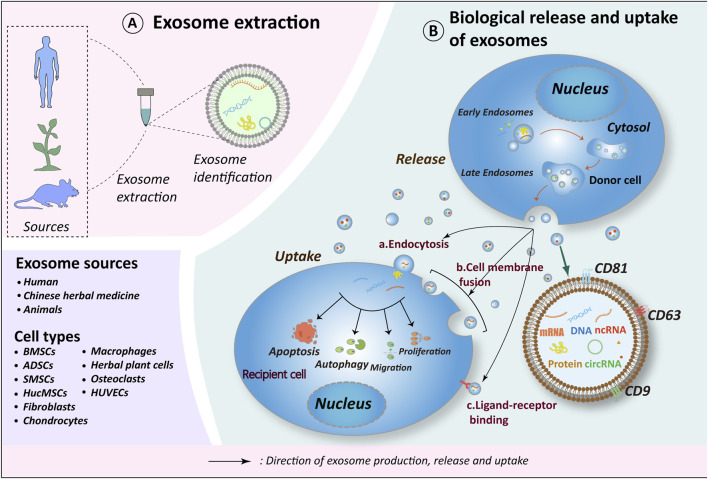
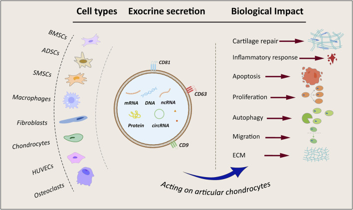
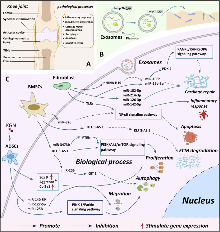
Osteoarthritis (OA) is a debilitating disease that predominantly impacts the hip, hand, and knee joints. Its pathology is defined by the progressive degradation of articular cartilage, formation of bone spurs, and synovial inflammation, resulting in pain, joint function limitations, and substantial societal and familial burdens. Current treatment strategies primarily target pain alleviation, yet improved interventions addressing the underlying disease pathology are scarce. Recently, exosomes have emerged as a subject of growing interest in OA therapy. Numerous studies have investigated exosomes to offer promising therapeutic approaches for OA through diverse in vivo and in vitro models, elucidating the mechanisms by which exosomes from various cell sources modulate the cartilage microenvironment and promote cartilage repair. Preclinical investigations have demonstrated the regulatory effects of exosomes originating from human cells, including mesenchymal stem cells (MSC), synovial fibroblasts, chondrocytes, macrophages, and exosomes derived from Chinese herbal medicines, on the modulation of the cartilage microenvironment and cartilage repair through diverse signaling pathways. Additionally, therapeutic mechanisms encompass cartilage inflammation, degradation of the cartilage matrix, proliferation and migration of chondrocytes, autophagy, apoptosis, and mitigation of oxidative stress. An increasing number of exosome carrier scaffolds are under development. Our review adopts a multidimensional approach to enhance comprehension of the pivotal therapeutic functions exerted by exosomes sourced from diverse cell types in OA. Ultimately, our aim is to pinpoint therapeutic targets capable of regulating the cartilage microenvironment and facilitating cartilage repair in OA.

Keywords: cartilage microenvironment; cartilage repair; exosomes; mechanism of action; osteoarthritis.


Conflict of interest statement

The authors declare that the research was conducted in the absence of any commercial or financial relationships that could be construed as a potential conflict of interest.

Figures

FIGURE 1
FIGURE 1
Exosome biorelease and uptake process. (A) Exosomes originate from diverse sources, with current research emphasizing those derived from human tissues, plants (including herbs), and animal tissues. The cell types of exosomes exhibiting therapeutic potential for OA include BMSCs, ADSCs, SMSCs, HucMSCs, Fibroblasts, Chondrocytes, Macrophages, Osteoclasts, HUVECs and herbs. (B) The biogenesis of exosomes occurs in four distinct stages: 1. Endocytosis, during which the cell membrane invaginates to form early endosomes; 2. Packaging of contents, wherein early endosomes further mature into late endosomes; 3. Fusion of late endosomes to form multivesicular bodies (MVBs); 4. MVBs subsequently fuse with the cell membrane to release their contents into the extracellular compartment, thereby forming exosomes. Recipient cells internalize exosomes via endocytosis, direct fusion, or ligand-receptor interactions. Through the release of materials encapsulated within exosomes (e.g., proteins, nucleic acids, lipids, metabolites), exosomes exert influence on the metabolic processes of recipient cells, including cell proliferation, migration, apoptosis, and autophagy, thereby further impacting the OA process. Schematic drawing reference from Chen J. et al. (2022). BMSCs, bone marrow mesenchymal stem cells; ADSCs, adipose mesenchymal stem cells; SMSCs, synovial mesenchymal stem cells; HucMSCs, human umbilical cord mesenchymal stem cells; HUVECs, human umbilical vein endothelial cells.
FIGURE 2
FIGURE 2
Flowchart and summary of exosomes from different sources. BMSCs, bone marrow mesenchymal stem cells; ADSCs, adipose-derived mesenchymal stem cells; SMSCs, synovial mesenchymal stem cells; HUVECs, human umbilical vein endothelial cells; ECM, extracellular matrix; mRNA, coding RNA; DNA, deoxyribonucleic acid; ncRNA, non-coding RNA; circRNA, circular RNA.
FIGURE 3
FIGURE 3
Summary of cartilage microenvironment regulation and cartilage repair mechanisms by different cellular exosomes. (A) A depicts the observed changes in injury and knee joint structure in knee OA cases, offering insights into the structural impacts of the condition (B) B illustrates the process of plasmid transfection of exosomes and genetic modification to enhance the targeting of exosomes, indicating potential avenues for novel treatment approaches (C) C delineates the specific molecular mechanisms through which exosomes from various cellular origins exert their biological effects on chondrocytes, providing a detailed understanding of the biological interactions involved in the disease. BMSCs, bone marrow mesenchymal stem cells; ADSCs, adipose mesenchymal stem cells; KGN, kartogenin; miR, microRNA.

Similar articles

Cited by

References

    1. Adam M. S., Zhuang H., Ren X., Zhang Y., Zhou P. (2024). The metabolic characteristics and changes of chondrocytes in vivo and in vitro in osteoarthritis. Front. Endocrinol. (Lausanne) 15, 1393550. 10.3389/fendo.2024.1393550 - DOI - PMC - PubMed
    1. Alahdal M., Huang R., Duan L., Zhiqin D., Hongwei O., Li W., et al. (2021). Indoleamine 2, 3 dioxygenase 1 impairs chondrogenic differentiation of mesenchymal stem cells in the joint of osteoarthritis mice model. Front. Immunol. 12, 781185. 10.3389/fimmu.2021.781185 - DOI - PMC - PubMed
    1. Alcaraz M. J., Compañ A., Guillén M. I. (2019). Extracellular vesicles from mesenchymal stem cells as novel treatments for musculoskeletal diseases. Cells 9 (1), 98. 10.3390/cells9010098 - DOI - PMC - PubMed
    1. Andaloussi E. L. S., Mager I., Breakefield X. O., Wood M. J. (2013). Extracellular vesicles: biology and emerging therapeutic opportunities. Nat. Rev. Drug Discov. 12 (5), 347–357. 10.1038/nrd3978 - DOI - PubMed
    1. Aziz A., Ganesan N. K., Kamarul T., Mobasheri A., Sharifi A. (2024). The interplay between dysregulated metabolites and signaling pathway alterations involved in osteoarthritis: a systematic review. Ther. Adv. Musculoskelet. Dis. 16, 1759720X241299535. 10.1177/1759720X241299535 - DOI - PMC - PubMed

LinkOut - more resources