. 2025 Mar 14:18:335-353.
doi: 10.2147/OTT.S506582. eCollection 2025.

Multi-Omics Analysis Revealed That TAOK1 Can Be Used as a Prognostic Marker and Target in a Variety of Tumors, Especially in Cervical Cancer

Affiliations

Multi-Omics Analysis Revealed That TAOK1 Can Be Used as a Prognostic Marker and Target in a Variety of Tumors, Especially in Cervical Cancer

Li Ning et al. Onco Targets Ther. .

Abstract

Background: Thousand and One Kinase 1 (TAOK1), a member of the MAPK kinase family, plays a crucial role in processes like microtubule dynamics, DNA damage response, and neurodevelopment. While TAOK1 is linked to tumorigenesis, its oncogenic role across cancers remains unclear. This study aims to explore the relationship between TAOK1 expression, prognosis, and immune function in various cancers.

Methods: We analyzed TAOK1 expression in multiple cancers using TCGA, GEO, CCLE, and other bioinformatics databases. The correlation between TAOK1 expression and immune cell infiltration was assessed with the ESTIMATE algorithm. We also examined associations with tumor stemness, DNA methylation, gene copy number alterations, and drug sensitivity. The oncogenic role of TAOK1 was further evaluated in vitro with SiHa and A2780 cells and in vivo with TAOK1 overexpression in SiHa cells.

Results: TAOK1 is a key prognostic biomarker in various cancers and its high expression is associated with poor prognosis. It showed a significant negative correlation with immune cell infiltration and immune checkpoints. GSEA identified its involvement in key tumour pathways, highlighting the therapeutic potential of inhibiting the TAOK1 gene. The high expression of TAOK1 is associated with DNA methylation and gene copy number variation, and in addition its upstream regulator, EP300, is closely associated with TAOK1 expression. In vitro cellular experiments demonstrated that inhibition of TAOK1 reduced the proliferation of SiHa and A2780 cells, whereas overexpression of TAOK1 in SiHa cells promoted growth. These findings were further validated in vivo by nude mouse tumourigenicity assay and human tissue immunohistochemistry.

Conclusion: TAOK1 serves as a promising prognostic biomarker and potential therapeutic target, especially for cervical cancer. These results support its clinical potential in cancer prognosis and treatment strategies.

Keywords: TAOK1; immunoinfiltration analysis; methylation; pan-cancer analysis; prognosis.


Conflict of interest statement

The authors declare no competing interests in this work.

Figures

Figure 1
Figure 1
Flowchart of pan-cancer analysis of TAOK1.
Figure 2
Figure 2
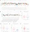
Examination of variations in mRNA and protein expression levels of TAOK1 among diverse tumor types. (A) The analysis of TAOK1 mRNA expression levels across 33 tumor types using data from The Cancer Genome Atlas (TCGA) combined with the Genotype-Tissue Expression (GTEx) database. Normal tissue samples are denoted in blue, while tumor samples are indicated in red. ns, not significant. (B) Protein expression levels of TAOK1 were analyzed across 14 types of tumors utilizing data from the Clinical Proteomic Tumor Analysis Consortium (CPTAC) database. (C) TAOK1 expression in four Gene Expression Omnibus (GEO) datasets was assessed: GSE43767, pertaining to adenocarcinoma (ADC); (D) GSE64951, focusing on esophageal squamous cell carcinoma (ESCC); (E) GSE174570, concerning liver cancer; and (F) GSE26712, related to ovarian cancer. Statistical significance is denoted as ns, not significant; * P < 0.05; ** P < 0.01; *** P < 0.001; ****P < 0.0001.
Figure 3
Figure 3
Prognostic analysis of TAOK1. (A) The forest plot presents the results of univariate Cox regression analysis of TAOK1, examining OS, DSS, DFI, and PFI. Heatmap illustrates the analysis of OS (B) and PFI (C) using six TAOK1 transcripts across 33 cancer types from TCGA dataset. The correlation between TAOK1 and clinicopathological stage is explored in multiple tumor types, including ACC (D), BLCA (E), LUSC (F), MESO (G), and SKCM (H).
Figure 4
Figure 4
The immune correlation analysis of TAOK1. (A) Heatmaps were generated to visually represent the relationship between TAOK1 and different immune infiltrating cells and quantified using the XCELL algorithm. Color gradients from purple to red denote correlation coefficients. A scatter plot demonstrates Spearman correlation analysis based on the XCELL algorithm, depicting the relationship between TAOK1 expression in tumors and (B) tumor fibroblast infiltration, (C) stromal score, (D) microenvironment score, and (E) regulatory T cell (Tregs) infiltration. (F) Heatmap depicting the correlation between TAOK1 and 11 immune checkpoints. (G) Correlation analysis between TAOK1 and tumor stemness, assessed via DNA and RNA stemness indices. * P < 0.05; ** P < 0.01; *** P < 0.001.
Figure 5
Figure 5
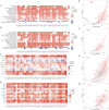
The correlation analysis between TAOK1 pathways and biological processes. (A) Heatmaps were generated via GSEA to illustrate the pathways associated with the KEGG and TAOK1. (B) Heatmap illustrating biological processes correlated with TAOK1. (C) Heatmap presenting an analysis of the association between TAOK1 and genes implicated in ferroptosis. (D) Heatmap showcasing a correlation analysis between TAOK1 and genes associated with cuproptosis. Correlation analyses between TAOK1 and cell cycle-related genes (E) as well as cell adhesion-related genes (F) in CESC. Correlation analyses between TAOK1 and cell cycle-related genes (G) along with cell adhesion-related genes (H) in OV. Normalized Enrichment Score (NES) is depicted by a color gradient ranging from purple to red; * P < 0.05; ** P < 0.01; *** P < 0.001.
Figure 6
Figure 6
Mutational analysis of DNA copy number variation and DNA methylation in TAOK1. (A) Analysis of TAOK1 mutation frequencies in tumors utilizing data from the TCGA database. (B) Investigation of DNA copy number variation in TAOK1. (C) Examination of methylation mutations in TAOK1, with the red box indicating the promoter region ID of TAOK1. * P < 0.05; ** P < 0.01; *** P < 0.001.
Figure 7
Figure 7
The impact of TAOK1 on the proliferation of A2780 and SiHa cell lines. (A and B) Validation of mRNA and protein expression levels of TAOK1 siRNA in A2780 and SiHa cells by RT-qPCR and Western Blot. (C) Proliferation levels of A2780 and SiHa cells were detected at 24 h, 48 h and 72 h after TAOK1 inhibition using Cell Counting Kit-8 (CCK-8) assay. (D and E) The effects of TAOK1 inhibition on the cell cycle of A2780 and SiHa cells were analysed using flow cytometry. (F) Overexpression levels of TAOK1 in SiHa cell lines were verified using RT-qPCR. (G) Proliferation level of SiHa cells after TAOK1 overexpression was assessed by CCK-8. (H) The effect of TAOK1 overexpression on SiHa cell cycle was analysed by flow cytometry. (I) The expression levels of TAOK1 in tumour tissues and their adjacent normal tissues of cervical cancer patients were detected by immunohistochemistry (IHC), and representative images are shown. Control group: adjacent normal tissues; tumour group: cervical cancer tissues. (J) BALB/c nude mice (5 mice per group) were subcutaneously inoculated with SiHa cells stably expressing Vector and TAOK1. ns, not significant; * p < 0.05 indicates statistically significant difference; ** P < 0.01; *** P < 0.001; ****P < 0.0001.
Figure 8
Figure 8
The correlation analysis between TAOK1 and EP300. (A) Transcription factors upstream of TAOK1 were analyzed using three databases and their correlations were examined. The mRNA expression levels of 10 transcription factors were assessed after transfection of TAOK1 with siRNA in A2780 (B) and SiHa (C). Correlation analysis of TAOK1 with EP300 was conducted based on the TCGA database (D) and the Cancer Cell Line Encyclopedia (CCLE) database (E). (F) Scatter plots depicting the correlation analysis between TAOK1 and EP300 in multiple tumors.

References

    1. Siegel RL, Giaquinto AN, Jemal A. Cancer statistics, 2024. CA Cancer J Clin. 2024;74(1):12–49. doi:10.3322/caac.21820 - DOI - PubMed
    1. Lu Z, Chen Y, Liu D, et al. The landscape of cancer research and cancer care in China. Nat Med. 2023;29(12):3022–3032. doi:10.1038/s41591-023-02655-3 - DOI - PubMed
    1. Ferrari F, Giannini A. Approaches to prevention of gynecological malignancies. BMC Women’s Health. 2024;24(1):254. doi:10.1186/s12905-024-03100-4 - DOI - PMC - PubMed
    1. Qi L, Li G, Li P, et al. Twenty years of Gendicine® rAd-p53 cancer gene therapy: the first-in-class human cancer gene therapy in the era of personalized oncology. Genes Dis. 2024;11(4):101155. doi:10.1016/j.gendis.2023.101155 - DOI - PMC - PubMed
    1. Zi M, Maqsood A, Prehar S, et al. The mammalian Ste20-like kinase 2 (Mst2) modulates stress-induced cardiac hypertrophy. J Biol Chem. 2014;289(35):24275–24288. doi:10.1074/jbc.M114.562405 - DOI - PMC - PubMed

LinkOut - more resources