Review
. 2025 Jan 23;4(1):lnaf004.
doi: 10.1093/lifemedi/lnaf004. eCollection 2025 Feb.

Exploration of the mechanism and therapy of ovarian aging by targeting cellular senescence

Affiliations
Review

Exploration of the mechanism and therapy of ovarian aging by targeting cellular senescence

Weicheng Tang et al. Life Med. .

Abstract

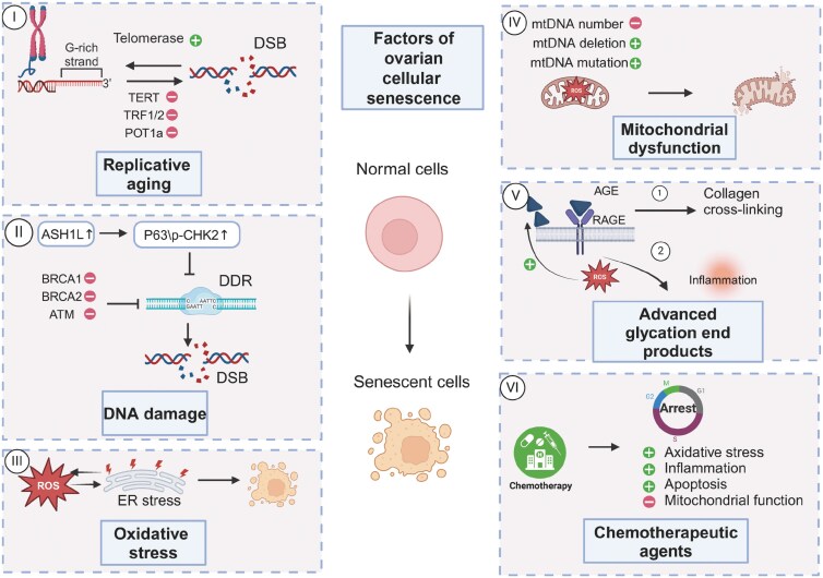
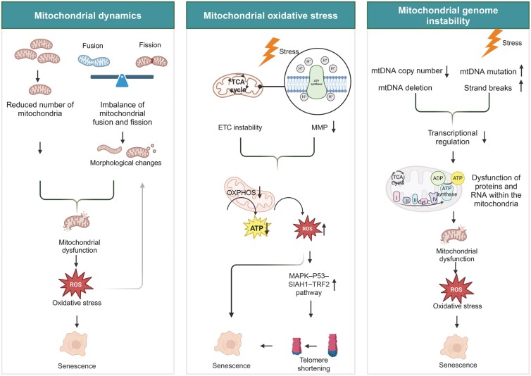
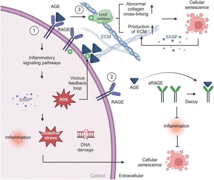
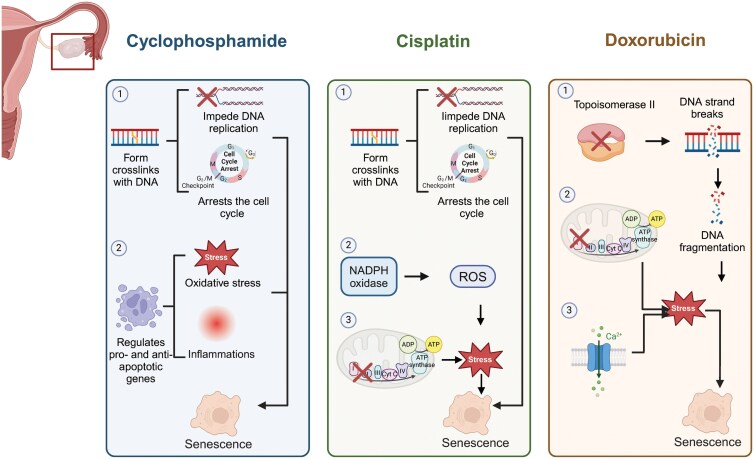
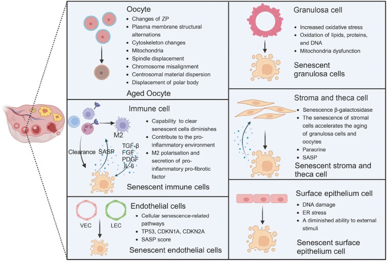
The ovary is a crucial gonadal organ that supports female reproductive and endocrine functions. Ovarian aging can result in decreased fertility and dysfunction across multiple organs. Research has demonstrated that cellular senescence in various cell types within the ovary can trigger a decline in ovarian function through distinct stress responses, resulting in ovarian aging. This review explores how cellular senescence may contribute to ovarian aging and reproductive failure. Additionally, we discuss the factors that cause ovarian cellular senescence, including the accumulation of advanced glycation end products, oxidative stress, mitochondrial dysfunction, DNA damage, telomere shortening, and exposure to chemotherapy. Furthermore, we discuss senescence in six distinct cell types, including oocytes, granulosa cells, ovarian theca cells, immune cells, ovarian surface epithelium, and ovarian endothelial cells, inside the ovary and explore their contribution to the accelerated ovarian aging. Lastly, we describe potential senotherapeutics for the treatment of ovarian aging and offer novel strategies for ovarian longevity.

Keywords: cellular senescence; granulosa cells; oocyte; ovarian aging; senotherapy.


Conflict of interest statement

The authors declare that there is no conflict of interest that could be perceived as prejudicing the impartiality of this review.

Figures

Figure 1.
Figure 1.
Mechanisms of cellular senescence in the ovary.Normal cells evolve into senescent cells through a series of mechanisms, including telomere shortening, DNA damage, the accumulation of oxidative stress, mitochondrial dysfunction, the presence of advanced glycation end products, and exposure to chemotherapy. Consequently, these mechanisms collectively contribute to the cellular transition towards senescence, highlighting the multifactorial nature of cellular senescence.
Figure 2.
Figure 2.
Mechanisms of mitochondrial dysfunction in ovarian cellular senescence.Mitochondria contribute to ovarian cellular senescence by modulating mitochondrial dynamics, mitochondrial oxidative stress, mitochondrial genome stability. Firstly, mitochondrial numbers decrease with age and an imbalance between mitochondrial fusion and fission results in abnormal mitochondrial morphology. All of the above can lead to mitochondrial dysfunction, resulting in the accumulation of oxidative stress and promoting cellular senescence. In addition, accumulated oxidative stress can affect mitochondrial morphology, which can further lead to mitochondrial dysfunction. Secondly, the instability of the mitochondrial electron transport chain coupled with the disruption of membrane potential can cause mitochondrial oxidative phosphorylation dysfunction. This, in turn, impairs ATP production and leads to an overproduction of ROS. On one side, the accumulation of ROS directly accelerates cellular aging; on the other side, it can induce telomere shortening through signaling pathways, thus promoting cellular aging. Thirdly, mitochondrial genome instability inhibits the regulation of mitochondrial transcription, thereby affecting the function of proteins in the mitochondrial electron transport chain. Consequently, this dysregulation of mitochondrial function results in oxidative stress, which subsequently leads to cellular senescence.
Figure 3.
Figure 3.
Mechanisms of AGEs induced ovarian cellular senescence.Firstly, AGEs, upon binding to its receptors (RAGE), triggers inflammatory signaling pathways, leading to the upregulation of ROS and SASPs. This cascade results in inflammation, oxidative stress and DNA damage, ultimately leading to cellular senescence. Additionally, ROS can enhance AGEs formation, and the interaction of AGEs with RAGE can further upregulate RAGE expression, perpetuating a vicious cycle. Secondly, another AGE receptor sRAGE, prevents excess ligand from binding to RAGE, thereby protecting cells from damage caused by the AGEs-induced inflammatory cascade. Thirdly, AGEs promote cellular senescence by upregulating abnormal collagen cross-linking and ECM formation in the ovaries. The SASPs produced by senescent cells also promotes ECM accumulation, thereby exacerbating cellular senescence.
Figure 4.
Figure 4.
Mechanisms of chemotherapy-induced ovarian cellular senescence.CPM induces cellular aging through two primary mechanisms. On one hand, they form cross-links within DNA, thereby preventing DNA replication and arresting the cell cycle. On the other hand, these drugs regulate pro-apoptotic and anti-apoptotic genes, causing oxidative stress and inflammation, which further promotes cellular senescence. Besides forming cross-links within DNA, cisplatin facilitates ROS accumulation via the NADPH pathway and disrupt mitochondrial electron transport chain complexes, resulting in oxidative stress damage and then cellular senescence. Doxorubicin can inhibit topoisomerase II, leading to DNA breakage and fragmentation. The accumulated DNA fragments trigger cellular senescence. Additionally, these drugs can interfere with intracellular redox processes or disrupt intracellular Ca2+ homeostasis, affecting mitochondrial function and inducing oxidative stress, thereby promoting cellular senescence.
Figure 5.
Figure 5.
The process of cellular senescence in various ovarian cells.In the context of oocyte aging, some alterations within the zona pellucida, abnormalities in the plasma membrane structure and changes in the cytoskeletal architecture are observed. Granulosa cells enter a state of cellular senescence through processes such as increased oxidative stress and mitochondrial dysfunction. Stromal cells undergo cellular senescence via SASP, paracrine actions, and interactions with granulosa cells and oocytes. Following the senescence of immune cells, their capacity to eliminate aging cells diminishes, leading to a pernicious cycle through the further secretion of pro-inflammatory and pro-fibrotic factors. Epithelial cells experience cellular senescence as a consequence of DNA damage and endoplasmic reticulum stress. Endothelial cells, including vascular and lymphatic endothelial cells (VEC and LEC), were found to exhibit increased expression of cellular senescence pathways, SASP score, and senescence markers. ZP, zona pellucida.

Similar articles

References

    1. Ata B, Seli E.. Strategies for controlled ovarian stimulation in the setting of ovarian aging. Semin Reprod Med 2015;33:436–48. - PubMed
    1. Minkin MJ. Menopause: hormones, lifestyle, and optimizing aging. Obstet Gynecol Clin North Am 2019;46:501–14. - PubMed
    1. Couzin-Frankel J. Reproductive biology. Faulty DNA repair linked to ovarian aging in mice and humans. Science 2013;339:749. - PubMed
    1. Hong W, Wang B, Zhu Y, et al.Female germline stem cells: aging and anti-aging. J Ovarian Res 2022;15:79. - PMC - PubMed
    1. Sang Q, Ray PF, Wang L.. Understanding the genetics of human infertility. Science 2023;380:158–63. - PubMed

LinkOut - more resources