. 2025 Mar 5:16:1508047.
doi: 10.3389/fphar.2025.1508047. eCollection 2025.

Amentoflavone protects against cisplatin-induced acute kidney injury by modulating Nrf2-mediated oxidative stress and ferroptosis and partially by activating Nrf2-dependent PANoptosis

Affiliations

Amentoflavone protects against cisplatin-induced acute kidney injury by modulating Nrf2-mediated oxidative stress and ferroptosis and partially by activating Nrf2-dependent PANoptosis

Yan Zhang et al. Front Pharmacol. .

Abstract

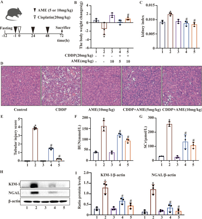
Background: Cisplatin is a widely used drug for the treatment of solid organ cancer, but its renal toxicity cannot be ignored. Amentoflavone (AME), a natural flavonoid compound, has remarkable pharmacological effects, including anti-inflammatory and antioxidative effects. The effect and mechanism of AME on cisplatin-induced acute kidney injury (CI-AKI) remain unclear.

Methods: We investigated the effect of AME on CI-AKI using the HK-2 cell line and C57BL/6 mice. Renal function, tissue damage, and molecular markers were assessed to explore the effects of AME on oxidative stress and cell death pathways.

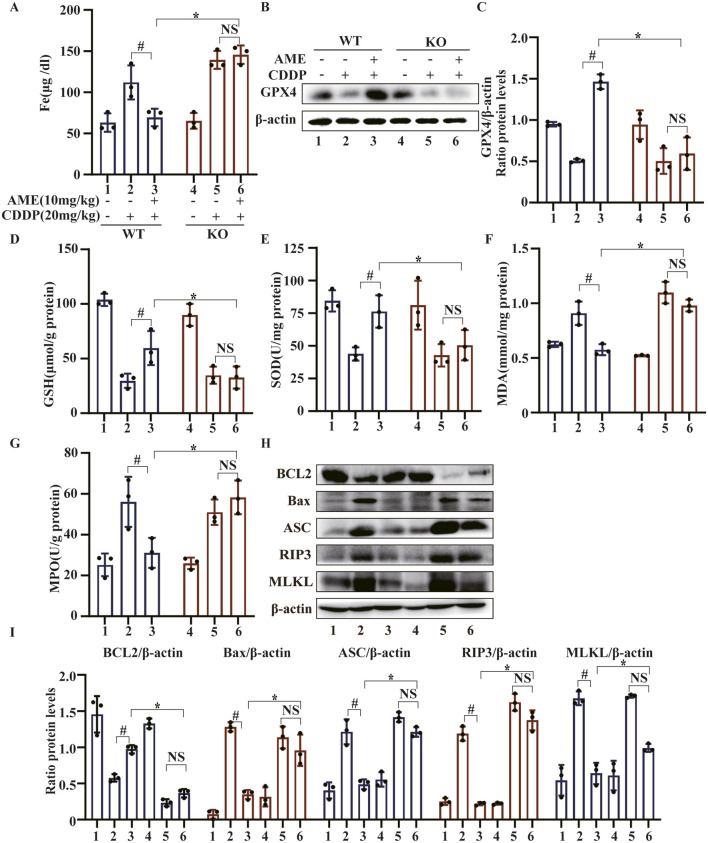
Results: In vitro, AME significantly suppressed the cytotoxic effects of cisplatin on HK-2 cells. Furthermore, AME significantly inhibited cisplatin-induced ferroptosis and PANoptosis (apoptosis, pyroptosis and necroptosis). In mice with acute kidney injury induced by a single intraperitoneal injection of cisplatin, the daily administration of AME during AKI effectively improved renal function and alleviated renal tubular injury, characterized by the normalization of blood urea nitrogen (BUN) and serum creatinine (SCr) levels; it also inhibited cisplatin-induced renal ferroptosis and PANoptosis. AME is a natural antioxidant that activates the Nrf2 antioxidant pathway both in vivo and in vitro. In Nrf2 knockout mice and knockdown cells, the protective effect of AME against cisplatin-induced nephrotoxicity disappeared. However, after Nrf2 knockout, the effect of AME on ferroptosis completely disappeared, and that on PANoptosis partially disappeared.

Conclusion: Amentoflavone has a protective effect on cisplatin-induced acute kidney injury via a mechanism related to the Nrf2-dependent antioxidant pathway and the regulation of ferroptosis and PANoptosis.

Keywords: CI-AKI; Nrf2; PANoptosis; amentoflavone; ferroptosis; oxidative stress.


Conflict of interest statement

The authors declare that the research was conducted in the absence of any commercial or financial relationships that could be construed as a potential conflict of interest.

Figures

FIGURE 1
FIGURE 1
Effects of amentoflavone on cisplatin-induced acute kidney injury (A) The experimental design and protocol for cisplatin treatment or combined treatment with amentoflavone involved fasting C57BL/6 mice for 12 h. (B) The change in body weight was calculated by subtracting the weight before the initial treatment from the weight before euthanasia. (C) The kidney index was calculated as the ratio of kidney weight to weight before euthanasia. (D, E) Representative kidney tissue sections were stained with H&E, and kidney injury scores were determined. (F) BUN and (G) SCr levels were measured. All the data are presented as means ± SEMs (n = 5 per group). (H, I) Kidney tissue was homogenized in lysis buffer to extract protein, and Western blotting was performed to assess the protein expression of proximal tubular injury markers, including KIM-1 and NGAL. The experiments were repeated three times. * indicates a significant difference (p < 0.05) between the control group and the CDDP group, and # indicates a significant difference (p < 0.05) between the CDDP group and the AME treatment groups. NS, not significant.
FIGURE 2
FIGURE 2
Effects of amentoflavone on cisplatin-induced ferroptosis and the PANoptosis pathway in vivo (A–B) The expression of xCT and GPX4 in kidney tissue was determined using immunoblotting. (C–F) The levels of proteins related to apoptosis (BCL2, Bax, and Caspase-3), pyroptosis (GSDMD, ASC, and Caspase-1), and necroptosis (RIP3 and MLKL) were measured. The experiments were repeated three times. * indicates a significant difference (p < 0.05) between the control group and the CDDP group, and # indicates a significant difference (p < 0.05) between the CDDP group and the AME treatment groups. NS, not significant.
FIGURE 3
FIGURE 3
Effects of amentoflavone on oxidative pathway-related proteins in the kidney tissues of cisplatin-treated mice. (A, D) Representative Western blots demonstrating the impact of amentoflavone treatment on the levels of oxidative pathway-related proteins, including Nrf2, HO-1, NQO1, and GCLC, in mouse kidney tissues. (B–C) The production of GSH and MDA in renal tissues was measured in the mice. The experiments were repeated three times. * indicates a significant difference (p < 0.05) between the control group and the CDDP group, and # indicates a significant difference (p < 0.05) between the CDDP group and the AME treatment groups. NS, not significant.
FIGURE 4
FIGURE 4
Amentoflavone reduces oxidative stress and lipid peroxidation induced by cisplatin in human kidney 2 (HK-2) cells. (A) HK-2 cells were treated with different concentrations of amentoflavone (1 or 2 μM) for 1 h and then with cisplatin (20 μM) for 24 h. Cell viability was assessed using the CCK-8 assay. (B, C) The levels of oxidative pathway-related proteins, including Nrf2, HO-1, and NQO1, in HK-2 cells were evaluated using Western blot analysis to determine the effect of amentoflavone. (D) HK-2 cells were stained with an ROS fluorescent probe (1 μM) for 30 min, and the resulting fluorescence was detected using a fluorescence microscope. (E) ROS fluorescence quantification analysis. (F) Liperfluo staining and confocal microscopy were used to assess lipid peroxidation in HK-2 cells. (G) Liperfluo fluorescence quantification analysis. * indicates a significant difference (p < 0.05) between the control group and the CDDP group, and # indicates a significant difference (p < 0.05) between the CDDP group and the AME treatment groups. NS, not significant.
FIGURE 5
FIGURE 5
Amentoflavone inhibits ferroptosis and PANoptosis in HK-2 cells. (A, B) The iron content in HK-2 cells was assessed by FerroOrange staining. (C, D) The protein levels of xCT and GPX4. (E, F) The levels of proteins related to apoptosis (BCL2, Bax, and Caspase-3). (G, H) The levels of proteins involved in pyroptosis (GSDMD, ASC, and Caspase-1). (I, J) The levels of proteins related to necroptosis (RIP3 and MLKL). The experiments were repeated three times. * indicates a significant difference (p < 0.05) between the control group and the CDDP group, and # indicates a significant difference (p < 0.05) between the CDDP group and the AME treatment groups. NS, not significant.
FIGURE 6
FIGURE 6
The impact of amentoflavone on oxidative stress and lipid peroxidation in HK-2 cells was found to be dependent on the expression of the Nrf2 gene. (A) A Cell Counting Kit-8 (CCK-8) assay was performed to assess the survival rate of the cells. The levels of (B) GSH and (C) MDA in HK-2 cells were measured using appropriate kits. (D) Lipid peroxidation in HK-2 cells was assessed through Liperfluo staining and confocal microscopy. (E) LiperFluo fluorescence quantification analysis. (F) ROS levels in HK-2 cells were measured using DCFH-DA. (G) ROS fluorescence quantification analysis. (H) The iron content in HK-2 cells was assessed through FerroOrange staining. (I) FerroOrange fluorescence quantification analysis. The experiments were repeated three times. WT indicates normal HK-2 cells, and si-Nrf2 indicates Nrf2 knockdown cells. # indicates a significant difference (p < 0.05) between the CDDP group and the AME treatment groups, and * indicates a significant difference (p < 0.05) between Nrf2 WT and KO mice after AME administration. NS, not significant.
FIGURE 7
FIGURE 7
The impact of amentoflavone on cisplatin-induced AKI is contingent on Nrf2 in vivo. (A) The level of Nrf2 expression in renal tissue was assessed detected using immunofluorescence (IF) staining. Nrf2 is labeled red, LTL is labeled green, and nuclei are labeled blue (DAPI). (B) Body weight change was calculated by subtracting the weight before the first treatment from the weight before euthanasia. (C) The kidney index was calculated as the kidney weight divided by the weight before euthanasia. (D) BUN and (E) SCr levels were measured. (F, G) Kidney injury scores were calculated, and representative kidney tissue sections were stained with H&E. (H, I) Proximal tubular injury markers, including KIM-1 and NGAL, were assessed using Western blotting to determine protein expression. The experiments were repeated three times. WT indicates wild-type mice, and KO indicates Nrf2 knockout mice. # indicates a significant difference (p < 0.05) between the CDDP group and the AME treatment groups, and * indicates a significant difference (p < 0.05) between Nrf2 WT and KO mice after AME administration. NS, not significant.
FIGURE 8
FIGURE 8
The effects of amentoflavone occur through Nrf2-dependent ferroptosis and partially through Nrf2-dependent PANoptosis. (A) Iron metabolism in mice was assessed by measuring the serum iron ion content. (B,C) Western blotting was used to assess the protein expression level of GPX4 in renal tissues. Renal tissues were collected from the mice to measure (D) GSH, (E) SOD, (F) MDA, and (G) MPO levels. (H, I) The protein levels of BCL2, Bax, ASC, and MLKL were assessed in renal tissue via Western blot analysis. The experiments were repeated three times. WT indicates wild-type mice, and KO indicates Nrf2 knockout mice. # indicates a significant difference (p < 0.05) between the CDDP group and the AME treatment groups, and * indicates a significant difference (p < 0.05) between Nrf2 WT and KO mice after AME administration.

References

    1. Anandhan A., Dodson M., Shakya A., Chen J., Liu P., Wei Y., et al. (2023). NRF2 controls iron homeostasis and ferroptosis through HERC2 and VAMP8. Sci. Adv. 9 (5), eade9585. 10.1126/sciadv.ade9585 - DOI - PMC - PubMed
    1. Bedoui S., Herold M. J., Strasser A. (2020). Emerging connectivity of programmed cell death pathways and its physiological implications. Nat. Rev. Mol. Cell Biol. 21 (11), 678–695. 10.1038/s41580-020-0270-8 - DOI - PubMed
    1. Bersuker K., Hendricks J. M., Li Z., Magtanong L., Ford B., Tang P. H., et al. (2019). The CoQ oxidoreductase FSP1 acts parallel to GPX4 to inhibit ferroptosis. Nature 575 (7784), 688–692. 10.1038/s41586-019-1705-2 - DOI - PMC - PubMed
    1. Bollong M. J., Lee G., Coukos J. S., Yun H., Zambaldo C., Chang J. W., et al. (2018). A metabolite-derived protein modification integrates glycolysis with KEAP1-NRF2 signalling. Nature 562 (7728), 600–604. 10.1038/s41586-018-0622-0 - DOI - PMC - PubMed
    1. Cui Y., Zhang Z., Zhou X., Zhao Z., Zhao R., Xu X., et al. (2021). Microglia and macrophage exhibit attenuated inflammatory response and ferroptosis resistance after RSL3 stimulation via increasing Nrf2 expression. J. Neuroinflammation 18 (1), 249. 10.1186/s12974-021-02231-x - DOI - PMC - PubMed

LinkOut - more resources