. 2025 Mar 20;135(10):e180900.
doi: 10.1172/JCI180900. eCollection 2025 May 15.

Erythrocyte-derived extracellular vesicles induce endothelial dysfunction through arginase-1 and oxidative stress in type 2 diabetes

Affiliations

Erythrocyte-derived extracellular vesicles induce endothelial dysfunction through arginase-1 and oxidative stress in type 2 diabetes

Aida Collado et al. J Clin Invest. .

Abstract

Red blood cells (RBCs) induce endothelial dysfunction in type 2 diabetes (T2D), but the mechanism by which RBCs communicate with the endothelium is unknown. This study tested the hypothesis that extracellular vesicles (EVs) secreted by RBCs act as mediators of endothelial dysfunction in T2D. Despite a lower production of EVs derived from RBCs of T2D patients (T2D RBC-EVs), their uptake by endothelial cells was greater than that of EVs derived from RBCs of healthy individuals (H RBC-EVs). T2D RBC-EVs impaired endothelium-dependent relaxation, and this effect was attenuated following inhibition of arginase in EVs. Inhibition of vascular arginase or oxidative stress also attenuated endothelial dysfunction induced by T2D RBC-EVs. Arginase-1 was detected in RBC-derived EVs, and arginase-1 and oxidative stress were increased in endothelial cells following coincubation with T2D RBC-EVs. T2D RBC-EVs also increased arginase-1 protein in endothelial cells following mRNA silencing and in the endothelium of aortas from endothelial cell arginase-1-knockout mice. It is concluded that T2D-RBCs induce endothelial dysfunction through increased uptake of EVs that transfer arginase-1 from RBCs to the endothelium to induce oxidative stress and endothelial dysfunction. These results shed important light on the mechanism underlying endothelial dysfunction mediated by RBCs in T2D.

Keywords: Cardiology; Cardiovascular disease; Diabetes; Endothelial cells; Vascular biology.


Figures

Figure 1
Figure 1. EVs are released by human RBCs.
Representative TEM images of EVs derived from RBCs from healthy subjects (H RBC-EVs) and patients with T2D (T2D RBC-EVs) negatively stained or immunostained with gold-labeled anti-CD63 antibody. Red arrows point at positive signals (black dots) for CD63-gold beads (A, n = 3). EVs were captured by anti-CD9–coated latex beads, detected either by anti-CD9, anti-CD63, or anti-CD81. The MFI was measured by flow cytometry. The dotted line indicates a signal above 1, which was considered as a positive signal (B, n = 3). Quantification of EV concentration (C, n = 9). Distribution of the average particle size of H RBC-EVs and T2D RBC-EVs (D, n = 9). Values are expressed as mean ± SD (B and D) and median ± interquartile range (Q1–Q3) (C). *P < 0.05, Mann-Whitney U test.
Figure 2
Figure 2. Increased uptake of the RBC-derived EVs from T2D patients by endothelial cells.
Representative immunofluorescence images depicting PKH67 (green) in the absence or presence of heparin (Hep) in HCtAECs (A, n = 6). Quantitative analyses of the integrated density for PKH67-labeled EVs after 24 hours of coincubation with HCtAECs in the absence or presence of heparin (B, n = 6). Values are expressed as mean ± SD. *P < 0.05; **P < 0.01, 1-way ANOVA.
Figure 3
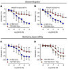
Figure 3. T2D RBC-EVs induce endothelial dysfunction, rescued by uptake inhibition.
EDR evoked by ACh in mouse aortas following 18 hours of coincubation with H RBC-EVs and T2D RBC-EVs. EVs were isolated by sequential ultracentrifugation (A, n = 4 and B, n = 3–9) or membrane affinity columns (C, n = 6–9). EDR evoked by ACh in mouse aortas following 18 hours of coincubation with T2D RBC-EVs and heparin (D, n = 7). Values are expressed as mean and SD. *P < 0.05; **P < 0.01, repeated-measures 2-way ANOVA.
Figure 4
Figure 4. Arginase-1 is present in RBC-derived EVs and mediates endothelial dysfunction.
Western blot images of arginase-1 (35 kDa) and GAPDH (36 kDa) in isolated EVs derived from H-RBCs and T2D-RBCs and quantification of the expression normalized to GAPDH (A, n = 5). EDR evoked by ACh in mouse aortas following 18 hours of coincubation of T2D RBC-EVs with the arginase inhibitor ABH (B, n = 6). EDR evoked by ACh in mouse aortas following 18 hours of coincubation of T2D RBC-EVs with the antioxidant NAC (C, n = 5). Values are expressed as median ± interquartile range (Q1–Q3) (A) and mean and SD (B and C). *P < 0.05, repeated measures 2-way ANOVA (B).
Figure 5
Figure 5. T2D RBC-EVs induce endothelial dysfunction through vascular arginase-1.
Immunohistochemical images (A, n = 6) and quantification (B, n = 6) of arginase-1 in aortic rings and in human IMAs (C and D, n = 4) following 18 hours of coincubation with H RBC-EVs and T2D RBC-EVs. Inserts show IgG controls. L indicates lumen, black arrows endothelial cells, and red arrows smooth muscle cells. mRNA levels of arginase-1 (ARG1) in HCtAEC after coincubation with H RBC-EVs and T2D RBC-EVs for 8 hours (E, n = 6) and 24 hours (F, n = 5–6). Immunofluorescence (G, n = 5–6) and quantification (H, n = 5–6) of arginase-1 (green) in HCtAECs following 24 hours of coincubation with H RBC-EVs and T2D RBC-EVs. Arginase activity in HCtAECs incubated with medium (control), H RBC-EVs, and T2D RBC-EVs for 24 hours (I, n = 6). EDR evoked by ACh in mouse aortas following 18 hours of coincubation with T2D RBC-EVs with and without the administration of ABH to the vessels for 1 hour in the organ baths (J, n = 5). Parentheses indicate that the inhibitor was added in the organ baths for 1 hour following the 18 hours of coincubation with T2D RBC-EVs. Values are expressed as median ± interquartile range (Q1–Q3) (B and DF), mean ± SD (H and I), and mean and SD (J). *P < 0.05; **P < 0.01; ***P < 0.001, Mann-Whitney U test (B and DF), 1-way ANOVA (H and I), and repeated measures 2-way ANOVA (J).
Figure 6
Figure 6. T2D RBC-EVs induce endothelial dysfunction through the transfer of arginase-1 to the endothelium.
Immunofluorescence (A, n = 5) and quantification (B, n = 5) of arginase-1 (green) in HCtAECs transfected with scramble or siRNA for arginase-1 (ARG1) and coincubated with T2D RBC-EVs for 24 hours. EDR evoked by ACh in mouse aortic rings from Arg1fl/fl/Tie2Cretg/– mice following 18 hours of coincubation with H RBC-EVs and T2D RBC-EVs (C, n = 6–7). Immunohistochemical images (D, n = 6) and quantification (E, n = 6) of arginase-1 in aortas from Arg1fl/fl/Tie2Cretg/– mice following 18 hours of coincubation with H RBC-EVs and T2D RBC-EVs. Inserts show IgG controls. L indicates lumen, black arrows endothelial cells, and red arrows smooth muscle cells. Immunofluorescence images of arginase-1 (green) and CD31 (red) in aortas from Arg1fl/fl/Tie2Cretg/– mice following coincubation with H RBC-EVs and T2D RBC-EVs. Nuclei were stained with Hoechst (blue) (F, n = 5). EDR evoked by ACh in mouse aortas from Arg1fl/fl/Tie2Cretg/– mice following 18 hours of coincubation with T2D RBC-EVs with and without the administration of ABH to the vessels for 1 hour in the organ baths (G, n = 6). Parentheses indicate that the inhibitor was added in the organ baths for 1 hour following the 18 hours of coincubation with T2D RBC-EVs. Values are expressed as mean ± SD (B), mean and SD (C and G), and median ± interquartile range (Q1–Q3) (E). *P < 0.05; **P < 0.01, 1-way ANOVA (B), repeated measures 2-way ANOVA (C and G), and Mann-Whitney U test (E).
Figure 7
Figure 7. T2D RBC-EVs induce endothelial dysfunction through ROS.
Immunohistochemical images (A, n = 7) and quantification (B, n = 7) of 4-HNE in mouse aortic rings following 18 hours of coincubation with H RBC-EVs and T2D RBC-EVs. Immunohistochemical images (C, n = 4) and quantification (D, n = 4) of 4-HNE in IMAs following 18 hours of coincubation with H RBC-EVs and T2D RBC-EVs. Inserts show IgG controls. L indicates lumen, black arrows endothelial cells, and red arrows smooth muscle cells. mRNA levels of NOX4 after coincubation of HCtAECs with H RBC-EVs and T2D RBC-EVs for 8 hours (E, n = 6) and 24 hours (F, n = 6). EDR evoked by ACh in mouse aortas following 18 hours of coincubation with T2D RBC-EVs with and without administration of NAC to the vessels for 1 hour in the organ baths (G, n = 6). EDR evoked by ACh in mouse aortas following 18 hours of coincubation with T2D RBC-EVs with and without administration of GLX481304 (NOX2/4 inhibitor) to the vessels for 1 hour in the organ baths (H, n = 6). Parentheses indicate that the inhibitor was added in the organ baths for 1 hour following the 18 hours of coincubation with T2D RBC-EVs. Values are expressed as median ± interquartile range (Q1–Q3) (B and DF), and mean and SD (G and H). *P < 0.05; **P < 0.01 using Mann-Whitney U test (B, D, and F), and repeated measures 2-way ANOVA (G and H).

Comment in

  • Endothelial dysfunction in patients with type 2 diabetes: the truth is in the blood

References

    1. Paneni F, et al. Diabetes and vascular disease: pathophysiology, clinical consequences, and medical therapy: part I. Eur Heart J. 2013;34(31):2436–2443. doi: 10.1093/eurheartj/eht149. - DOI - PMC - PubMed
    1. Mahdi A, et al. The effect of glycemic control on endothelial and cardiac dysfunction induced by red blood cells in type 2 diabetes. Front Pharmacol. 2019;10:861. doi: 10.3389/fphar.2019.00861. - DOI - PMC - PubMed
    1. Gimbrone MA, Jr Endothelial cell dysfunction and the pathobiology of atherosclerosis. Circ Res. 2016;118(4):620–636. doi: 10.1161/CIRCRESAHA.115.306301. - DOI - PMC - PubMed
    1. Zhou Z, et al. Erythrocytes from patients with type 2 diabetes induce endothelial dysfunction via arginase I. J Am Coll Cardiol. 2018;72(7):769–780. doi: 10.1016/j.jacc.2018.05.052. - DOI - PubMed
    1. Yang J, et al. Red blood cells in type 2 diabetes impair cardiac post-ischemic recovery through an arginase-dependent modulation of nitric oxide synthase and reactive oxygen species. JACC Basic Transl Sci. 2018;3(4):450–463. doi: 10.1016/j.jacbts.2018.03.006. - DOI - PMC - PubMed

MeSH terms