. 2025 Mar 20;21(3):e1011593.
doi: 10.1371/journal.pgen.1011593. eCollection 2025 Mar.

A defining member of the new cysteine-cradle family is an aECM protein signalling skin damage in C. elegans

Affiliations

A defining member of the new cysteine-cradle family is an aECM protein signalling skin damage in C. elegans

Thomas Sonntag et al. PLoS Genet. .

Abstract

Apical extracellular matrices (aECMs) act as crucial barriers, and communicate with the epidermis to trigger protective responses following injury or infection. In Caenorhabditis elegans, the skin aECM, the cuticle, is produced by the epidermis and is decorated with periodic circumferential furrows. We previously showed that mutants lacking cuticle furrows exhibit persistent immune activation (PIA), providing a valuable model to study the link between cuticle damage and immune response. In a genetic suppressor screen, we identified spia-1 as a key gene downstream of furrow collagens and upstream of immune signalling. spia-1 expression oscillates during larval development, peaking between each moult together with patterning cuticular components. It encodes a secreted protein that localises to furrows. SPIA-1 shares a novel cysteine-cradle domain with other aECM proteins. SPIA-1 mediates immune activation in response to furrow loss and is proposed to act as an extracellular signal activator of cuticle damage. This research provides a molecular insight into intricate interplay between cuticle integrity and epidermal immune activation in C. elegans.


Conflict of interest statement

The authors have declared that no competing interests exist.

Figures
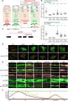
Fig 1
Fig 1. Loss of spia-1 suppresses furrow collagen AMP induction.
(A) Design of the suppressor screen. The strain carries the frIs7 transgene, containing an AMP transcriptional reporter (nlp-29p::GFP) and a control transgene (col-12p::DsRed) constitutively expressed in the epidermis. Under standard growth conditions, worms only express the control transgene and are red at all stages (left). RNAi inactivation of any furrow collagen gene, like dpy-7, leads to the expression of nlp-29p::GFP in a PMK-1/STA-2 dependent manner: worms appear “green” at all stages (middle). The strain used for the suppressor screen additionally bears the frIs30 construct to express a gain of function of GPA-12 in the epidermis, only from the young adult stage (col-19p::GPA-12gf). In this strain, inactivation of a gene downstream of GPA-12 eliminates the expression of nlp-29p::GFP in both larvae and adults (Nipi phenotype [7]), whereas inactivation of any gene acting upstream of, or in parallel to, GPA-12, inhibits the expression of nlp-29p::GFP in the larvae but not in the adult due to the activation of GPA-12 (red larvae, green adults, right). This rescue is total if the targeted gene acts upstream of GPA-12 (A, dcar-1), but only partial if it acts in parallel to GPA-12 (B/C). (B) Quantification of the green fluorescence in worms carrying frIs7 and frIs30 constructs in different mutant backgrounds, upon dpy-7 RNAi, in L4 and young adult stages (n>25). Statistics were made by comparing to the corresponding wt control; **p < 0.01; ***p < 0.001; ****p < 0.0001. (C) Structure of the spia-1 genomic locus. The location of the fr179 mutation is indicated with an arrowhead, the extent of the syb7920 deletion is shown with a red line. Exons are shown as black boxes, introns as solid lines. UTR are represented as white boxes; the blue region shows the sequence encoding the signal peptide of spia-1. (D) spia-1(fr179) suppresses the induction of nlp-29p::GFP in young adult worms after RNAi inactivation of furrow collagen genes. Wild-type and spia-1(fr179) mutants carrying the frIs7 transgene were treated with the indicated RNAi bacteria, with sta-1 used as a control (see Mat&Methods). Red and green fluorescences were visualised simultaneously in all images. Representative images of young adults from one of three experiments are shown; scale bar, 200 µm; see S1C and S1D Fig for quantification with the COPAS Biosort. (E) Oscillation of nlp-29p::GFP expression from L4 to adulthood. Representative confocal images of different mutant strains carrying the frIs7 transgene, red and green fluorescences were visualised simultaneously. The L4 stage is subdivided into sub-stages with the shape of the vulva [23]; n>5, scale bar, 10 µm. (F) Proposed schematic illustration of nlp-29p::GFP oscillation shown in E. Expression levels are estimated and not to scale.
Fig 2
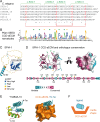
Fig 2. SPIA-1 is a secreted protein containing a novel cysteine-cradle domain.
(A) In C. elegans, 6 proteins share a common and previously uncharacterised domain in their C-terminal region, of which the amino acid sequences are depicted. This domain contains 4 invariant cysteine residues predicted to form two disulfide bridges (red) connecting 4 α-helices (green) and was named the cysteine cradle domain (CCD-aECM). (B) The sequence logo derived from the Pfam (PF23626) SEED alignment shows residues of the domain conserved across nematode homologues. The relative size of the residue letters indicates their frequency in the aligned sequences of the Pfam SEED. Arrows point to the aromatic residues lining the groove (green) and other highly conserved amino acids (yellow). (C) SPIA-1 structural model predicted with AlphaFold2 [27,65], rendered in cartoon and coloured according to the Predicted Local Distance Difference Test score (pLDDT), which indicates how well a predicted protein structure matches protein data bank structure information and multiple sequence alignment data: dark blue >90, light blue <90 & >70, yellow <70 & >50, orange <50. The CCD-aECM domain is framed in black. (D) Amino acid sequence (bottom) and AlphaFold prediction of the CCD-aECM rendered in a cartoon, with the side-chains shown as sticks (top left) or in surface with a 90° rotation (top right), and coloured according to the Consurf conservation scores [67] based on SPIA-1 orthologs alignment. Arrows point to the aromatic residues lining the groove (green), aliphatic residues that are in contact (black), and other highly conserved amino acids (yellow). Numbers indicate the position of the amino acid in the full-length SPIA-1. The predicted structural model of SPIA-1 CCD-aECM is also shown on S1 Movie. (E) The AlphaFold prediction model of Y34B4A.10 (left) is rendered in cartoon and coloured in rainbow (blue to red indicating the path of the polypeptide chain from N- to C-terminal end). Residues from the α-helix that are predicted to engage in hydrophobic interactions are shown as sticks. The same model rendered in surface (right) demonstrates how the N-terminal α-helix of Y34B4A.10 (cyan) is predicted to bind to the CCD-aECM groove of this protein (orange). (F) Simplified illustration of the proposed model for the interaction of the CCD-aECM with a ligand.
Fig 3
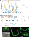
Fig 3. spia-1 is expressed in the epidermis and oscillatory between each moult.
(A) AMP and aECM gene expression oscillates between each moult, with expression levels from an RNA-seq time course with 1-hour intervals between 5 and 40 hours after hatching, reanalysed from (Meeuse et al. 2020) [37]. (B) Between each moult, a timeline of gene expression is represented, with dpy-6 starting each cycle. Peak phase data reanalysed from (Meeuse et al. 2020). (C-G) Expression pattern of spia-1 transcript in worms carrying the frEx631[pSO22(spia-1p::GFP), myo-2p::mCherry] transgene. Representative confocal images, n>5, of (C) 2-fold embryo, (D) L1 larva, (E, F) L4 larva, and (G) adult head. The signal is visible in the epidermis (e) at all stages, and also in head socket cell (so) and a neuron (n), but not in the seam cell (s), nor the vulva (v). Grey colour in (F) was acquired with a transmitted detection module; scale bar, 10 µm.
Fig 4
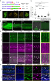
Fig 4. SPIA-1 localises to furrows.
(A) Position of the insertion of GFP in each translational reporter with their expression pattern and rescue activities. For the KI strain, mNG is inserted at the same place as in GFP-int. (B-C) spia-1 mutation suppresses nlp-29p::GFP overexpression in dpy-7 worms. The rescue of this suppression has been tested in spia-1(syb7920) young adults with the extra-chromosomal gene producing SPIA-1 tagged with GFP in Nter, Cter or internal position, in three independent experiments. (B) Representative images of one experiment; scale bar, 500 µm. (C) Relative green fluorescence is quantified (n=58-79); ****p < 0.0001. (D) Representative confocal images of the SPIA-1::sfGFP reporter (GFP-int) in 3-fold embryo, L1, L2, L4 vulval lumen and adult. We used a laser power ~2 times higher in adults compared to other stages (see S3A Fig). White arrows and arrowhead indicate signal in furrows and in vulval lumen, respectively; n>5, scale bar, 5 µm. (E) The L4 stage is subdivided into sub-stages in relation to the shape of the vulva, as previously described [23]. SPIA-1::sfGFP and mCherry::LPR-3 are observed in parallel. ~7 times magnification of the areas contained in the dashed rectangles are provided on the far right; scale bar, 10 µm (left), 2 µm (magnified area). (F) Representative confocal images of L4.4 (top) and L4.9 (bottom) larvae expressing DPY-2::BFP, SPIA-1::mNG-int, and NUC-1::mCherry. ~2.5 times magnification of the areas contained in the dashed rectangles are provided on the far right. Both single channels and the merge are shown; n>5, scale bar, 5 µm.
Fig 5
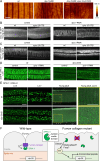
Fig 5. SPIA-1 acts downstream of furrow collagens.
(A) AFM topography of the cuticle in wild-type, dpy-7(e88) and dpy-7(e88); spia-1(syb7920) young adult worms; n>3, scale bar, 1 µm. (B) spia-1(fr179) does not suppress COL-19::GFP abnormal pattern following dpy-7 RNAi. Representative images of wt or spia-1(fr179) young adults carrying COL-19::GFP treated with sta-1 or dpy-7 RNAi bacteria; n>5, scale bar, 10 µm. (C) spia-1 does not suppress the absence of furrows following dpy-3 RNAi; representative images of wt or spia-1(fr179) worms carrying DPY-7::sfGFP treated with sta-1 or dpy-3 RNAi bacteria; scale bar, 10 µm. (D) spia-1 does not suppress the abnormal sfGFP::LPR-3 in dpy-2(e8); representative images of wt or dpy-2(e8) L4.6 worms carrying sfGFP::LPR-3 treated with control (sta-1) or spia-1 RNAi bacteria; scale bar, 10 µm. (E) SPIA-1::mNG-int is mislocalised in dpy-2(e8) mutants. Representative confocal images of the cuticle plane of wt or dpy-2(e8) L4.4, L4.7, or young adult worms carrying SPIA-1::mNG-int. We used a laser power ~2 times higher in adults compared to other stages, see S3A Fig. A ~2.5 times magnification of the areas contained in the dashed rectangles is provided on the far right; n>5, scale bar, 10 µm. (F) Cartoon presenting the proposed model for SPIA-1 activity in wild-type or furrow-less adults. Not to scale. SPIA-1 could bind directly or indirectly to furrow collagens. In the absence of furrow collagens, SPIA-1 may become unbound, allowing it to directly or indirectly activate signalling in the epidermis.

Update of

References

    1. Sundaram MV, Pujol N. The Caenorhabditis elegans cuticle and precuticle: a model for studying dynamic apical extracellular matrices in vivo. Genetics. 2024;227(4):iyae072. doi: 10.1093/genetics/iyae072 - DOI - PMC - PubMed
    1. Adams J, Pooranachithra M, Jyo E, Zheng S, Goncharov A, Crew J. Nanoscale patterning of collagens in C. elegans apical extracellular matrix. Nature Communications. 2023;14(1):7506. PMID: 37980413 - PMC - PubMed
    1. Cox GN, Kusch M, Edgar RS. Cuticle of Caenorhabditis elegans: its isolation and partial characterization. J Cell Biol. 1981;90(1):7–17. doi: 10.1083/jcb.90.1.7 - DOI - PMC - PubMed
    1. McMahon L, Muriel JM, Roberts B, Quinn M, Johnstone IL. Two sets of interacting collagens form functionally distinct substructures within a Caenorhabditis elegans extracellular matrix. Mol Biol Cell. 2003;14(4):1366–78. doi: 10.1091/mbc.e02-08-0479 - DOI - PMC - PubMed
    1. Lažetić V, Fay DS. Molting in C. elegans. Worm. 2017;6(1):e1330246. doi: 10.1080/21624054.2017.1330246 - DOI - PMC - PubMed

MeSH terms

Substances