Review
. 2025 Mar 21;14(1):7.
doi: 10.1186/s13619-025-00225-1.

Revolutionizing bone healing: the role of 3D models

Affiliations
Review

Revolutionizing bone healing: the role of 3D models

Raffaella De Pace et al. Cell Regen. .

Abstract

The increasing incidence of bone diseases has driven research towards Bone Tissue Engineering (BTE), an innovative discipline that uses biomaterials to develop three-dimensional (3D) scaffolds capable of mimicking the natural environment of bone tissue. Traditional approaches relying on two-dimensional (2D) models have exhibited significant limitations in simulating cellular interactions and the complexity of the bone microenvironment. In response to these challenges, 3D models such as organoids and cellular spheroids have emerged as effective tools for studying bone regeneration. Adult mesenchymal stem cells have proven crucial in this context, as they can differentiate into osteoblasts and contribute to bone tissue repair. Furthermore, the integration of composite biomaterials has shown substantial potential in enhancing bone healing. Advanced technologies like microfluidics offer additional opportunities to create controlled environments for cell culture, facilitating more detailed studies on bone regeneration. These advancements represent a fundamental step forward in the treatment of bone pathologies and the promotion of skeletal health. In this review, we report on the evolution of in vitro culture models applied to the study of bone healing/regrowth, starting from 2 to 3D cultures and microfluids. The different methodologies of in vitro model generation, cells and biomaterials are presented and discussed.

Keywords: 3D in vitro model; Biomaterial; Bone regeneration; Cell-ECM interaction; Microfluidic; Stem cell.


Conflict of interest statement

Declarations. Ethics approval and consent to participate: Not applicable. Consent for publication: Not applicable. Competing interests: The authors declare no potential conflicts of interest.

Figures
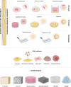
Fig. 1
Fig. 1
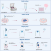
Bone regeneration research: from 2 to 3D in vitro models. Historically, 2D in vitro models were used to study bone physiology and pathology. Monolayer cultures involve cells growing in a single layer on a flat surface, while co-culture models facilitate communication between different cell types. Co-cultures can be direct, allowing physical interaction, or indirect, using transwell membranes to separate cells while permitting medium exchange. 3D models better mimic the morphology and metabolic processes of native bone tissue, promoting osteogenic differentiation. Growing cells on biomaterials creates a complex 3D microenvironment with cell–matrix interactions and mechanical properties comparable to bone. Advanced 3D models include bone organoids cultured in scaffolds that support their growth and self-organization, and spheroids which are aggregates of cells forming a sphere. Microfluidic-based spheroid/organoid-on-chips incorporate miniaturized cell-culturing environments with microchannels and compartments that replicate the natural cell environment, with potential for studying bone regeneration and orthopedic diseases. 2D culture is cheap, simple, and standardized, but lacks the complexity of native tissues. 3D cultures and microfluidics offer higher physiological relevance and throughput, but are more complex, costly, and technically challenging. Bone research models use osteoblastic and osteoclastic mammalian cell cultures from humans and animals, malignant osteosarcoma cell lines, virus immortalized osteoblasts, and human stem cells, particularly induced pluripotent stem cells and mesenchymal stem cells, are promising sources for osteoblast progenitors. Biomaterials like polymers, ceramics, and composites are ideal for bone grafting, while Matrigel and hydrogels are great for encapsulating spheroids and organoids due to their natural ECM-like characteristics
Fig. 2
Fig. 2
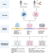
Hierarchical diagram of the models for studying bone tissue. The diagram uses a pyramidal structure to show the logical progression and increasing complexity among the different approaches for studying bone tissue. The Biomaterials form the base of the pyramid. They provide essential physical and chemical support for cell growth, differentiation, and the creation of three-dimensional microenvironments. Transwell and Co-culture Systems represent an intermediate step in complexity. They facilitate cell-to-cell interactions and mutual influence through soluble and physical signals, creating a more realistic microenvironment. Bone Spheroids offer an advanced three-dimensional model, enabling the study of cell-extracellular matrix interactions and simulating the bone microenvironment in vitro. Bone Organoids are the higher level in terms of biological simulation. They replicate more complex structures and functions, such as bone formation and regeneration. Spheroids-on-chip integrate spheroids into a dynamic system that simulates in vivo conditions, such as nutrient flow and mechanical signals. Organoids-on-chip represent the most advanced level. It combines organoids with microfluidic technologies to recreate highly specific physiological microenvironments, providing a platform for complex and personalized studies. The transition from 2 to 3D models and ultimately to chip-based systems is driven by the need to better replicate the biological environment of bone, enabling more realistic models for studying bone regeneration and developing innovative therapies. Each step in the progressive evolution of techniques used to study bone—from bidimensional models to advanced technologies like organoids, spheroids, and chip systems—is based on identifying specific limitations of the previous model and addressing them through innovative approaches
Fig. 3
Fig. 3
Signaling pathways of TGF-β and BMP. The members of the TGFβ and BMP families are involved in bone development, extracellular matrix and cartilage maturation. TGF-β and BMP bind to the extracellular domains of specific receptors and require SMAD proteins for signal transduction within cells. TGF-β and BMP act through heterodimer receptors made up of kinase proteins type I and II. After ligand binding to the receptor, the receptor forms homodimeric complexes, which can auto phosphorylate serine/threonine residues. This triggers a cascade of events involving the phosphorylation of the SMAD protein. The TGF-β pathway requires SMAD2 and SMAD3 which react with SMAD4 to create a heterocomplex. This complex enters the nucleus and controls transcription by binding to target gene promoters including SOX9 and other genes involved in the chondrogenesis mechanism. SMAD7 is an adaptor protein that recruits ubiquitin ligases, called Smurfs and binds them to the TGF-β receptor complex to promote its degradation through proteasomal and lysosomal pathways. Therefore, Smad7 plays a crucial role in a negative feedback cycle to control TGF-β activity. The BMP2 signal depends on SMAD1, 5 and 8. They bind SMAD4 to move into the core, where they induce the expression of RUNX2 and other genes leading to differentiation of osteoblasts and osteocytes
Fig. 4
Fig. 4
Stages of bone fracture healing. After a bone fracture, an inflammatory response occurs that lasts for two weeks. This phase starts an intricate network of proinflammatory signals and growth factors. Polymorphonucleate (PMN) cells and macrophages are recruited to endocyte microdebris and micro-organisms derived from the fracture. The damage to the blood vessels results in edema. After 2–3 weeks from the fracture, endocondral bone formation occurs. During this process, the MSCs are recruited in the injured site and begin to differentiate into chondroblasts (condrogenesis), which proliferate into chondrocytes, resulting in soft calluses. Chondrocytes synthesize and secrete the cartilage matrix, containing type II collagen and proteoglycans. Between the third and sixth week, the cartilage undergoes hypertrophy and mineralization in a spatially organized way. New MSCs are recruited which differentiate into osteoblasts, leading to the formation of interwoven bone (hard callus). Mineralized bone formation is induced by the signaling of factors such as BMP, TGF-β 2 and -β 3 in the cartilaginous callus. The last phase of bone remodeling begins 8 weeks after fracture and can last up to 2 years. Communication between osteoclasts and osteoblasts, during this phase, mediates the replacement of the braided bone with lamellar bone through two key activities: removal of the bone (resorption) by the resulting osteoclasts of the hematopoietic line and formation of the bone matrix by the mesenchymal line osteoblasts
Fig. 5
Fig. 5
Techniques used for spheroids generation. In the pellet culture technique centrifugal force concentrates the cells on the bottom of the tube. The proximity of the single cells at the tube bottom maximizes cell–cell adhesions. After the cell pellets are resuspended in spheroid formation cell culture medium. Cells in the medium are added to a 96-well U-bottom plate with a cell-repellent surface. Liquid overlay culture technique, also called “static suspension culture”, forms spheroids by interrupting the adhesion of cells on non-adherent culture plates. Agarose gel or agar substrate is commonly used to create non-adherent culture layers. By encouraging cell–cell adhesive molecules, cells naturally form spheroids above the non-adherent surface. Droplet-shaped spheroids are generated using the hanging drop culture technique, which efficiently generates specific size spheroids. To achieve appropriate cell density, technique begins with a monolayer cell culture, from which cells are prepared as a suspension with culture media. The cell suspension is then pipetted into wells of a mini plate. After it is completely upside-down. Surface tension keeps the cell suspension drops fixed to the mini plate on the inverted surface. This approach uses the simultaneous action of surface tension and gravitational force to create spheroids as droplets. Magnetic particles are used in magnetic levitation-based culture. During cell growth, cells are combined with magnetic particles and exposed to magnetic force. Cells remain levitation against gravity. This state causes a change in the mass and cell’s shape and encourages cell–cell contact, resulting in cell aggregation
Fig. 6
Fig. 6
Main components of bone organoid production. Bone organoids, derived from stem cells, allow studying how cell–cell and cell-ECM interactions in 3D affect bone differentiation. iPSC organoids are suitable for researching primitive organ biology and physiology. iPSCs are generated by reprogramming fibroblasts with nuclear transcription factors, then adding osteoblast-specific factors for osteogenic differentiation. ASCs, like bone marrow or adipose-derived MSCs, produce single-cell type organoids without needing different differentiation conditions. Bioactive materials, like Matrigel and hydrogels, are key components. Matrigel is a solubilized extract of the basement membrane from Engelbreth-Holm-Swarm mouse sarcoma. Its high levels of collagen and growth factors promote cell adhesion. However, its application is restricted in various contexts because it originates from animals, has unpredictable composition, may be contaminated with xenobiotic contamination and exhibits batch-to-batch variability. Hydrogel is made up of polymers, which can be either natural or synthetic, and can be tailored to have specific chemical and physical properties, creating an optimized environment for cell culture. Its modifiable mechanical, physical, and biological characteristics enable a controlled response from cells. Natural hydrogels closely resemble the extracellular matrix (ECM) because they contain its primary components. Common examples of natural hydrogels include polysaccharides and proteins like collagen, hyaluronic acid, gelatin, chitosan, and alginate. On the other hand, synthetic hydrogels are composed of hydrophilic polymers such as polyethylene glycol, polyvinyl alcohol, polylactic acid, and polyacrylamide

References

    1. Abbasi, Fatemeh, Mohammad Hossein Ghanian, Hossein Baharvand, Bahman Vahidi, e Mohamadreza Baghaban Eslaminejad. 2018. «Engineering Mesenchymal Stem Cell Spheroids by Incorporation of Mechanoregulator Microparticles». Journal of the Mechanical Behavior of Biomedical Materials 84 (agosto):74–87. 10.1016/j.jmbbm.2018.04.026. - PubMed
    1. Abu-El-Rub E, Khaswaneh RR, Almahasneh FA, Almazari R, e Ayman Alzu’bi. Adipose Tissue and Bone Marrow-Derived Mesenchymal Stem Cells Are Not Really the Same: Investigating the Differences in Their Immunomodulatory, Migratory, and Adhesive Profile. Biochemical Genetics, Marzo. 2024. 10.1007/s10528-024-10724-6. - PubMed
    1. Achilli T-M, Meyer J, e Jeffrey R Morgan. Advances in the Formation, Use and Understanding of Multi-Cellular Spheroids. Expert Opin Biol Ther. 2012;12(10):1347–60. 10.1517/14712598.2012.707181. - PMC - PubMed
    1. Aibar-Almazán A, Voltes-Martínez A, Castellote-Caballero Y, Afanador-Restrepo DF, Del Carmen M, Carcelén-Fraile, e Elena López-Ruiz. Current Status of the Diagnosis and Management of Osteoporosis. Int J Mol Sci. 2022;23(16):9465. 10.3390/ijms23169465. - PMC - PubMed
    1. Akiva A, Melke J, Ansari S, Liv N, Van Der Meijden R, Van Erp M, Zhao F, et al. An Organoid for Woven Bone. Adv Func Mater. 2021;31(17):2010524. 10.1002/adfm.202010524.

LinkOut - more resources