. 2025 Mar 20;16(1):2754.
doi: 10.1038/s41467-025-57866-8.

Structure and unusual binding mechanism of the hyaluronan receptor LYVE-1 mediating leucocyte entry to lymphatics

Affiliations

Structure and unusual binding mechanism of the hyaluronan receptor LYVE-1 mediating leucocyte entry to lymphatics

Fouzia Bano et al. Nat Commun. .

Abstract

Immune surveillance involves the continual migration of antigen-scavenging immune cells from the tissues to downstream lymph nodes via lymphatic vessels. To enable such passage, cells first dock with the lymphatic entry receptor LYVE-1 on the outer surface of endothelium, using their endogenous hyaluronan glycocalyx, anchored by a second hyaluronan receptor, CD44. Why the process should require two different hyaluronan receptors and by which specific mechanism the LYVE-1•hyaluronan interaction enables lymphatic entry is however unknown. Here we describe the crystal structures and binding mechanics of murine and human LYVE-1•hyaluronan complexes. These reveal a highly unusual, sliding mode of ligand interaction, quite unlike the conventional sticking mode of CD44, in which the receptor grabs free hyaluronan chain-ends and winds them in through conformational re-arrangements in a deep binding cleft, lubricated by a layer of structured waters. Our findings explain the mode of action of a dedicated lymphatic entry receptor and define a distinct, low tack adhesive interaction that enables migrating immune cells to slide through endothelial junctions with minimal resistance, while clinging onto their hyaluronan glycocalyx for essential downstream functions.


Conflict of interest statement

Competing interests: The authors declare no competing interests.

Figures
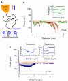
Fig. 1
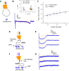
Fig. 1. The nanomechanics of LYVE-1•HA are distinct from CD44•HA and other biomolecular bonds.
A Schematic illustration of the dynamic force spectroscopy (DFS) setup to probe receptor•HA interactions (full wild-type ectodomains: hLYVE-1 Δ238 with a His10 single tag and hCD44 Δ267 with a biotin/His10 dual tag) with an HA chain (840 kDa) anchored via the reducing end (red diamond). Owing to the sharp AFM tip and the low HA grafting density, only one or at most a few HA tails can interact simultaneously with receptors on the substrate. B CD44•HA bonds are conventional sticking bonds. The representative force vs. distance curve (pink —tip approach, green—tip retract) shows a sequence of unbinding events representing the sequential and independent rupture of multiple CD44•HA bonds. Each unbinding event is well-fitted with a worm-like chain model with a 4.1 nm persistence length (red lines), confirming a single HA chain is being stretched. Three representative retract curves (inset, with y axes offset for clarity) illustrate the stochastic nature of CD44•HA bond formation and sequential rupture along the HA chain. See Ref. for a detailed analysis of CD44•HA bond mechanics. C LYVE-1•HA bonds are sliding bonds. The representative force vs. distance curve (pink—tip approach, blue—tip retract) reveals bond mechanics are unlike those of CD44•HA bonds. Three representative retract curves (right inset, with y-axis offset for clarity) illustrate the deterministic nature of LYVE-1•HA interactions, indicating that multiple receptors must act in concert on each HA chain. The gross shape and magnitude of the experimental retract curves are reproduced by a toy reductionist model (left inset; see Supplementary Fig. 2) that assumes HA stochastically moves in steps of one disaccharide and with a zero-force rate constant k = 103 s−1 across the LYVE-1 binding sites and only detaches from a receptor once the chain end is reached. All data are representative of three independent experiments with distinct HA-coated probes and receptor-coated substrates per condition. Source data are provided as a Source Data file.
Fig. 2
Fig. 2. LYVE-1 discriminates HA chains with a free end from closed loops and binds preferentially to the non-reducing terminus.
A Schematic illustration of the setup to probe interactions with closed HA loops where the low LYVE-1 receptor density (hLYVE-1 full wild-type ectodomain) enables probing of single bonds. Key results representative of three independent experiments with distinct HA-coated probes and LYVE-1 coated substrates are shown in (B, C), see Supplementary Figs. 7, 8 for a detailed analysis of the full dataset. B Representative force vs. distance curve (pink—tip approach, blue—tip retract) with a worm-like chain model fit (red line) to the unbinding event. The inset shows effective persistence lengths Lp,eff vs. effective contour lengths Lc,eff for three selected retract velocities (1, 4 and 12 μm/s, covering instantaneous loading rates as indicated with colour code as mean ± SD; a total of n = 285 data points are shown). The effective persistence lengths scatter just above 2 nm, consistent with the simultaneous and parallel stretching of two equal-sized HA chain segments. The effective contour lengths scatter around discrete values (vertical arrowheads indicate means), consistent with the stochastic probing of a small set of loops of distinct size. C Mean rupture forces as a function of instantaneous loading rate (in semi-log presentation; mean ± SD; a total of n = 482 rupture events is included here, see Supplementary Fig. 7C for numbers resolved by loading rate) with a Bell-Evans model fit (black line; best fit (±1 σ confidence interval) parameters for the zero-force unbinding rate koff and the barrier width xβ are displayed). D, F Setups to probe interactions with HA (320 kDa) immobilised via either the reducing (arrowhead; HA-b) or non-reducing (sphere; b-HA) ends, and E, G their representative force curves, offset along the y-axis for clarity (pink—tip approach, blue—tip retract). Both orientations show a response characteristic of deterministic binding to HA tails as in Fig. 1C. Note the magnitude of the force response is threefold weaker for HA with a free-reducing end (G), indicating preferential binding to the non-reducing end. Forces displayed in (G) were increased by a factor of 1.9 to account for a proportionally reduced HA coverage of b-HA over HA-b (see Supplementary Fig. 11). Source data are provided as a Source Data file.
Fig. 3
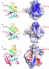
Fig. 3. X-ray crystallographic structures of unbound mLYVE-1 and hLYVE-1 HABDs compared to the mCD44 HABD.
A mLYVE-1, B hLYVE-1, C mCD44. On the left in each case, shown as a ribbon representation with the three disulfides present in each HABD shown in ball-and-stick format, as well as the glycan sidechains at mAsn129/hAsn130 (lefthand in this view) and mAsn52/hAsn53 (righthand in this view). Glycans were not present in the CD44 crystal structure as the protein was expressed and purified from E. coli. Prominent β strands within the structures are labelled, and coloured rainbow wise from the amino terminal to the carboxy terminal, save for β0 (wheat coloured in each case) and β7 and β8 in CD44 (pink). On the right in each case is a surface electrostatics representation scaled −5.0 kBT/e (red) to +5.0 kBT/e (blue) computed using APBS and with the HA binding site identified previously, boxed. Backbone variations between CD44 and LYVE-1 were mLYVE-1 RMSD 0.92 Å for 90 Cα pairs, and 5.2 Å across all 113 pairs; hLYVE-1 RMSD 0.87 Å for 89 Cα pairs, and 4.04 Å across all 115 pairs. The greater variance in the all-pairs comparisons is due principally to the long loop between β5 and β6.
Fig. 4
Fig. 4. Structures of the free and HA-bound complexes of mouse and human LYVE-1 compared.
Surface representations of the unbound (left) and HA-bound (right) structures of mLYVE-1 (A) and hLYVE-1 (B), with key residues in the HA binding site labelled. Significant conformational changes are limited to the HA binding groove. See the main text for more details.
Fig. 5
Fig. 5. Key residues and water-mediated interactions holding HA within the mLYVE-1 binding surface.
Close-ups of the HA binding groove in the mouse receptor with key gatekeeper and ligand residues labelled. In each case, orthogonal views are shown as indicated by the arrow. A mLYVE-1 apoprotein structure, B, C mLYVE-1•HA8 complex. In B, C water molecules resolved and involved in either bridging interaction between HA and LYVE-1 or bridging two or more atomic sites on HA are shown as aquamarine spheres, and hydrogen bonds are indicated by dashed yellow lines. Water molecules are labelled w1-w17 (of which w1–w10 are present for mLYVE-1) – see text for more details and Supplementary Table 2. Atomic surfaces are all protein atoms in (B) and hydrophobic contacts are rendered as a surface in (C). See also Supplementary Movie 1.
Fig. 6
Fig. 6. Key residues and water-mediated interactions holding HA within the hLYVE-1 binding surface.
Close-ups of the HA binding groove in the human receptor with key gatekeeper and ligand residues labelled. In each case, orthogonal views are shown as indicated by the arrow. A hLYVE-1 apoprotein structure, B hLYVE-1•HA10 complex. Resolved water molecules involved in HA: hLYVE-1 or HA:HA interactions are shown as aquamarine spheres with numbering as in Fig. 5, and hydrogen bonds are similarly indicated by dotted yellow lines – see text and Supplementary Table 2 for more details. In C, the ten bound waters considered an index in mLYVE-1 are extended with seven additional waters observed only in the human receptor. Atomic surfaces are all protein atoms in (B), and hydrophobic contacts are rendered as a surface in (C). See also Supplementary Movie 2.
Fig. 7
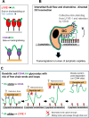
Fig. 7. Hypothetical model of how a sliding mode of HA adhesion/de-adhesion to LYVE-1 facilitates dendritic cell attachment and entry to lymphatic capillaries.
Individual panels show cartoon representations of: A the sequential engagement of an individual HA chain (green) by neighbouring LYVE-1 molecules (red) via end-on binding as opposed to engagement with CD44 via side-on binding, as determined by DFS. The water cushion (blue dots) in the LYVE-1 HABD observed by crystallography and calculated by MD is predicted to facilitate HA chain sliding, B a migrating DC adhering to the basolateral endothelium of an initial lymphatic capillary via its HA glycocalyx and transmigrating at a junction via sequential binding/unbinding of HA from LYVE-1 at the DC uropod, and C a more detailed model showing the proposed sequence of events during such unbinding of the DC HA glycocalyx from lymphatic endothelium (boxed area in B) by collective reverse sliding of HA through LYVE-1, together with stochastic bond rupture between HA and CD44 to leave the DC glycocalyx intact. For purposes of clarity, LYVE-1 is depicted as a monomer.

References

    1. Randolph, G. J., Ivanov, S., Zinselmeyer, B. H. & Scallan, J. P. The lymphatic system: integral roles in immunity. Annu. Rev. Immunol.35, 31–52 (2017). - PMC - PubMed
    1. Randolph, G. J., Angeli, V. & Swartz, M. A. Dendritic-cell trafficking to lymph nodes through lymphatic vessels. Nat. Rev. Immunol.5, 617–628 (2005). - PubMed
    1. Jackson, D. G. Leucocyte trafficking via the lymphatic vasculature—mechanisms and consequences. Front. Immunol. 10, 471 (2019). - PMC - PubMed
    1. Oliver, G., Kipnis, J., Randolph, G. J. & Harvey, N. L. The lymphatic vasculature in the 21(st) century: novel functional roles in homeostasis and disease. Cell182, 270–296 (2020). - PMC - PubMed
    1. Schineis, P., Runge, P. & Halin, C. Cellular traffic through afferent lymphatic vessels. Vascul. Pharmacol.112, 31–41 (2019). - PubMed

MeSH terms