. 2025 Mar 20;9(1):34.
doi: 10.1038/s41538-025-00400-3.

Protective effects of human breast milk-derived exosomes on inflammatory bowel disease through modulation of immune cells

Affiliations

Protective effects of human breast milk-derived exosomes on inflammatory bowel disease through modulation of immune cells

Ki-Uk Kim et al. NPJ Sci Food. .

Abstract

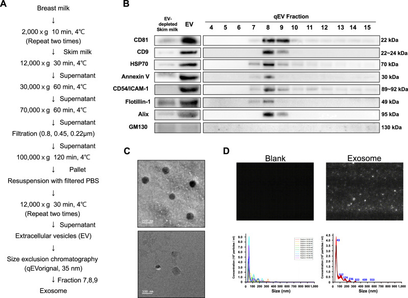
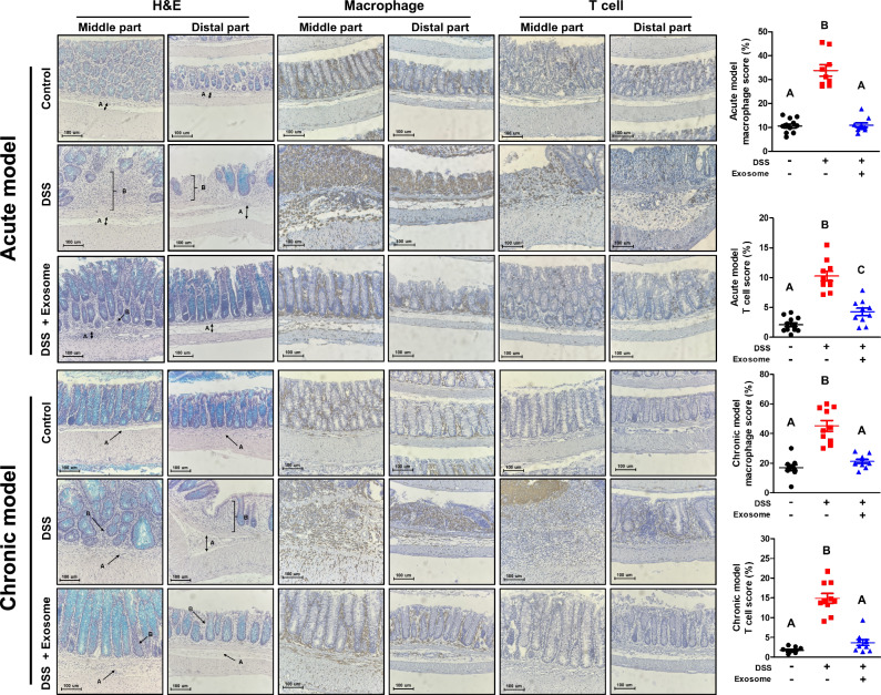
Human breast milk (HBM)-derived exosomes play a crucial role not only in infant nutrition but also in modulating inflammation, immunity, and epithelial cell protection. This study investigated how HBM-derived exosomes regulate immune cell development and function. The exosomes promoted the differentiation of naïve CD4+ T cells into Treg and Th2 cells while suppressing their differentiation into Th17 and Th1 cells. They also enhanced the proliferation of intestinal epithelial Caco-2 cells and reduced apoptosis in dextran sulfate sodium (DSS)-damaged caco-2 cells. In a DSS-induced colitis mouse model, the exosomes significantly alleviated disease severity, as evidenced by improvements in colon length, disease activity index, and histology grades. Furthermore, the exosomes normalized CD4+ T cell subsets in the spleen, mesenteric lymph nodes, and colon, restoring levels comparable to controls. These findings suggest that HBM-derived exosomes hold promise as a potential therapeutic strategy for inflammatory bowel disease by modulating T-cell responses and protecting intestinal epithelial cells.


Conflict of interest statement

Competing interests: The authors have declared that no conflict of interest exists.

Figures

Fig. 1
Fig. 1. Isolation and characterization of human breast milk (HBM)-derived exosome.
A Overview of HBM-derived exosome isolation using ultracentrifugation and size-exclusion chromatography (SEC). Samples of HBM were pooled to minimize sample variation (n = 10). Initially, 400 mL of HBM was subjected to centrifugation at 2000 × g. The upper layer (milk fat) and the pellet (cells and debris) were discarded. The middle portion, known as skim milk, was isolated and utilized for further processing. The final volume of the extracellular vesicle (EV) sample obtained from the skim milk was 3 mL. To sort exosomes, EVs were classified into 15 fractions according to the size by using the SEC (43.92 ± 4.28 μg/μL, n = 11). B characterization of EVs and exosomes were demonstrated by specific exosome markers, such as CD81, CD9, HSP70, Annexin V, CD54/ICAM-1, Flotillin-1, and Alix, and nonspecific exosome marker, GM130, using western blot. C, D Morphology and size were identified by using transmission electron microscopy (TEM; the scale bar is 200 and 100 nm) and nanoparticle tracking analysis (NTA; median of exosome size = 43 nm, the concentration of exosome = 1.39 × 108 particles/mL).
Fig. 2
Fig. 2. Anti-inflammatory effects of HBM-derived exosomes on RAW 264.7 cells.
AC Raw 264.7 cells were treated with HBM-derived exosomes for 24 h and activated with LPS for 20 h. A, B Cultured media were harvested, and expression levels of pro-inflammatory cytokines (TNF-α, IL-6, IL-1β) and other mediators (NO and PGE2) were measured using ELISA and NO assay. C Total protein was extracted from cells. Protein expression of iNOS and COX-2 was demonstrated by western blot. D Raw 264.7 cells were treated with HBM-derived exosomes for 24 h and activated with LPS for 15 min. Phosphorylation of JNK and ERK was identified by western blot. Data represent the means ± SEM (n ≥ 3). The significance of the data were examined by one-way ANOVA with Tukey’s post hoc test (P < 0.05).
Fig. 3
Fig. 3. Effect of HBM-derived exosomes on the differentiation of naïve CD4+ T cells.
AD Naïve CD4+ T cells were treated with the HBM-derived exosomes and differentiated into Th1, Th2, Th17, and Treg cells. Different cytokines (Th17: IL-17a, Treg: IL-10, Th1: IFN-γ, Th2: IL-4) and specific intracellular markers (Th17: RORγt, Treg: Foxp3, Th1: t-bet, Th2: GATA-3) were observed by Flow cytometry. E Total protein was extracted from each differentiated CD4+ T cell, and transcription factors (Th17: STAT3 and p-STAT3, Treg: STAT5 and p-STAT5, Th1: STAT1 and p-STAT1, Th2: STAT6 and p-STAT6) were demonstrated by western blot. Data represent the means ± SEM (n ≥ 3). The significance of the data were examined by one-way ANOVA with Tukey’s post hoc test (P < 0.05).
Fig. 4
Fig. 4. Effects of HBM-derived exosomes on Caco-2 cells.
A The wound closed values were indicated by measuring the percentage of gap closed compared with the 0 h point for each sample. The percent gap closed was shown in a dose- and time-dependent manner as a graph. Microscopic images were captured using a JuLI Stage Real-Time Cell History Recorder at the indicated time points. B Caco-2 cells were treated with the HBM-derived exosomes (1 mg/mL), and total protein was extracted. Protein expression of tight junction proteins (ZO-1, OCLD, CLDN3) were measured by western blot. C Caco-2 cells were pretreated with HBM-derived exosomes for 1 day and damaged for 1 day using DSS. Annexin V/PI staining was used to measure the apoptosis and viability. Data represent the means ± SEM (n ≥ 3). The significance of the data were examined by one-way ANOVA with Tukey’s post hoc test. *P < 0.05; **P < 0.01; ***P < 0.001; ****P < 0.0001.
Fig. 5
Fig. 5. Therapeutic effects of HBM-derived exosomes in acute DSS-induced colitis.
HBM-derived exosomes were orally administrated to mice with acute DSS-induced colitis. AC Various parameters such as body weight, disease activity index (DAI) score, and colon length were measured. D Diverse immune cells, including macrophages, regulatory T cells (Treg cells), Th17 cells, Th1 cells, and Th2 cells, was analyzed in different organs, including the spleen, mesenteric lymph nodes, and colon. Flow cytometry was used as the method to quantify these immune cell populations. Significance of the data were examined by one-way ANOVA with Tukey’s post hoc test. Control (n = 10), DSS group (n = 10), DSS + exosome group (n = 10). Data were presented as mean ± SEM (for A, B) and as individual samples represented by dots with mean values shown as bars (for C, D). *P < 0.05.
Fig. 6
Fig. 6. Therapeutic effects of HBM-derived exosomes in chronic DSS-induced colitis.
HBM-derived exosomes were orally administrated to mice with chronic DSS-induced colitis AC Various parameters such as body weight, disease activity index (DAI) score, and colon length were measured. D Diverse immune cells (macrophage, Treg cell, Th17 cell, Th1 cell, and Th2 cell) in different organs (spleen, mesenteric lymph node, and colon) were measured by flow cytometry. Significance of the data were examined by one-way ANOVA with Tukey’s post hoc test. Control (n = 10), DSS group (n = 10), DSS + exosome group (n = 10). Data were presented as mean ± SEM (for A, B) and as individual samples represented by dots with mean values shown as bars (for C, D). *P < 0.05; **P < 0.01.
Fig. 7
Fig. 7. The histology of the colon in mice with DSS-induced colitis upon exosome treatment.
Hematoxylin & eosin staining and immunohistochemistry was performed to identify the morphological changes in the colon and to detect the infiltration of macrophages and T cells. Significance of the data were examined by one-way ANOVA with Tukey’s post hoc test (P < 0.05). The scale bar is 100 µm. A: extensive edema of the submucosal layer, B: inflammatory cell infiltration. Samples of all groups (n = 10).

References

    1. Kim, K. U., Kim, W. H., Jeong, C. H., Yi, D. Y. & Min, H. More than nutrition: therapeutic potential of breast milk-derived exosomes in cancer. Int. J. Mol. Sci.21, 7327 (2020). - PMC - PubMed
    1. Nutrition, E. C. et al. Breast-feeding: a commentary by the ESPGHAN committee on nutrition. J. Pediatr. Gastroenterol. Nutr.49, 112–125 (2009). - PubMed
    1. Ip, S. et al. Breastfeeding and maternal and infant health outcomes in developed countries. Evid. Rep. Technol. Assess. 1–186 (2007). - PMC - PubMed
    1. Yu, J. C. et al. Innate immunity of neonates and infants. Front. Immunol.9, 1759 (2018). - PMC - PubMed
    1. Lasser, C. et al. Human saliva, plasma and breast milk exosomes contain RNA: uptake by macrophages. J. Transl. Med.9, 9 (2011). - PMC - PubMed

LinkOut - more resources