. 2025 Mar 20;23(1):226.
doi: 10.1186/s12951-025-03312-2.

Selenium nanoparticles activate selenoproteins to mitigate septic lung injury through miR-20b-mediated RORγt/STAT3/Th17 axis inhibition and enhanced mitochondrial transfer in BMSCs

Affiliations

Selenium nanoparticles activate selenoproteins to mitigate septic lung injury through miR-20b-mediated RORγt/STAT3/Th17 axis inhibition and enhanced mitochondrial transfer in BMSCs

Wan-Jie Gu et al. J Nanobiotechnology. .

Abstract

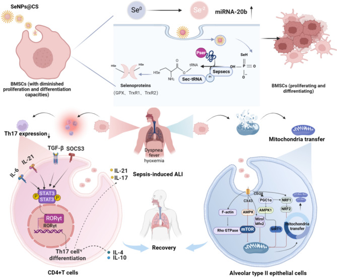
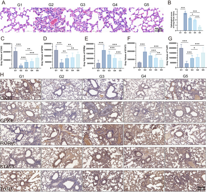
Sepsis-induced acute lung injury (ALI) remains a critical clinical challenge with complex inflammatory pathogenesis. While bone marrow mesenchymal stem cells (BMSCs) demonstrate therapeutic potential through anti-inflammatory and cytoprotective effects, their age-related functional decline limits clinical utility. This study developed chitosan-functionalized selenium nanoparticles (SeNPs@CS, 100 nm) to rejuvenate BMSCs through miR-20b-mediated selenoprotein biosynthesis. Mechanistic investigations revealed that SeNPs@CS-treated BMSCs exhibited enhanced mitochondrial transfer capacity, delivering functional mitochondria to damaged alveolar epithelial cells (AECII) for cellular repair. Concurrently, miR-20b upregulation suppressed the RORγt/STAT3/Th17 axis, reducing pro-inflammatory Th17 cell differentiation in CD4+ T lymphocytes. The dual-target mechanism integrates immunomodulation via Th17 pathway inhibition with mitochondrial rejuvenation therapy, representing a paradigm-shifting approach for ALI management. These engineered BMSCs mitigated inflammatory markers in murine models, demonstrating superior efficacy to conventional BMSC therapies. Our findings establish SeNPs@CS-modified BMSCs as a novel therapeutic platform combining nanotechnology-enhanced stem cell engineering with precision immunometabolic regulation, providing new avenues for the treatment of sepsis-induced ALI.

Keywords: Acute lung injury; BMSCs; Mitochondrial transfer; RORγt/STAT3; Selenium; Th17.


Conflict of interest statement

Declarations. Ethics approval and consent to participate: All animal experimental care and operating procedures were performed in accordance with the Guidelines for Care and Use of Laboratory Animals and approved by by the the Animal Ethical and Welfare Committee (Approval number: IACUC-MIS2023075). Consent for publication: Not applicable. Competing interests: The authors declare no competing interests.

Figures

Scheme 1
Scheme 1
Schematic illustration of SeNPs to mitigate septic lung injury through miR-20b-mediated RORγt/STAT3/Th17 axis inhibition and enhanced mitochondrial transfer in BMSCs
Fig. 1
Fig. 1
Characterization of SeNPs@CS. (A, B) Representative TEM; (A) Images of SeNPs@CS. (C) Particle size distribution of SeNPs@CS. (D) Zeta potential of CS and SeNPs@CS. (E) UV spectra of CS and Se@CS NPs. (F) IR spectra of CS and SeNPs@CS. (G-I) XPS spectra of SeNPs@CS: (G) Survey spectrum, (H) C 1s spectrum, and (I) Se 3d spectrum
Fig. 2
Fig. 2
(A) Mechanistic studies on the promotion of BMSCs proliferation and differentiation by SeNPs@CS. (B) Detection of BMSCs proliferation using CCK-8 assay. (C) q-PCR analysis for the expression of miRNA-20b. (D) Determination of mitochondrial ATP content. (E) Measurement of mitochondrial OCR content. (F, G, H, I) Flow cytometry analysis for BMSCs markers CD44, CD105, and CD29. (J, K) Determination of mitochondrial membrane potential. (L, M) Detection of mitochondrial Ca2+ content. (N, O) Quantification of mitochondrial number. (P) Western blot analysis for the expression of selenium-containing proteins GPX4, TrxR1, and TrxR2. G1: Control; G2: SeNPs@CS 5 µM; G3: SeNPs@CS 10 µM; G4: SeNPs@CS 20 µM; G5: SeNPs@CS 40 µM. ***(P < 0.001), **(P < 0.01), *(P < 0.05)
Fig. 3
Fig. 3
Bioinformatic analysis of the targeting relationship between miRNA-20b and RORγt/STAT3 and their inflammatory levels. (A) Schematic diagram of the miRNA-20b and RORγt/STAT3 pathway. (B) Venn diagram representation. (C) Dual-luciferase reporter gene assay for miRNA-20b and STAT3. (D, E, F, G) Qualitative and quantitative analysis of Th17 cell markers in different groups. (H, I, J, K, L, M, N, O) Expression levels of TGF-β, IL-17, IL-6, IL-21, Socs3, IL-10, IL-2, and IL-4. G1: CD4 + T; G2: OE-RORγtCD4 + T; G3: BMSCs + RORγtCD4 + T; G4: OE-miRNA-20b-BMSCs + OE-RORγtCD4 + T; G5: SeNPs@CS + OE-miRNA-20b-BMSCs + OE-RORγt CD4 + T. ***(P < 0.001), **(P < 0.01), *(P < 0.05)
Fig. 4
Fig. 4
SeNPs@CS synergizes with BMSCs to promote mitochondrial repair in damaged AECII cells. (A, B) Qualitative and quantitative detection of mitochondrial membrane potential. (C) Detection of mitochondrial ATP content. (D, E) Qualitative and quantitative detection of mitochondrial calcium ion content. (F) Detection of mitochondrial OCR content. (G, H) Qualitative and quantitative detection of cell apoptosis by flow cytometry. (I) Detection of mitochondrial NAD+ content. (J) Transmission electron microscopy (TEM) examination of mitochondrial structure. G1: LPS; G2: BMSCs; G3: OE-miR-20b-BMSCs; G4:SeNPs@CS + OE-miR-20b-BMSCs. ***(P < 0.001), **(P < 0.01), *(P < 0.05)
Fig. 5
Fig. 5
Detection of the mitochondrial exchange laws. (A) Mitochondrial transfer was detected using WB, q-PCR, and flow cytometry. (B, C, D, E, F, G) WB was used for qualitative and quantitative analysis of Cx43, PGC1α, Miro1, CD38, and F-αctin. (H, I, J, K, L) Theexpression levels of Cx43, PGC1α, Miro1, F-αctin, and CD38 were determined by q-PCR. (M) Flow cytometry was utilized for quantitative analysis of mitochondrial transfer in BMSCs. (N)flow cytometry was employed for quantitative assessment of mitochondrial transfer in AECII cells. G1: LPS; G2:BMSCs; G3: OE-miR-20b-BMSCs; G4: SeNPs@CS + OE-miR-20b-BMSCs. ***(P < 0.001), **(P < 0.01), *(P < 0.05)
Fig. 6
Fig. 6
Flow cytometry and ELISA were used to detect the expression of Th17 and pro/anti-inflammatory factors. (A, B, C, D) Flow cytometry detection of Th17 expression in alveolar lavage fluid. (E, F, G, H) Flow cytometry detection of Th17 expression in peripheral blood. (I, J, K, L, M) ELISA detection of pro-inflammatory factor expression in peripheral blood. (N, O, P) ELISA detection of anti-inflammatory factor expression in peripheral blood. G1: Sham; G2: Model; G3: BMSCs; G4: OE-miR-20b-BMSCs; G5: SeNPs@CS + OE-miR-20b-BMSCs. ***(P < 0.001), **(P < 0.01), *(P < 0.05)
Fig. 7
Fig. 7
Explore the therapeutic mechanism in the animals. (A) Detection of selenoenzymes and mitochondrial transfer-related genes and proteins in lung tissue. (B) WB analysis was conducted to detect the expression levels of RORγt, STAT3, GPX4, TrxR1, TrxR2, Cx43, PGC1α, Miro1, and CD38. (C, D, E) ELISA assay was performed to measure the expression of ROS, SOD, and MDA. (F, G, H, I, J, K, L, M) q-PCR was used to assess the mRNA levels of RORγt, STAT3, GPX4, TrxR1, Cx43, PGC1α, Miro1, and CD38. (N) The wet/dry weight ratio of the lungs was calculated. (O) Survival curves were generated to assess the survival rate of mice. G1: Sham; G2: Model; G3: BMSCs; G4: OE-miR-20b-BMSCs; G5: SeNPs@CS + OE-miR-20b-BMSCs. ***(P < 0.001), **(P < 0.01), *(P < 0.05)
Fig. 8
Fig. 8
Pathological histochemical detection in vivo. (A, B) HE of mouse lung tissue was carried out. (C, D, E, F, G, H) Immunohistochemical detection of Cx43, GPX4, RORγt, STAT3, and TrxR1 expression in mouse lung tissue. G1: Sham; G2: Model; G3: BMSCs; G4: OE-miR-20b-BMSCs; G5: SeNPs@CS + OE-miR-20b-BMSCs. ***(P < 0.001), **(P < 0.01), *(P < 0.05)

References

    1. Ericson JE, Agthe AG, Weitkamp JH. Late-Onset sepsis: epidemiology, microbiology, and controversies in practice. Clin Perinatol. 2025;52(1):33–45. - PubMed
    1. Xu JQ, Zhang WY, Fu JJ, Fang XZ, Gao CG, Li C, et al. Viral sepsis: diagnosis, clinical features, pathogenesis, and clinical considerations. Mil Med Res. 2024;11(1):78. - PMC - PubMed
    1. Prescott HC, Angus DC. Enhancing recovery from sepsis: A review. JAMA. 2018;319(1):62–75. - PMC - PubMed
    1. Rudd KE, Johnson SC, Agesa KM, Shackelford KA, Tsoi D, Kievlan DR, et al. Global, regional, and National sepsis incidence and mortality, 1990–2017: analysis for the global burden of disease study. Lancet. 2020;395(10219):200–11. - PMC - PubMed
    1. Sun B, Lei M, Zhang J, Kang H, Liu H, Zhou F. Acute lung injury caused by sepsis: how does it happen? Front Med (Lausanne). 2023;10:1289194. - PMC - PubMed

MeSH terms