. 2025 Mar 20;20(1):34.
doi: 10.1186/s13024-025-00813-4.

A reciprocal relationship between markers of genomic DNA damage and alpha-synuclein pathology in dementia with Lewy bodies

Affiliations

A reciprocal relationship between markers of genomic DNA damage and alpha-synuclein pathology in dementia with Lewy bodies

David J Koss et al. Mol Neurodegener. .

Abstract

Background: DNA damage and DNA damage repair (DDR) dysfunction are insults with broad implications for cellular physiology and have been implicated in various neurodegenerative diseases. Alpha-synuclein (aSyn), a pre-synaptic and nuclear protein associated with neurodegenerative disorders known as synucleinopathies, has been associated with DNA double strand break (DSB) repair. However, although nuclear aSyn pathology has been observed in cortical tissue of dementia with Lewy body (DLB) cases, whether such nuclear pathology coincides with the occurrence of DNA damage has not previously been investigated. Moreover, the specific types of DNA damage elevated in DLB cases and the contribution of DNA damage towards Lewy body (LB) formation is unknown.

Methods: DNA damage and aSyn pathology were assessed in fixed lateral temporal cortex from clinically and neuropathologically confirmed DLB cases and controls, as well as in cortical tissue from young 3-month-old presymptomatic A30P-aSyn mice. Frozen lateral temporal cortex from DLB and control cases was subject to nuclear isolation, western blotting, aSyn seed amplification and proteomic characterisation via mass spectrometry.

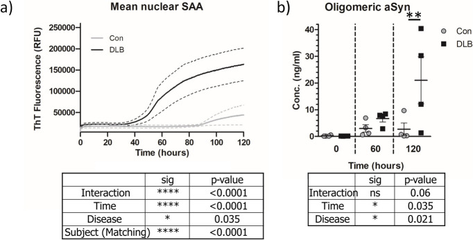
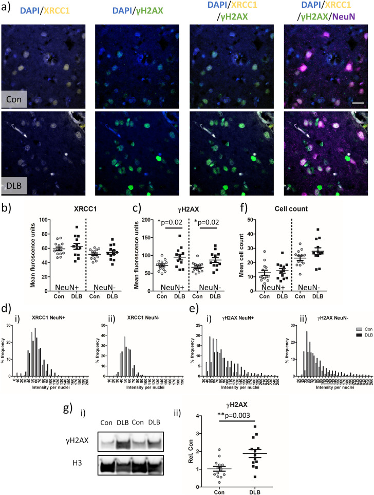
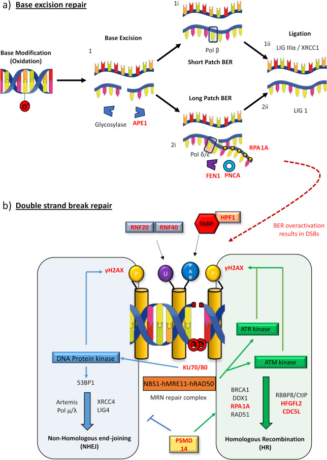
Results: We detected seed-competent nuclear aSyn, and elevated nuclear serine-129 phosphorylation in DLB temporal cortex, alongside the accumulation of DSBs in neuronal and non-neuronal cellular populations. DNA damage was also present in cortical tissue from presymptomatic A30P mice, demonstrating it is an early insult closely associated with pathogenic aSyn. Strikingly, in postmortem DLB tissue, markers of genomic DNA damage-derived cytoplasmic DNA (CytoDNA) were evident within the majority of LBs examined. The observed cellular pathology was consistent with nuclear upregulation of associated DDR proteins, particularly those involved in base excision repair and DSB repair pathways.

Conclusions: Collectively our study demonstrates the accumulation of seed-competent pathological nuclear associated aSyn, alongside nuclear DNA damage and the potential involvement of DNA damage derived cytoDNA species in cytoplasmic aSyn pathology. Ultimately, our study supports the hypothesis of a reciprocal relationship between aSyn pathology and nuclear DNA damage and highlights a potential underlying role for DNA damage in pathological mechanisms relevant to DLB, as well as other synucleinopathies, opening novel possibilities for diagnosis and treatment.

Keywords: Alpha-synuclein; DNA damage; synucleinopathy; Parkinson’s disease; Dementia with Lewy bodies.


Conflict of interest statement

Declarations. Ethics approval and consent to participate: The use of human tissue throughout this study was in accordance with Newcastle University Ethics Board (The Joint Ethics Committee of Newcastle and North Tyneside Health Authority, reference: 08/H0906/136). Mice from which tissue was obtained were housed and subject to procedures in accordance with the UK Animals (Scientific Procedures) Act 1986 and European Union directive 2010/63EU. Consent for publication: N/A. Competing interests: N/A.

Figures

Fig. 1
Fig. 1
DLB nuclear extracts are seed competent. a Thioflavin T fluorescence traces from seed amplification assay (SAA) of nuclear extractions from controls (Con) and dementia with Lewy body (DLB) cases (n = 4, per group). Mean trace per group ± SEM (dotted lines) is shown. b SAA products taken at 20, 60 120h of reaction time were quantified for oligomeric aSyn concentration as detected via Syn-O2 antibody ELISA. Data shown as scatter plot of aSyn oligomer concentration per disease group at relevant time points. Data shown as Mean ± SEM. Outcome of 2 way ANOVA shown in corresponding plots and significant Bonferroni post-test indicated. * = p < 0.05, ** = p < 0.01, **** = p < 0.0001
Fig. 2
Fig. 2
Double strand breaks are increased in temporal cortex nuclei of DLB cases. a Example confocal images (63 × objective) from control (Con) and cases of dementia with Lewy bodies (DLB) showing single strand break (XRCC1) and double strand break (γH2AX) associated immuno-fluorescence in neurons (NeuN) and non-neuronal cells. Nuclei are co-stained with DAPI. Quantification of mean b) XRCC1 c) γH2AX fluorescence signal, alongside f) nuclei based cell counts from Con (n = 13 cases) and DLB (n = 12 cases) are presented. Additionally, di-ii) frequency distribution of XRCC1 and ei-ii) γH2AX per nuclei are shown. Each measure output is reported for neuronal (NeuN +) and non-neuronal (NeuN-) populations. gi Western blot images of γH2AX and loading control Histone H3 and gii) quantification of loading adjusted γH2AX signal in Con and DLB cases (n = 14 and 13 respectively; g.ii) are also shown. Data are expressed in scatterplots with mean ± SEM (in b,c,f and g) and as percentage frequency of immunofluorescence intensity per bins of 10 units width (in d + e). Statistical outcome of Mann–Whitney non-parametric tests shown in appropriate plots, * = p < 0.05 and ** = p < 0.01. Scale = 20 µm
Fig. 3
Fig. 3
Double strand breaks are increased at 3 months of age in an A30P mouse model of synucleinopathy. a Immunofluorescence confocal images (63 × objective lens) of XRCC1, γH2AX, NeuN and nuclei co-stained with DAPI from the somatosensory cortex of wild type (WT) and A30P mice. Quantification of neuronal (NeuN +) and non-neuronal (NeuN -) mean b) XRCC1 and c) γH2AX signal in WT (n = 5) and A30P mice (n = 4). Frequency distribution of d) XRCC1 and e) γH2AX per nuclei are shown in NeuN + (i) and NeuN – (ii) nuclei are also shown alongside f) cell count, as per nuclei number. Data are expressed in scatterplots with mean ± SEM (in b, c and f) and as percentage of immunofluorescence intensity binned with 10 unit widths (d and e). Statistical outcome of Mann–Whitney non-parametric tests shown in appropriate plots, * = p < 0.05. Scale = 20 µm
Fig. 4
Fig. 4
Phosphorylated aSyn increases in concert with double strand breaks and correlate with each other in the nuclear compartment. a Example confocal images (63 × objective) from control (Con) and cases of dementia with Lewy bodies (DLB) showing alpha-synuclein phosphorylated at serine 129 (pS129), double strand break (γH2AX) immuno-fluorescence in neurons (NeuN) and non-neuronal cells.. Intra-nuclear yH2AX and ps129 signal were confirmed via b) Z-stack reconstructed orthogonal views of digitally zoomed neuronal nuclei images captures from DLB cases. Quantification of c) nuclear pS129 (pS129 aSynNuc) and d) γH2AX fluorescence from Con (n = 13 cases) and DLB (n = 12 cases) are presented. ei-ii Frequency distribution of ps129 (per nuclei) are shown as well as fi-ii) correlative levels of ps129 aSynNuc with levels of γH2AX, per case, with spearman's correlation (r) reported. Measures are reported for neuronal (NeuN +) and non-neuronal (NeuN-) populations. Data are expressed in scatterplots with mean ± SEM (in c, d) and as percentage frequency of immunofluorescence intensity per bins of 10 units width (in e). Statistical outcome of Mann–Whitney non-parametric tests and Spearman’s (r) correlation shown in appropriate plots, * = p < 0.05, ** = p < 0.01 and ** = p < 0.001. Scale = 20 µm
Fig. 5
Fig. 5
Nuclear material is a core constituent of cortical Lewy bodies. a Example widefield images (i. 20 × objective) cases of dementia with Lewy bodies (DLB) showing Lewy bodies (LBs) as detected via alpha-synuclein phosphorylated at serine 129 (pS129), γ H2AX and b) 53BP-1 immuno-fluorescence, with nuclei are co-stained with DAPI. Digitally zoomed images (ii) show LBs immunoreactivity (arrowhead) and Lewy neurites (arrows) in greater magnification. Note the colocalization of γH2AX with extranuclear pS129 LBs (a) and absence of colocalization of 53BP-1 with LBs (b). c Confocal image of YH2AX and d) H3K27me3 reactive LBs. Quantification of cii) γH2AX and dii) H3K27me3 positive (γH2AX +) and negative (γH2AX -) LBs in examined cases. Data shown as percentage LBs positive and negative for γH2AX and H3K27me3. Scale bar in a.i + b.i = 100 µm, in a.ii + bii = 50 µm and in c + d = 20 µm
Fig. 6
Fig. 6
Altered nuclear proteome of DLB temporal cortex tissue. a Upregulated, downregulated and unchanged nuclear proteins in temporal cortex of dementia with Lewy body cases (DLB), compared to controls (Con). Data provided as absolute numbers and percentages of total identified proteins. b Z-score heatmap of altered proteins between Con and DLBs. c Uniprot keyword enrichment of the 89 proteins established as elevated in nuclear proteome of DLB compared to controls. Fold enrichment and significance is shown as per false discovery rate for each individual keyword. Keywords are grouped into clusters as reported per the DAVID bioinformatic database. Dotted lines denote the boundaries of clusters, with cluster enrichment (C.Enrich) from background reference database shown within each cluster. d STRING map of protein interactome of upregulated proteins. Prominent functional category of “Cellular response to DNA damaging stimulus” is highlighted (red) with individual proteins associated with each term denoted by colour and hub of strong interaction associated highlighted by background colour. Physical and functional interactions are shown with the weighting of line indicating confidence in interactions. e STRING interaction map and f) Z-score heat map of upregulated proteins associated with “Cellular response to DNA damaging stimulus” plotted in isolation
Fig. 7
Fig. 7
Upregulated protein components of DDR in DLB and principal pathways. a Base excision repair (BER) diagram. Following oxidation (as shown), alkylation or deamination, modified bases are excised via glycosylase activity and deoxyribose-phosphate DNA backbone cut by APEX1 [1]. Repair proceeds as short patch (i) or long patch repair (ii). In short patch BER, the single nucleotide gap is repaired by β-polymerase (2.i) and the DNA ligase IIIα-XRCC1 complex (2.ii). Long patch BER, activated in cases of excess oxidation, produces a 2–8 nucleotide sequence by δ/ε polymerases, which is bound to replication protein 1 A (RPA1A). Excessive sequence is then cleavage by FEN1, facilitated by proliferating nuclear protein A (PCNA) (2.ii), before ligated by DNA ligase I [99, 100] (3.ii). When overactive, BER can result in double strand breaks (DSBs) [–95]. b Non-homologous end-joining (NHEJ) and homologous recombination (HR) DSBs repair pathways. In NHEJ, DSBs are sensed by DNA–protein kinase (DNA-PK) regulatory subunits KU70/KU80, activating DNA-PK, enabling histone phosphorylation generating DSB signal γH2AX. Surrounding chromatin DSB is relaxed, in part due to histone ubiquitination (U) via ubiquitin ligases including ring finger 20/40 (RNF20/40) [56]. NHEJ components are recruited to the DSB, including the 53BP1, Artemis nuclease, DNA polymerase µ/λ and XRCC4/DNA ligase 4 [96]. HR is initiated via the DSB sensing of the MRN repair complex, activating Ataxia-telangiectasia mutated (ATM) and ataxia telangiectasia and Rad3-related (ATR) protein kinases, generating γH2AX signal [3]. CDC5L is a regulator of ATR kinases [101]. DSBs strands are resected by MRE11 promoted by BRCA1 and RBBP8/CtIP [3], facilitated by HDGFL2 [102], RNA–DNA hybrids are cleared via DEAD Box 1 (DDX1) [103, 104], prior to RP1A coverage, RAD51 guided homologous DNA search and strand invasions prior to holiday junction formation and repair [3]. HR engagement over NHEJ is part co-ordinated by PSMD14, which restricts the accumulation of NHEJ inducer 53BP1 at damaged sites [105]. DDR protein upregulated in DLB nuclei highlighted in red

References

    1. De Bont R, van Larebeke N. Endogenous DNA damage in humans: a review of quantitative data. Mutagenesis. 2004;19(3):169–85. - PubMed
    1. Jackson AL, Loeb LA. The contribution of endogenous sources of DNA damage to the multiple mutations in cancer. Mutat Res. 2001;477(1–2):7–21. - PubMed
    1. Chatterjee N, Walker GC. Mechanisms of DNA damage, repair, and mutagenesis. Environ Mol Mutagen. 2017;58(5):235–63. - PMC - PubMed
    1. McKinnon PJ. Maintaining genome stability in the nervous system. Nat Neurosci. 2013;16(11):1523–9. - PMC - PubMed
    1. Lodato MA, Rodin RE, Bohrson CL, Coulter ME, Barton AR, Kwon M, et al. Aging and neurodegeneration are associated with increased mutations in single human neurons. Science. 2018;359(6375):555–9. - PMC - PubMed