

Napkin-ring sign plaques are associated with cerebral small vessel disease
- PMID: 40114260
- PMCID: PMC11927270
- DOI: 10.1186/s40001-025-02371-3
Napkin-ring sign plaques are associated with cerebral small vessel disease
Abstract
Background: Few studies have investigated the association between the carotid artery napkin-ring sign (NRS) and cerebral small vessel disease (CSVD). This study aimed to investigate whether carotid NRS plaque burden and CSVD are associated.
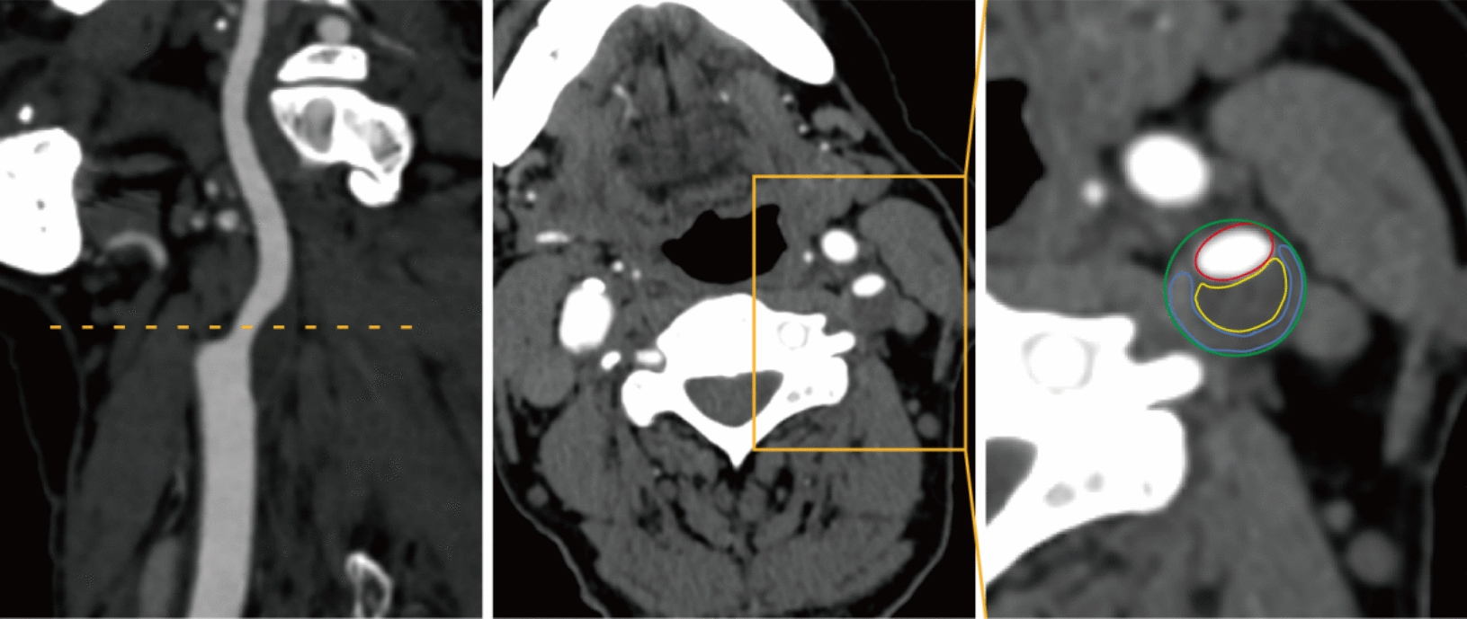
Methods: This retrospective, single-center, cross-sectional study following STROBE guidelines enrolled patients with symptoms or clinical suspicion of anterior circulation acute ischemic stroke (AIS). Plaques were evaluated using preoperative cervicocerebral computed tomography angiography (CTA). Imaging markers of CSVD, such as white matter hyperintensities (WMHs) and perivascular spaces (PVSs), were assessed.
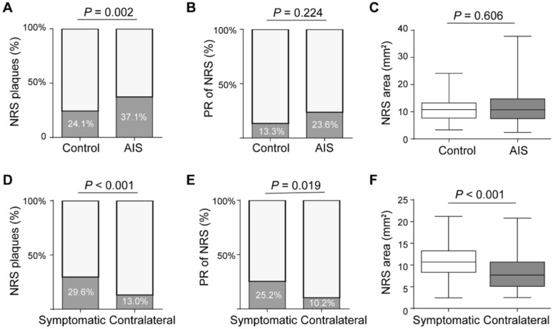
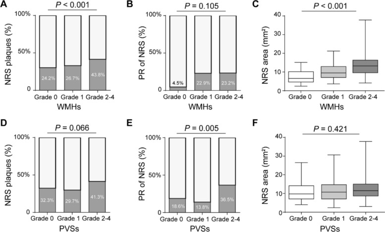
Results: A total of 575 patients (64.9 ± 8.0 years, 378 men) were evaluated. Patients with AIS had a higher percentage of total NRS plaques than those in the control group (144 (37.1%) vs. 45 (24.1%), P = 0.002), and the total NRS amount increased the risk of AIS after adjusting for confounding factors (odds ratio 1.717; 95%CI 1.141-2.584; P = 0.009). A higher WMHs grade was associated with the presence of NRS plaques (P < 0.001) and a higher total NRS area (P < 0.001). A higher PVSs grade was associated with positive remodeling (PR) on the NRS (P = 0.006).
Conclusions: An increased incidence of NRS plaques on CTA was associated with the occurrence of AIS, and the area and PR of NRS plaques were associated with the risk stratification of CSVD.
Keywords: Acute ischemic stroke; Cerebral small vessel disease; Napkin-ring sign plaque.
© 2025. The Author(s).
Conflict of interest statement
Declarations. Ethics approval and consent to participate: This study was approved by the ethical review committee of Taizhou Central Hospital waived of the requirement for informed consent and approved the study (ZFPH No: 2025L-01-06). All subject records and data were deidentified and anonymized prior to analysis. This study was conducted in accordance with the Declaration of Helsinki. Consent for publication: Not applicable. Competing interests: The authors declare no competing interests.
Figures
Similar articles
-
Increased incidence of napkin-ring sign plaques on cervicocerebral computed tomography angiography associated with the risk of acute ischemic stroke occurrence.Eur Radiol. 2024 Jul;34(7):4438-4447. doi: 10.1007/s00330-023-10404-w. Epub 2023 Nov 25. Eur Radiol. 2024. PMID: 38001250
-
Associations between carotid plaques and white matter hyperintensities in cerebral small vessel disease.J Clin Neurosci. 2024 Nov;129:110871. doi: 10.1016/j.jocn.2024.110871. Epub 2024 Oct 20. J Clin Neurosci. 2024. PMID: 39433006
-
Carotid vulnerable plaque coexisting with cerebral small vessel disease and acute ischemic stroke: a Chinese Atherosclerosis Risk Evaluation study.Eur Radiol. 2022 Sep;32(9):6080-6089. doi: 10.1007/s00330-022-08757-9. Epub 2022 Apr 2. Eur Radiol. 2022. PMID: 35364716
-
Association between carotid artery perivascular fat density and cerebral small vessel disease.Aging (Albany NY). 2021 Jul 21;13(14):18839-18851. doi: 10.18632/aging.203327. Epub 2021 Jul 21. Aging (Albany NY). 2021. PMID: 34289452 Free PMC article.
-
Total cerebral small vessel disease burden and stroke outcomes in large vessel occlusion stroke receiving endovascular treatment: A systematic review and meta-analysis.J Clin Neurosci. 2024 May;123:179-185. doi: 10.1016/j.jocn.2024.04.003. Epub 2024 Apr 6. J Clin Neurosci. 2024. PMID: 38583374
References
-
- Bushnell C, Kernan WN, Sharrief AZ, et al. 2024 guideline for the primary prevention of stroke: a guideline from the American Heart Association/American Stroke Association. Stroke. 2024;55(12):e344–424. 10.1161/STR.0000000000000475. - PubMed
-
- Bos D, Wolters FJ, Darweesh SKL, et al. Cerebral small vessel disease and the risk of dementia: a systematic review and meta-analysis of population-based evidence. Alzheimers Dement. 2018;14(11):1482–92. 10.1016/j.jalz.2018.04.007. - PubMed
-
- Seifarth H, Schlett CL, Nakano M, et al. Histopathological correlates of the napkin-ring sign plaque in coronary CT angiography. Atherosclerosis. 2012;224(1):90–6. 10.1016/j.atherosclerosis.2012.06.021. - PubMed
MeSH terms
Grants and funding
LinkOut - more resources
Full Text Sources
Research Materials
