Case Reports
. 2025 Feb 28;14(2):639-648.
doi: 10.21037/tlcr-24-707. Epub 2025 Feb 27.

Unravelling the complexity of EGFR-mutated lung adenocarcinoma: a unique case report with histological transformations and co-alteration acquisition

Affiliations
Case Reports

Unravelling the complexity of EGFR-mutated lung adenocarcinoma: a unique case report with histological transformations and co-alteration acquisition

Anissa Boutahir et al. Transl Lung Cancer Res. .

Abstract

Background: Osimertinib, a third-generation tyrosine kinase inhibitor that targets epidermal growth factor receptor (EGFR), specifically inhibits both EGFR tyrosine kinase inhibitor-sensitive mutations and T790M resistance mutations. Despite initial positive responses to EGFR tyrosine kinase inhibitors, nearly all patients eventually experience disease progression. Mechanisms of resistance are classically divided into EGFR-dependent and EGFR-independent mechanisms, such as the activation of alternative pathways and histological changes. We report a case of histological transformation into large cell carcinoma associated with the subsequent acquisition of an anaplastic lymphoma kinase (ALK) rearrangement after osimertinib exposure.

Case description: A 67-year-old female with no smoking history presented with supraclavicular lymphadenopathy and asthenia, which led to a diagnosis of stage IVB lung adenocarcinoma. Next generation sequencing (NGS) identified an EGFR Ex19del mutation, which suggested the use of afatinib, as it was prescribed prior to osimertinib and was covered by insurance. Initial treatment with afatinib resulted in partial remission, followed by pulmonary progression without the EGFR-T790M mutation. Moreover, ALK and ROS1 were identified through immunohistochemistry (IHC), with ROS1 expression subsequently confirmed by fluorescence in situ hybridization (FISH); this prompted a switch to crizotinib, which was discontinued owing to further disease progression. Osimertinib was then administered, which resulted in a significant positive response; however, after six months pulmonary progression was observed. A subsequent biopsy indicated a transformation to large cell neuroendocrine carcinoma, which led to treatment with platinum-etoposide chemotherapy and, later, paclitaxel and osimertinib, both of which are partially effective. Finally, a new biopsy confirmed ALK positivity in a large cell neuroendocrine carcinoma that was still harbouring an EGFR exon 19 deletion, so alectinib was introduced.

Conclusions: To our knowledge, this case is the first reported incidence of transformation into large cell carcinoma coupled with a second acquisition of alterations in ALK. These findings underscore the necessity of monitoring patients with oncogenic addiction through both liquid biopsy for on-target mechanism detection and tissue sampling to detect histological transformations. These mechanisms can occasionally be combined, thereby providing comprehensive panels at each stage of tumour progression.

Keywords: Epidermal growth factor receptor (EGFR); anaplastic lymphoma kinase (ALK); case report; large-cell neuroendocrine carcinoma (LCNEC); transformation.


Conflict of interest statement

Conflicts of Interest: All authors have completed the ICMJE uniform disclosure form (available at https://tlcr.amegroups.com/article/view/10.21037/tlcr-24-707/coif). J.B.O. reports payment or honoraria for scientific presentations from Astra-Zeneca and Pierre Fabre; payment for expert testimony from Novartis and support for attending meetings and travel from Astra-Zeneca, all outside of the submitted work. G.D. reports payment or honoraria for lectures, presentations, speakers’ bureaus, manuscript writing or educational events from AstraZeneca, Chiesi, Sanofi, and GSK (personal fees), outside of the submitted work. J.A. reports grants from AMGEN, and French Innovative Research Fund; payment or honoraria for lectures, presentations, speakers’ bureaus, manuscript writing or educational events from Roche, Pfizer, MSD, Bristol-Myers Squibb, Novartis, AstraZeneca, Takeda, Sanofi, and Amgen; support for attending meetings and/or travel from Roche, Pfizer, MSD, Takeda, and Sanofi. The other authors have no conflicts of interest to declare.

Figures

Figure 1
Figure 1
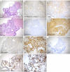
Biopsies revealed a metastatic lung adenocarcinoma, supported by immunohistochemical markers. The tumor was positive for TTF-1 and CK7, negative for neuroendocrine markers, and showed no ALK or ROS1 rearrangements by IHC, though ROS1 was positive by FISH. Biopsy of a supraclavicular lymph node showing metastatic tumour proliferation with a tubulo-glandular architecture, composed of cylindrical cells with clear, mucin-secreting cytoplasm (A, HE, ×100). The nuclei exhibit marked cytonuclear atypia, including anisokaryosis and hyperchromasia (B, HE, ×200). The tumour was positive for TTF-1 (C, IHC, ×200) and CK7 (D, IHC, ×200). It was negative for neuroendocrine markers (E, IHC, ×200), including CD56, synaptophysin, and chromogranin A (top to bottom). Bronchial biopsy of the primary tumour revealed that the tumour cells were arranged either as isolated cells or in three-dimensional structures (F, HE, ×200), with a markedly increased nuclear-cytoplasmic ratio. The nuclei were vesicular with prominent nucleoli. These cells focally infiltrated the bronchial wall within a reactive fibroinflammatory stroma (G, HE, ×200). The tumour was positive for TTF-1 (H, IHC, ×100) and CK7 (I, IHC, ×50) but negative for neuroendocrine markers (J, IHC, ×200), including CD56 and chromogranin A (top to bottom). ALK (K, IHC, ×200) and ROS1 (L, IHC, ×200) staining by IHC was negative, whereas ROS1 was positive by FISH analysis (M) (×400). IHC was performed with anti-ALK clone 1A4 (Diagomics) and anti-ROS1 clone D4D6 (Ozyme) antibodies on a Ventana instrument (Roche Diagnostics). Positive staining corresponded to the presence of a cytoplasmic signal for more than 10% of the tumour cells. FISH analysis was performed with the ZytoLight SPEC Dual Colour Break Apart Kit (Zytovision) for ALK and ROS1, with the split orange signals indicated by red arrowheads; a nucleus was interpreted as positive for translocation if the split orange and green signals were separated by more than two signal diameters. ALK, anaplastic lymphoma kinase; IHC, immunohistochemistry; FISH, fluorescence in situ hybridization; HE, hematoxylin and eosin; TTF-1, thyroid transcription factor-1.
Figure 2
Figure 2
Further biopsies revealed transformation into large-cell neuroendocrine carcinoma as suggested by loss of TTF-1 and CK7 expression, positivity for neuroendocrine markers CD56, synaptophysin, chromogranin A) and a high proliferative index with Ki67 >80%. This histologic transformation was accompanied by the acquisition of an ALK translocation, as evidenced by a 3+ staining pattern for ALK, with persistent absence of ROS1 expression. HE staining showed bronchial parenchyma infiltrated by poorly differentiated proliferating tumours with a carcinomatous architecture composed of confluent sheets of large cells (A, HE, ×50). These cells had hyperchromatic nuclei, sometimes with irregular contours and prominent nucleoli, which displayed a fine salt-and-pepper chromatin pattern. The cytoplasm was eosinophilic, sparse, and had well-defined borders (B, HE, ×100). The tumour cell sheets were separated by extensive areas of necrosis. The tumour cells were negative for TTF-1 (C, IHC, ×200) and CK7 (D, IHC, ×200) staining. Tumour cells were positive for neuroendocrine markers, including CD56 (moderate staining, E, IHC, ×200), synaptophysin (intense staining, F, IHC, ×200), and chromogranin A (moderate staining, G, IHC, ×200). More than 80% of the tumour cells were positive for Ki67 (H, IHC, ×200). ROS1 staining remained negative by IHC (I, IHC, ×100), while ALK staining was positive by IHC, with a score of 3+ (J, IHC, ×100 on the left, ×200 on the right). ALK, anaplastic lymphoma kinase; HE, haematoxylin and eosin; IHC, immunohistochemistry; TTF-1, thyroid transcription factor-1.
Figure 3
Figure 3
Evolution of the tumour response over time in response to various therapies. This figure illustrates the temporal evolution of the tumour response categorized by different therapeutic interventions, accompanied by histopathological evaluations and molecular biology findings from tissue samples and/or liquid biopsies. Molecular results have been differentiated between those obtained from tissue analyses and those from ctDNA. Icons placed alongside the NGS analyses indicate the source of the sample analyzed. The detection of EGFR mutations in cell-free DNA was performed with the TaqMan PrimePCR™ ddPCR mutation assay (Bio-Rad) via digital PCR on a QX200 AutoDG droplet digital system (Bio-Rad). NGS, next generation sequencing; EGFR, epidermal growth factor receptor; ALK, anaplastic lymphoma kinase; IHC, immunohistochemistry; FISH, fluorescence in situ hybridization; ctDNA, circulating tumor DNA; PDL1, programmed death-ligand 1; LCNEC, large-cell lung cancer neuroendocrine carcinoma; PCR, polymerase chain reaction; TTF-1, thyroid transcription factor-1.

References

    1. Soria JC, Ohe Y, Vansteenkiste J, et al. Osimertinib in Untreated EGFR-Mutated Advanced Non-Small-Cell Lung Cancer. N Engl J Med 2018;378:113-25. 10.1056/NEJMoa1713137 - DOI - PubMed
    1. Carlisle JW, Ramalingam SS. Role of osimertinib in the treatment of EGFR-mutation positive non-small-cell lung cancer. Future Oncol 2019;15:805-16. 10.2217/fon-2018-0626 - DOI - PubMed
    1. Del Re M, Crucitta S, Gianfilippo G, et al. Understanding the Mechanisms of Resistance in EGFR-Positive NSCLC: From Tissue to Liquid Biopsy to Guide Treatment Strategy. Int J Mol Sci 2019;20:3951. 10.3390/ijms20163951 - DOI - PMC - PubMed
    1. Marcoux N, Gettinger SN, O'Kane G, et al. EGFR-Mutant Adenocarcinomas That Transform to Small-Cell Lung Cancer and Other Neuroendocrine Carcinomas: Clinical Outcomes. J Clin Oncol 2019;37:278-85. 10.1200/JCO.18.01585 - DOI - PMC - PubMed
    1. Hendriks LE, Kerr KM, Menis J, et al. Oncogene-addicted metastatic non-small-cell lung cancer: ESMO Clinical Practice Guideline for diagnosis, treatment and follow-up. Ann Oncol 2023;34:339-57. 10.1016/j.annonc.2022.12.009 - DOI - PubMed

Publication types

LinkOut - more resources