Review
. 2025 Mar 6:5:1298054.
doi: 10.3389/fradi.2025.1298054. eCollection 2025.

The advancement and utility of multimodal imaging in the diagnosis of degenerative disc disease

Affiliations
Review

The advancement and utility of multimodal imaging in the diagnosis of degenerative disc disease

Eric M Teichner et al. Front Radiol. .

Abstract

Degenerative disc disease (DDD) is a common spinal condition characterized by the deterioration of intervertebral discs, leading to chronic back pain and reduced mobility. While magnetic resonance imaging (MRI) has long been the standard for late-stage DDD diagnosis, its limitations in early-stage detection prompt the exploration of advanced imaging methods. Positron emission tomography/computed tomography (PET/CT) using 18F- fluorodeoxyglucose (FDG) and 18F-sodium fluoride (NaF) has shown promise in identifying metabolic imbalances and age-related spinal degeneration, thereby complementing CT grading of the disease. The novel hybrid imaging modality PET/MRI provides new opportunities and are briefly discussed. The complex pathophysiology of DDD is dissected to highlight the role of genetic predisposition and lifestyle factors such as smoking and obesity. These etiological factors significantly impact the lumbosacral region, manifesting in chronic low back pain (LBP) and potential nerve compression. Traditional grading systems, like the Pfirrmann classification for MRI, are evaluated for their limitations in capturing the full spectrum of DDD. The potential to identify early disease processes and predict patient outcomes by the use of artificial intelligence (AI) is also briefly mentioned. Overall, the manuscript aims to spotlight advancements in imaging technologies for DDD, emphasizing their implications in refining both diagnosis and treatment strategies. The role of ongoing and future research is emphasized to validate these emerging techniques and overcome current limitations for more effective early detection and treatment.

Keywords: artificial intelligence (AI); degenerative disc disease (DDD); early diagnosis; imaging technologies; magnetic resonance imaging (MRI); pathophysiology; positron emission tomography/computed tomography (PET/CT); treatment strategies.


Conflict of interest statement

The authors declare that the research was conducted in the absence of any commercial or financial relationships that could be construed as a potential conflict of interest.

Figures

Figure 1
Figure 1
Degenerative disc disease (DDD) occurs when spinal discs are herniated and the nucleus pulposus is protruded due to mechanical stress, resulting in a compromised structure that induces pain radiating downward. Created with Biorender.
Figure 2
Figure 2
Pfirrmann grading system for disc degeneration on sagittal T2 weighted images. (A) Grade I, bright and homogeneous disc with clear distinction between nucleus pulposus and annulus fibrosis. Normal disc height. (B) Grade II, inhomogeneous disc with horizontal dark band. Nucleus and annulus are clearly differentiated. Preserved disc height. (C) Grade III, dark disc with unclear distinction between nucleus and annulus. Disc height is usually normal. (D) Grade IV, dark and heterogeneous disc with decreased disc height. (E) Grade V, dark and collapsed disc with no distinction between the nucleus and annulus. Reproduced with permission from Abdalkader et al., (77).
Figure 3
Figure 3
Morphologic and compositional imaging findings of lumbar intervertebral discs of an asymptomatic volunteer (A,B), a patient with nonspecific low back pain (C,D), and a patient with radiculopathy (E,F). (A,C,E): Sagittal T2-weighted (T2w) images show the absence of morphologic signs of relevant IVD degeneration (A), substantial dehydration at the L4/L5 segment (C) and the L5/S1 segment (C,E) accompanied by extrusion at the L4/L5 segment (E) (B,D,F) Sagittal glycosaminoglycan Chemical Exchange Saturation Transfer (gagCEST) images with overlaid color-coded maps to visualize the GAG contents of the IVD segments. Low GAG content is depicted in blue, and high GAG content is depicted in red. The unit of scale on the right is gagCEST effect in %. The lowest values are found in the patient with non-specific low back pain (nsLBP), while the highest values are seen in the asymptomatic volunteer. Reproduced with permission from Frenken et al., (57).
Figure 4
Figure 4
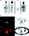
18F-FDG PET/CT images of lumbosacral spine show increased 18F-FDG uptake in region of facet joint, corresponding to abnormal findings on CT (arrows). (A) Coronal, sagittal, axial, and maximum-intensity-projection (MIP) PET images. (B) CT, attenuation-corrected, fused, and nonattenuation-corrected PET images. AC, attenuation-corrected PET image; FUSED, fused CT and PET images; NAC, nonattenuation-corrected PET image. This figure was originally published in JNM. Rosen RS, et al. J Nucl Med. 2006;47:1274–1280. © SNMMI. Reproduced with permission. (59).
Figure 5
Figure 5
Radiological techniques for degenerative disc disease (1) x-Ray radiographs, (2) computed tomography (CT), (3) magnetic resonance imaging (MRI), (4) positron emission tomography (PET).

References

    1. Donnally III CJ, Hanna A, Varacallo M. Lumbar degenerative disk disease. StatPearls. Treasure Island, FL: StatPearls Publishing; (2023). http://www.ncbi.nlm.nih.gov/books/NBK448134/ (accessed August 31, 2023). - PubMed
    1. Kos N, Gradisnik L, Velnar T. A brief review of the degenerative intervertebral disc disease. Med Arch. (2019) 73:421–4. 10.5455/medarh.2019.73.421-424 - DOI - PMC - PubMed
    1. Kirnaz S, Capadona C, Wong T, Goldberg JL, Medary B, Sommer F, et al. Fundamentals of intervertebral disc degeneration. World Neurosurg. (2022) 157:264–73. 10.1016/j.wneu.2021.09.066 - DOI - PubMed
    1. Emch TM, Modic MT. Imaging of lumbar degenerative disk disease: history and current state. Skeletal Radiol. (2011) 40:1175–89. 10.1007/s00256-011-1163-x - DOI - PubMed
    1. Zhou X, Cipriano P, Kim B, Dhatt H, Rosenberg J, Mittra E, et al. Detection of nociceptive-related metabolic activity in the spinal cord of low back pain patients using 18F-FDG PET/CT. Scand J Pain. (2017) 15:53–7. 10.1016/j.sjpain.2016.11.017 - DOI - PubMed

LinkOut - more resources