

Light and Electron Microscope Observations of the Male Reproductive Organs of Adult Julodis ehrenbergii Laporte (Coleoptera, Buprestidae)
- PMID: 40115986
- PMCID: PMC12214042
- DOI: 10.1002/jemt.24850
Light and Electron Microscope Observations of the Male Reproductive Organs of Adult Julodis ehrenbergii Laporte (Coleoptera, Buprestidae)
Abstract
This paper was the first preliminary description of the male reproductive organs in Julodis ehrenbergii Laporte (Coleoptera, Buprestidae) using light and scanning electron microscope techniques. The structure has a couple of testes, a couple of vasa deferentia, a couple of accessory glands, a couple of spermatophoral glands, and a single ejaculatory duct opening in the aedeagus. Each testis is made up of many elongated testicular follicles. Our histological observation showed cysts at different stages of development in the testicular follicles. In other words, spermatocytes, spermatids, and spermatozoa, respectively, are formed from the spermatogonia located at the distal end of the testicular follicles. Mature sperm bundles were observed in the lumen of the vasa deferentia. The male accessory glands are curved and thin tube-shaped. There are dense secretion granules in the cytoplasm of the accessory glands' epithelial cells. The spermatophoral glands are roughly oval-like structures and have three lobes on one side. The ejaculatory duct is characterized by the presence of the cuticle layer on the apical surface of the cells. The findings were compared with the male reproductive structures of some other species in the Coleoptera order. Some characteristics, including the number and shape of testicular follicles and accessory glands, among the species of the Coleoptera order, show differences. These features help allow future comparisons with the male reproductive organs of other Coleoptera species.
Keywords: jewel beetles; light microscopy; scanning electron microscopy; spermatophoral glands; testicular follicles.
© 2025 The Author(s). Microscopy Research and Technique published by Wiley Periodicals LLC.
Conflict of interest statement
The authors declare no conflicts of interest.
Figures


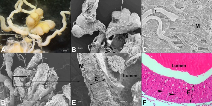
 : trachea. (B) Testicular follicle (F) is marked in red and is seen with its distal region closed (SEM).
  : trachea. (C) The cross section of the testicular follicles (SEM). (D) The longitudinal section of the testicular follicles in LM (HE staining). *: vasa deferentia, dz: differentiation zone, mz: maturation zone, gz: growth zone.


 : nucleus.
 : long spines on the cuticle layer.
Similar articles
-
New findings on the male reproductive system and spermatozoa of Aedes aegypti (Diptera: Culicidae).Parasit Vectors. 2025 Jul 1;18(1):246. doi: 10.1186/s13071-025-06808-w. Parasit Vectors. 2025. PMID: 40598390 Free PMC article.
-
Histomorphology of the Hindgut of Adult Julodis ehrenbergii Laporte, 1835 (Coleoptera: Buprestidae).Microsc Microanal. 2025 Sep 3;31(5):ozaf079. doi: 10.1093/mam/ozaf079. Microsc Microanal. 2025. PMID: 40899913
-
The histomorphological structure of the male reproductive system of maize leaf weevil Tanymecus dilaticollis Gyllenhal, 1834 (Coleoptera: Curculionidae).Microsc Res Tech. 2019 Aug;82(8):1345-1352. doi: 10.1002/jemt.23286. Epub 2019 May 14. Microsc Res Tech. 2019. PMID: 31087461
-
A rapid and systematic review of the clinical effectiveness and cost-effectiveness of paclitaxel, docetaxel, gemcitabine and vinorelbine in non-small-cell lung cancer.Health Technol Assess. 2001;5(32):1-195. doi: 10.3310/hta5320. Health Technol Assess. 2001. PMID: 12065068
-
Systemic pharmacological treatments for chronic plaque psoriasis: a network meta-analysis.Cochrane Database Syst Rev. 2021 Apr 19;4(4):CD011535. doi: 10.1002/14651858.CD011535.pub4. Cochrane Database Syst Rev. 2021. Update in: Cochrane Database Syst Rev. 2022 May 23;5:CD011535. doi: 10.1002/14651858.CD011535.pub5. PMID: 33871055 Free PMC article. Updated.
References
-
- Araújo, V. A. , Munhoz I. L. A., and Serrão J. E.. 2021. “Morphology of the Male Reproductive Tract in the Water Scavenger Beetle Tropisternus collaris Fabricius, 1775 (Coleoptera: Hydrophilidae).” Revista Brasileira de Entomologia 65, no. 2: e20210012.
-
- Bal, N. , Özdikmen H., Amutkan Mutlu D., and Suludere Z.. 2022. “Ultrastructure of Aedeagus, Spermatheca and Ovipositor of Julodis ehrenbergii Laporte (Coleoptera: Buprestidae: Julodinae) by Scanning Electron Microscope.” Microscopy Research and Technique 85, no. 12: 3882–3894. - PubMed
-
- Barker, G. M. 1989. “Functional Anatomy of the Reproductive System of Listronotus bonariensis (Kuschel).” New Zealand Entomologist 12, no. 1: 34–42.
-
- Calder, A. A. 1990. “Gross Morphology of the Soft Parts of the Male and Female Reproductive Systems of Curculionoidea (Coleoptera).” Journal of Natural History 24, no. 2: 453–505.
-
- Carrillo‐Ruiz, H. , Martínez M. I., and Morón M. A.. 2008. “Comparative Study of the Reproductive System of Two Species of Hoplia (Coleoptera: Scarabaeidae: Hopliinae).” Proceedings of the Entomological Society of Washington 110, no. 3: 778–788.
MeSH terms
LinkOut - more resources
Full Text Sources
