. 2025 Mar;14(3):e70062.
doi: 10.1002/jev2.70062.

Circulating Extracellular Vesicles as Putative Mediators of Cardiovascular Disease in Paediatric Chronic Kidney Disease

Affiliations

Circulating Extracellular Vesicles as Putative Mediators of Cardiovascular Disease in Paediatric Chronic Kidney Disease

Felix Behrens et al. J Extracell Vesicles. 2025 Mar.

Abstract

Cardiovascular disease (CVD) is the leading cause of mortality in chronic kidney disease (CKD). However, the pathogenesis of CVD in CKD remains incompletely understood. Endothelial extracellular vesicles (EC-EVs) have previously been associated with CVD. We hypothesized that CKD alters EV release and cargo, subsequently promoting vascular remodelling. We recruited 94 children with CKD, including patients after kidney transplantation and healthy donors, and performed EV phenotyping and functional EV analyses in the absence of age-related comorbidities. Plasma EC-EVs were increased in haemodialysis patients and decreased after kidney transplantation. Thirty microRNAs were less abundant in total CKD plasma EVs with predicted importance in angiogenesis and smooth muscle cell proliferation. In vitro, CKD plasma EVs induced transcriptomic changes in angiogenesis pathways and functionally impaired angiogenic properties, migration and proliferation in ECs. High shear stress, as generated by arterio-venous fistulas, and uremic toxins were considered as potential drivers of EV release, but only the combination increased EV generation from venous ECs. The resulting EVs recapitulated miRNA changes observed in CKD in vivo. In conclusion, CKD results in the release of EVs with altered miRNA profiles and anti-angiogenic properties, which may mediate vascular pathology in children with CKD. EVs and their miRNA cargo may represent future therapeutic targets to attenuate CVD in CKD.

Keywords: angiogenesis; cardiovascular disease; chronic kidney disease; microRNAs; shear stress; uremic toxins.


Conflict of interest statement

The authors declare no conflicts of interest.

Figures

FIGURE 1
FIGURE 1
Plasma extracellular vesicle concentrations increase in peritoneal dialysis patients. (A) 94 children and adolescents were enrolled in the paediatric CKD study, which consisted of five groups in the cross‐sectional study: (i) healthy donors, (ii) CKD G3‐G5 patients not requiring kidney replacement therapy, (iii) patients on peritoneal dialysis (PD) or (iv) haemodialysis (HD) and (v) patients who had already received a kidney transplant (KTx) and had stable graft function. During the study period 12 patients underwent KTx and longitudinal follow‐up after KTx was performed. (B) EVs of similar shape and size could be visualized in the plasma of all CKD patient groups by negative staining transmission electron microscopy, scale bar 400 nm. Nanoparticle tracking analysis (NTA) revealed that plasma EV size was not affected by CKD, as shown by (C) similar size distribution (data given as mean of all patients ± SEM), (D) similar mean EV size and (E) no changes in EV size after KTx. (F) Total plasma EV concentrations were increased in PD patients as compared to healthy donors and KTx recipients. (G) Longitudinal EV concentration did not change after KTx. HD haemodialysis patients. P values according to Kruskal–Wallis test and Dunn's post hoc test.
FIGURE 2
FIGURE 2
CD31+ endothelial EVs are increased in haemodialysis patients, whereas CD3+ T cell and CD68+ macrophage EVs are increased in CKD patients without dialysis. Specialized multicolour EV flow cytometry was performed on plasma from all patients in the paediatric CKD cohort. Cross‐sectional (A) and longitudinal (B) comparison of EVs of different cellular origin: CD31+ endothelial (EC‐) EVs were higher in haemodialysis (HD) patients as compared to kidney transplant (KTx) recipients in cross‐sectional comparison and decreased in longitudinal follow‐up of CKD patients after receiving KTx. CD68+ macrophage‐derived (Mac‐) EVs and CD3+ T cell‐derived (T‐) EVs were both increased in CKD without dialysis as compared to healthy donors and, in the case of Mac‐EVs, also compared to KTx recipients. Longitudinal analyses showed no significant changes in Mac‐EVs and T‐EVs. PD peritoneal dialysis patients. P values according to (A) Kruskal–Wallis test and Dunn's post hoc test, (B) Wilcoxon test.
FIGURE 3
FIGURE 3
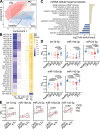
EV microRNA cargo is altered in CKD patients on dialysis and restored after kidney transplantation. Small RNA sequencing was performed on EVs from 75 patients of the paediatric CKD cohort. (A) Partial least‐squares discriminant analysis (PLS‐DA) showed a clear differentiation of EV microRNAs (miRNAs) between patients with normal kidney function (healthy donors, kidney transplant recipients (KTx)), and dialysis patients (haemodialysis (HD) and peritoneal dialysis (PD) patients) with CKD without dialysis in between, corresponding to the clinical state of intermediate reduced kidney function. (B) 31 miRNAs were differentially regulated in CKD, PD and/or HD patients as compared to healthy donors and/or KTx patients as shown in the heatmap of group comparisons according to DESeq2 for miRNAs that were also confirmed by LongDat. (C) Cellular target processes of altered miRNAs in CKD patients were predicted using TargetScanHuman miRNA target gene identification and PANTHER gene set enrichment analyses. Most specific gene ontology (GO) terms of hierarchical trees with fold enrichment ≥1.5 or ≤0.5 and FDR‐corrected p < 0.05 according to Fisher's exact test are shown. (D) RT‐qPCR confirmed that five selected miRNAs were reduced in the EVs of dialysis patients and (E) KTx partially reversed these changes in longitudinal comparison according to RT‐qPCR. (B) *p < 0.1, **p < 0.01 by DESeq2 with FDR‐correction. (D) P values according to one‐way ANOVA and Sidak's post hoc test for normally distributed data or Kruskal–Wallis test and Dunn's post hoc test for non‐normally distributed data as appropriate. (E) P values according to paired Student's t‐test for normally distributed data or Wilcoxon test for non‐normally distributed data.
FIGURE 4
FIGURE 4
CKD EVs cause impaired angiogenesis at transcriptomic and functional levels. The effect of plasma EVs from healthy donors and CKD patients on endothelial function was assessed using bulk RNA sequencing of human aortic endothelial cells (HAoECs) and functional assays on human umbilical vein endothelial cells (HUVECs). (A) HAoECs were incubated with isolated plasma EVs from healthy donors, hemodialysis (HD) patients and kidney transplant (KTx) recipients for 18 h and RNA was isolated from HAoECs for bulk RNA sequencing. (B) Principal component analysis (PCA) of genes belonging to gene ontology (GO) terms ‘cellular response to platelet‐derived growth factor stimulus’ (GO:0036120), ‘G1/S transition of mitotic cell cycle’ (GO:0000082), ‘regulation of smooth muscle cell proliferation’ (GO:0048660), ‘regulation of angiogenesis’ (GO:0045765), ‘negative regulation of cell migration’ (GO:0030336) and ‘angiogenesis’ (GO:0001525), which were identified by microRNA target prediction, showed a clear separation of healthy donors, HD and KTx patients. (C) Heatmap of differentially enriched genes (DEGs) of these GO terms showed an independent clustering of patient groups with the most pronounced changes between HD and KTx patients and the majority of DEGs involved in angiogenesis. Differential abundance was tested using DESeq2 with FDR‐correction. Based on the prediction of miRNA targets and transcriptomic changes in HAoECs upon CKD EV treatment functional angiogenesis properties were tested in vitro. Matches of DEGs with miRNA target genes are highlighted in bold. HUVECs were seeded on Matrigel and incubated with healthy or HD EVs for 6 h and vascular tube formation was analysed. (D) Representative microscopic images of HUVECs after 6 h of incubation. (E) Quantification of angiogenic properties: manual quantification of vascular tube‐like structures showed decreased angiogenesis of HD EV‐treated cells, which was supported by automated quantification using ImageJ Angiogenesis Analyzer software indicating decreased vessel density upon HD EV treatment as compared to healthy EVs, as evidenced by an increased branching interval, although without reaching statistical significance, and significantly increased mesh size. Vascular tube formation results were confirmed by assessing endothelial (HUVEC) (F,G) migration and (H,I) proliferation (immunofluorescence for Ki‐67). (F) HUVECs were grown with cell culture inserts to obtain a standardized gap area. Representative microscopic images of HUVECs before (0 h) and after incubation with healthy/HD EVs (4 h), (G) reduced relative reduction of gap area upon HD EV exposure as compared to healthy EVs. (H) Representative microscopic images of proliferating HUVECs after incubation with healthy/HD EVs for 18 h, (I) reduced endothelial proliferation upon exposure to HD EVs compared to healthy EVs as measured by the proportion of Ki67+ nuclei. Scale bars (D,F) 100 µm (10×), (H) 25 µm (40×). P values according to Mann–Whitney U test.
FIGURE 5
FIGURE 5
Dysregulated tryptophan metabolism induces endothelial EV release during vascular mechanical stress. To assess the role of tryptophan (TRP)‐derived uremic toxins in EV release in CKD targeted plasma metabolomics from the paediatric CKD cohort was performed. (A) Principal component analysis (PCA) clearly differentiated patients with normal kidney function (healthy donors and kidney transplant (KTx) recipients) from dialysis patients (HD haemodialysis, PD peritoneal dialysis) with CKD without dialysis in between. (B) Cumulated mean plasma concentrations show a major shift of TRP metabolism towards decreased TRP levels and increased indole metabolites in CKD, most prominently indoxyl sulphate (IS) with highest concentrations in dialysis patients. (C) Formyl kynurenine (FKYN), IS and xanthurenic acid (XA) correlated positively with endothelial EV (EC‐EV) plasma concentrations. To address potential trigger factors for increased endothelial EV release in HD patients two potential mechanisms were analysed. (D) Acute effects of HD on EV release were analysed in an additional cohort of CKD patients comparing EV concentrations before and after one HD session with no significant changes in endothelial (EC‐) EV concentrations. (E) The influence of dialysis access was addressed comparing HD patients with central venous catheters and patients with arteriovenous fistula (AVF) in the original paediatric CKD cohort, which showed a higher mean EC‐EV concentration in patients with AVF, but without reaching statistical significance. To evaluate the role of abnormal arterial flow conditions with high shear stress on the venous side of the AVF in HD human saphenous vein endothelial cells (HSaVECs) in vitro under different flow and uraemia conditions in μ‐slides using a pumping system to achieve unidirectional laminar flow. (F) Flow cytometry showed increased EV release from HSaVECs as measured by CFSE staining under dual stimulation with arterial high shear stress (10 dyn/cm2) and uremic 50 µM indoxyl sulphate (IS) as compared to venous low shear stress (1 dyn/cm2) ± 50 µM IS. (G) EV miRNA RT‐qPCR from these experiments showed that the combination of high shear stress with uremic stimulus was sufficient to partially recapitulate the EV miRNA reduction observed in CKD patients, as indicated by significantly lower abundance of let‐7d‐5p and miR‐24‐3p in 10 dyn/cm2 + 50 µM IS as compared to 1 dyn/cm2 + vehicle, while 50 µM IS alone was also sufficient to lower let‐7d‐5p and miR‐19a‐3p. IAA indoleacetic acid, ILA indole lactate, I3PA indole‐3‐propionic acid, KA kynurenic acid, KYN kynurenine, TRP tryptophan, 3OH‐AN 3OH‐anthranilate, 5OCH3‐IA 5OCH3‐indoleacetate, 5OH‐IA 5OH‐indoleacetate. (C) r and p according to Pearson's correlations. (E) P value according to Mann–Whitney U test. (F,G) P values according to Kruskal–Wallis test and Dunn's post hoc test.

References

    1. Adams, D. , Gonzalez‐Duarte A., O'riordan W. D., et al. 2018. “Patisiran, an RNAi Therapeutic, for Hereditary Transthyretin Amyloidosis.” New England Journal of Medicine 379: 11–21. 10.1056/NEJMoa1716153. - DOI - PubMed
    1. Agarwal, V. , Bell G. W., Nam J. W., and Bartel D. P.. 2015. “Predicting Effective MicroRNA Target Sites in Mammalian mRNAs.” Elife: 4. 10.7554/eLife.05005. - DOI - PMC - PubMed
    1. Alique, M. , Bodega G., Corchete E., et al. 2020. “Microvesicles From Indoxyl Sulfate‐Treated Endothelial Cells Induce Vascular Calcification in Vitro.” Computational and Structural Biotechnology Journal 18: 953–966. 10.1016/j.csbj.2020.04.006. - DOI - PMC - PubMed
    1. Amabile, N. , Guérin A. P., Leroyer A., et al. 2005. “Circulating Endothelial Microparticles Are Associated With Vascular Dysfunction in Patients With End‐Stage Renal Failure.” Journal of the American Society of Nephrology 16: 3381–3388. 10.1681/ASN.2005050535. - DOI - PubMed
    1. Arinze, N. V. , Yin W., Lotfollahzadeh S., et al2022. .“Tryptophan Metabolites Suppress the Wnt Pathway and Promote Adverse Limb Events in Chronic Kidney Disease.” Journal of Clinical Investigation 132, 10.1172/JCI142260. - DOI - PMC - PubMed