. 2025 Mar 21;10(105):eadr0547.
doi: 10.1126/sciimmunol.adr0547. Epub 2025 Mar 21.

GM-CSF-mediated epithelial-immune cell cross-talk orchestrates pulmonary immunity to Aspergillus fumigatus

Affiliations

GM-CSF-mediated epithelial-immune cell cross-talk orchestrates pulmonary immunity to Aspergillus fumigatus

Kathleen A M Mills et al. Sci Immunol. .

Abstract

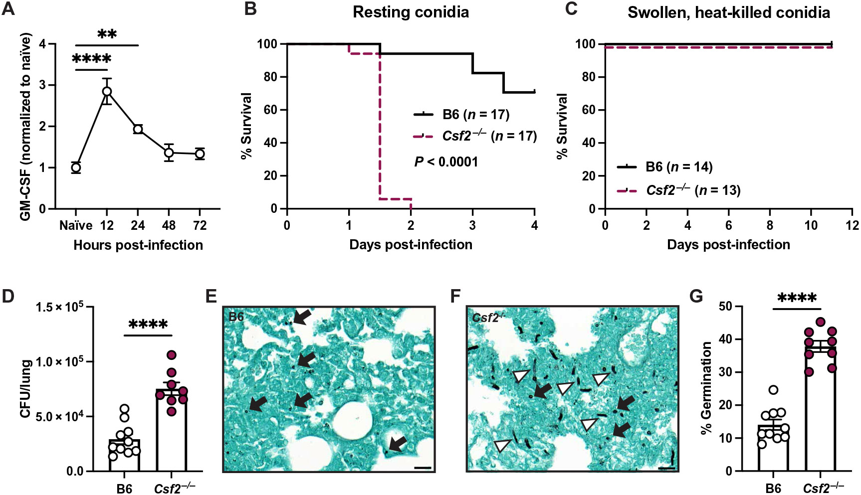
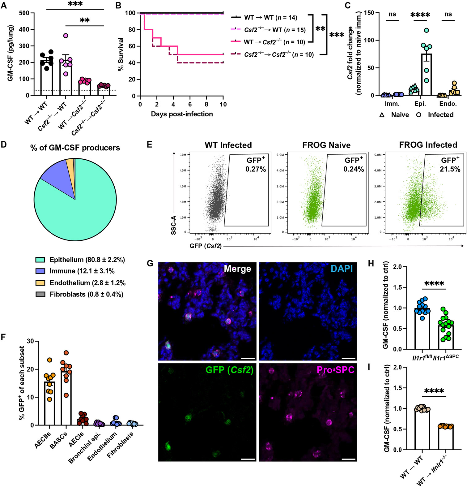
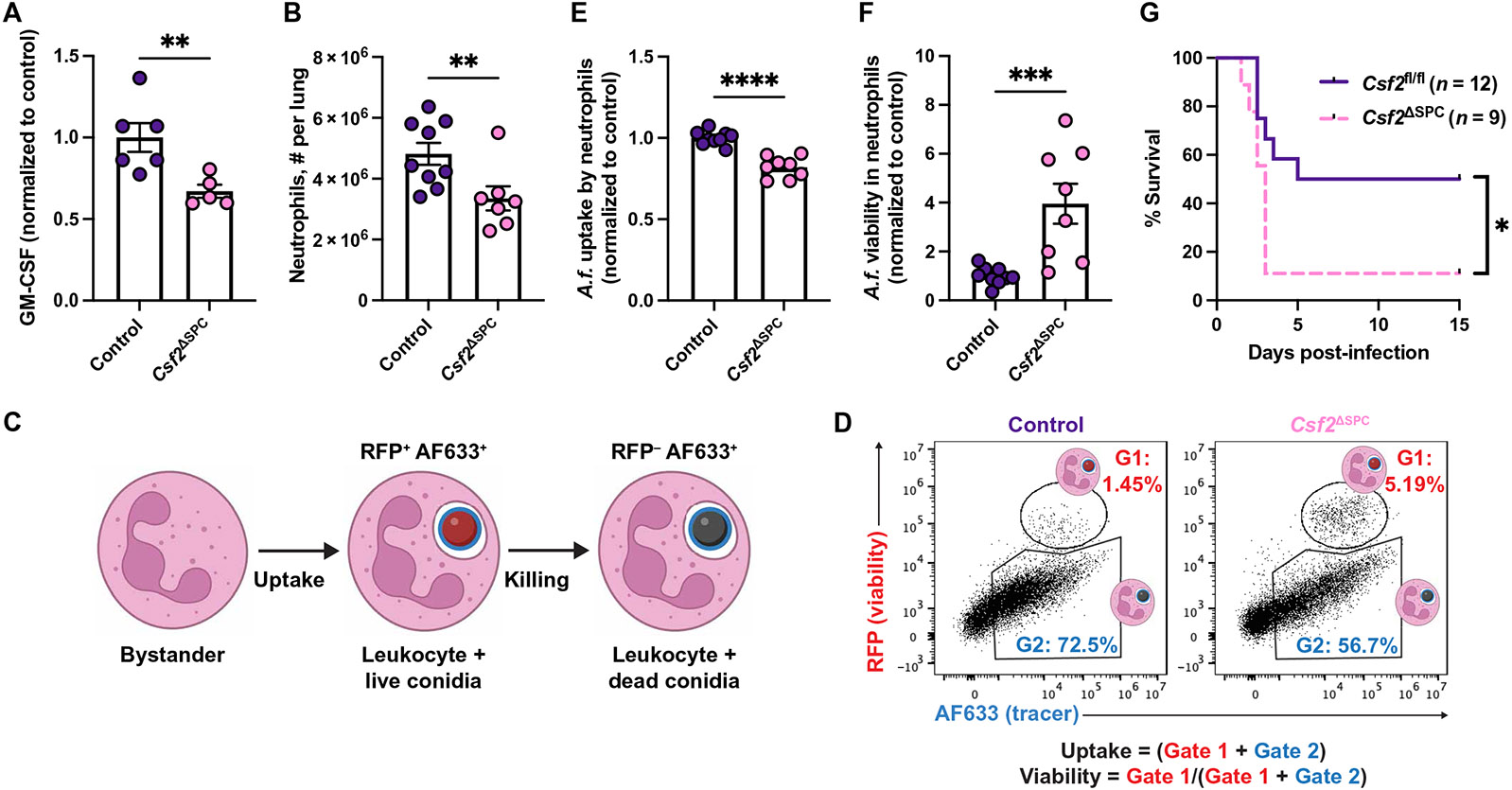
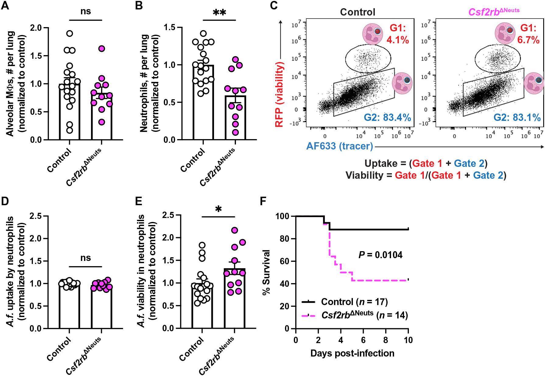
Aspergillus fumigatus causes life-threatening mold pneumonia in immunocompromised patients, particularly in those with quantitative or qualitative defects in neutrophils. Whereas innate immune cell cross-talk licenses neutrophil antifungal activity in the lung, the role of epithelial cells in this process is unknown. Here, we find that surfactant protein C (SPC)-expressing lung epithelial cells integrate infection-induced interleukin-1 and type III interferon signaling to produce granulocyte-macrophage colony-stimulating factor (GM-CSF) preferentially at local sites of fungal infection and neutrophil influx. Using in vivo models that distinguish the role of GM-CSF during acute infection from its homeostatic function in alveolar macrophage survival and surfactant catabolism, we demonstrate that epithelial-derived GM-CSF increases the accumulation and fungicidal activity of GM-CSF-responsive neutrophils, which is essential for host survival. Our findings establish SPC+ epithelial cells as a central player in regulating the quality and strength of neutrophil-dependent immunity against inhaled mold pathogens.


Figures

Fig. 1.
Fig. 1.. GM-CSF is required for host defense against A. fumigatus.
(A) GM-CSF levels measured by ELISA in supernatants from lung homogenates of naïve and A. fumigatus–infected (3 x 10⁷ conidia) B6 mice. (B and C) Survival of B6 and Csf2−/− mice after infection with 3 × 10⁷ to 4 × 10⁷ (B) resting or (C) swollen, heat-killed conidia. (D) CFU from lungs of B6 and Csf2−/− mice 48 hpi with 1.5 × 10⁷ resting conidia. (E and F) Representative lung sections from B6 and Csf2−/− mice infected with 1.5 × 10⁷ resting conidia 48 hpi stained with GMS. Black arrows indicate conidia, and white arrowheads indicate germlings. Scale bars, 20 μm. (G) Percentage of germinating conidia quantified from GMS staining of lung sections from B6 and Csf2−/− mice infected with 1.5 × 10⁷ resting conidia 48 hpi. Germinating conidia are defined as GMS+ regions with a maximum Feret diameter of >6 μm. Each symbol represents one mouse. Panel (A) was analyzed by Kruskal-Wallis test with Dunn’s multiple comparisons test. **adjusted P = 0.0021 and ****adjusted P < 0.0001. Panels (D) and (G) were analyzed by Mann-Whitney test. ****P < 0.0001. Panels (B) and (C) were analyzed by log-rank (Mantel-Cox) test. Panels (A) to (D) and (G) are all pooled from two experiments. See also fig. S1.
Fig. 2.
Fig. 2.. IL-1 and type III IFN signaling promote GM-CSF production during A. fumigatus infection.
(A to F) GM-CSF levels measured by ELISA in supernatants from lung homogenates from B6 mice and (A) Il1a/b−/−, (B) Il1r1−/−, (C) Myd88 −/−, (D) Ifnl2/3−/−, (E) Ifnlr1−/−, and (F) Stat1−/− mice 12 hpi with 3 × 10⁷ conidia. (G) IFNλ2/3 levels measured by ELISA in supernatants from lung homogenates from B6 and Il1r1−/− mice 12 hpi with 3 × 10⁷ conidia. (H) IL-1α and (I) IL-1β levels measured by ELISA in supernatants from lung homogenates from B6 and Stat1−/− mice 12 hpi with 3 × 10⁷ conidia. (J) GM-CSF levels measured by ELISA in supernatants from lung homogenates from naïve or infected B6 mice and infected Il1r1−/− Stat1−/− mice. [(A) to (I)] Two or three experiments pooled, (J) data from one experiment; each symbol represents one mouse. Significance determined by [(A) to (I)] Mann-Whitney test (*P = 0.0332, **P = 0.0021, ***P = 0.0002, and ****P < 0.0001) or (J) Kruskal-Wallis test with Dunn’s multiple comparisons test (**adjusted P = 0.0079). See also fig. S2. ns, not significant. ctrl, control.
Fig. 3.
Fig. 3.. IL-1 and Stat1 are dispensable for GM-CSF production and AM homeostasis in the steady state.
(A) Representative flow plots showing CD45+ live cells from lungs of uninfected B6, Csf2−/−, Il1r1−/−, Stat1−/−, and Il1r1−/− Stat1−/− mice. Black gate indicates alveolar macrophages (CD11c+ Siglec F+). (B) Number of alveolar macrophages (Mϕs) per lung in uninfected B6, Csf2−/−, Il1r1−/−, Stat1−/−, and Il1r1−/− Stat1−/− mice and (C) GM-CSF levels measured by ELISA in the lungs of uninfected B6, Il1r1−/−, and Stat1−/− mice. (D) GM-CSF levels measured by ELISA in the lungs of uninfected B6 and Il1r1−/− Stat1−/− mice. (B and C) Two to four experiments pooled. Each symbol represents one mouse. Significance determined by Kruskal-Wallis test with Dunn’s multiple comparisons test. ***adjusted P = 0.0002. (D) Data are from one experiment. Each symbol represents one mouse. Significance determined by Mann-Whitney test. PE, phycoerythrin.
Fig. 4.
Fig. 4.. GM-CSF is produced by SPC+ pulmonary epithelial cells.
(A) GM-CSF levels measured by ELISA in supernatants from lung homogenates from Csf2−/− bone marrow chimeras 12 hpi with 3 × 10⁷ conidia. Dotted line represents lower limit of detection of ELISA. (B) Survival of Csf2−/− chimeras after A. fumigatus infection with 5 × 10⁷ conidia. (C) Csf2 transcript levels measured by qPCR in lung immune, epithelial, or endothelial cells sorted by FACS (gating strategy in fig. S3A) from naïve B6 mice or B6 mice 6 hpi with 3 × 10⁷ conidia. (D) Pie chart displaying percentage of the indicated cell populations out of all GFP+ (Csf2+) cells in the lung after infection with 3 × 10⁷ conidia. Data analyzed by flow cytometry. Summary data from four mice are displayed as means ± SD. (E) Representative flow cytometry plots of AECIIs from infected (24 hpi with 3 × 10⁷ conidia) WT and naïve or infected FROG mice showing the percentage of GFP+ (Csf2+) cells. (F) Percentage of each indicated cell type that is GFP+ (Csf2+) as measured by flow cytometry in lungs of FROG mice 24 hpi with 3 × 10⁷ conidia. (G) Representative immunofluorescence images of FROG mouse lung 24 hpi with 3 × 10⁷ conidia, showing DAPI, GFP (Csf2), and Pro-SPC staining. Scale bars, 20 μM. (H and I) GM-CSF levels measured by ELISA in supernatants from lung homogenates of (H) Il1r1fl/fl and Il1r1ΔSPC mice or (I) Ifnlr1−/− bone marrow chimeric mice at 12 hpi with 3 × 10⁷ A. fumigatus conidia. [(A), (C), (F), (H), and (I)] Each symbol represents one mouse. Statistical analysis: (A) Kruskal-Wallis with Dunn’s multiple comparisons test, **adjusted P = 0.0027 and ***adjusted P = 0.0007; (B) log-rank (Mantel-Cox); (C) two-way analysis of variance (ANOVA); and [(H) and (I)] Mann-Whitney. **P = 0.0021, ***P = 0.0002, and ****P < 0.0001. Panels (B) to (D) and (I) are data from two pooled experiments. Panels (F) and (H) are data from three pooled experiments. See also fig. S3.
Fig. 5.
Fig. 5.. GM-CSF–producing epithelial cells form a signaling circuit in proximity to A. fumigatus and neutrophils.
(A) Schematic of hypothesis that GM-CSF–producing SPC+ cells are in closer proximity to conidia and neutrophils than GM-CSF–nonproducing SPC+ cells. The zoomed-in portion represents two alveoli in the same lung. The top alveolus is devoid of conidia and therefore lacks infection-induced GM-CSF production or neutrophil infiltration. The bottom alveolus has conidia present and subsequent GM-CSF production by SPC+ cells and neutrophil infiltration. (B) Representative image of FROG mouse lung 24 hpi with AF633+ conidia stained with DAPI, anti-GFP, and pro-SPC. White arrowheads indicate GFP+ SPC+ cells. Scale bar, 20 μM. (C) Distance from GM-CSF SPC+ or GM-CSF+ SPC+ cell to AF633+ conidia. Each dot represents one mouse and is the median of distance measurements from the indicated cell type to AF633+ conidia within that mouse. Dots are paired to indicate measurements made in the same mouse (n = 7 mice). Dots are superimposed on violin plots representing all cells analyzed in a SuperPlot (62). (D) Representative image of FROG mouse lung 24 hpi stained with DAPI, anti-GFP, pro-SPC, and Ly6G. White arrowheads indicate GFP+ SPC+ cells. Scale bar, 20 μM. (E) Distance from GM-CSF SPC+ or GM-CSF+ SPC+ cell to Ly6G+ cell. Each dot represents one mouse and is the median of distance measurements from the indicated cell type to Ly6G+ cells within that mouse. Dots are paired to indicate measurements made in the same mouse (n = 6 mice). Dots are superimposed on violin plots representing all cells analyzed. Panels (C) and (E) analyzed by Wilcoxon test. *P = 0.0332. See also fig. S4.
Fig. 6.
Fig. 6.. GM-CSF derived from SPC+ epithelial cells is required for neutrophil fungicidal activity.
(A) GM-CSF levels measured by ELISA in supernatants from lung homogenates from control and Csf2ΔSPC mice 12 hpi with 3 × 10⁷ conidia. (B) Neutrophil numbers in lungs of control and Csf2ΔSPC mice 24 hpi. (C and D) Schematic of FLARE conidia and fluorescence emission after uptake and killing by leukocytes. (E) Uptake of conidia by and (F) conidial viability in lung neutrophils, quantified using FLARE conidia and flow cytometry from infected control and Csf2ΔSPC mice 24 hpi. (G) Survival of Csf2fl/fl and Csf2ΔSPC mice after infection with 4 × 10⁷ to 6 × 10⁷ conidia. [(A), (B), and (E) to (G)] Data are from two pooled experiments. [(A), (B), (E), and (F)] Each symbol represents one mouse. Data were analyzed by [(A), (B), (E), and (F)] Mann-Whitney test or (G) log-rank (Mantel-Cox) test. *P = 0.0332, **P = 0.0021, ***P = 0.0002, and ****P < 0.0001. All control mice in (A) and half of the control mice in (B), (E), and (F) are Sftpc-Cre–positive mice given corn oil (vehicle for tamoxifen) because of low numbers of Cre-negative mice. All control mice in (G) are Cre-negative mice given tamoxifen. Individual experimental replicates for (B), (E), and (F) are in fig. S5, L to Q. Tamoxifen was administered as four doses on consecutive days [100 μl of a solution (40 mg/ml) dissolved in corn oil] via the intraperitoneal route. Mice were rested for 8 to 12 days after the last dose. See also fig. S5. A.f., A. fumigatus.
Fig. 7.
Fig. 7.. GM-CSF signaling on neutrophils is required for conidial killing and murine survival.
(A) Alveolar macrophage and (B) neutrophil numbers in the lungs of control and Csf2rbΔNeuts mice 24 hpi with 3 × 10⁷ conidia. (C) Representative flow cytometry plots of lung neutrophils from control and Csf2rbΔNeuts mice at 24 hpi with 3 × 10⁷ FLARE conidia showing AF633 and RFP. (D) Uptake of conidia by and (E) conidial viability in lung neutrophils 24 hpi. (F) Survival of control and Csf2rbΔNeuts mice after infection with 4 × 10⁷ to 6 × 10⁷ A. fumigatus conidia. [(A), (B), (D), and (E)] Two experiments pooled, significance calculated by Mann-Whitney test. (F) Three experiments pooled, analyzed by log-rank (Mantel-Cox) test. *P = 0.0332 and **P = 0.0021. See also fig. S6.

Update of

References

    1. Latgé J-P, Chamilos G, Aspergillus fumigatus and aspergillosis in 2019. Clin. Microbiol. Rev 33, e0014018 (2019). - PMC - PubMed
    1. Espinosa V, Jhingran A, Dutta O, Kasahara S, Donnelly R, Du P, Rosenfeld J, Leiner I, Chen C-C, Ron Y, Hohl TM, Rivera A, Sheppard DC, Inflammatory monocytes orchestrate innate antifungal immunity in the lung. PLOS Pathog. 10, e1003940 (2014). - PMC - PubMed
    1. Guo Y, Kasahara S, Jhingran A, Tosini NL, Zhai B, Aufiero MA, Mills KAM, Gjonbalaj M, Espinosa V, Rivera A, Luster AD, Hohl TM, During Aspergillus infection, monocyte-derived DCs, neutrophils, and plasmacytoid DCs enhance innate immune defense through CXCR3-dependent crosstalk. Cell Host Microbe 28, 104–116.e4 (2020). - PMC - PubMed
    1. Gerson SL, Talbot GH, Hurwitz S, Strom BL, Lusk EJ, Cassileth PA, Prolonged granulocytopenia: The major risk factor for invasive pulmonary aspergillosis in patients with acute leukemia. Ann. Intern. Med 100, 345–351 (1984). - PubMed
    1. Nivoix Y, Velten M, Letscher-Bru V, Moghaddam A, Natarajan-Amé S, Fohrer C, Lioure B, Bilger K, Lutun P, Marcellin L, Launoy A, Freys G, Bergerat J, Herbrecht R, Factors associated with overall and attributable mortality in invasive aspergillosis. Clin. Infect. Dis 47, 1176–1184 (2008). - PubMed

MeSH terms

Substances