Published Erratum

Conformational Antibodies to Proteolipid Protein-1 and Its Peripheral Isoform DM20 in Patients With CNS Autoimmune Demyelinating Disorders

Stefano Masciocchi et al. Neurol Neuroimmunol Neuroinflamm. 2025 May.
No abstract available


Figures

Figure 5
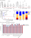
Figure 5. PLP1-IgG Titers in Serum and CSF, IgG Subclasses, and Complement-Dependent Cytotoxicity
(A) Serum and CSF PLP1-IgG titers tested in 41 serum and 24 CSF samples. The red dotted line indicates the cutoff for serum (1:40, left graph) and for CSF (1:5, right graph). (B) Serum:CSF PLP1-IgG titer ratio. The dotted arrow marks the 200:1 ratio expected in physiologic conditions. Values below the dotted line might associate with intrathecal synthesis. Blue dots represent patients positive in serum only, red dots patients positive in CSF only, and black dots patients positive in both serum and CSF. (C) PLP1-IgG subclasses in the whole cohort and in the 3 diagnostic groups of PLP1-IgG–positive patients. (D) PLP-IgG1 complement-dependent cytotoxicity. Data are normalized according to the amount of viable cells found in untreated cells (PLP1 cells, first 2 columns), which is considered 100%. The graph represents the finding after incubation with patient's sera. A reduction of viable cells is detected for 2 AQP4-IgG–positive serum samples (AQP4-01 and AQP4-02), which were used as positive controls, only when incubated with AQP4-transfected cells and in the presence of complement. Similarly, 3 of 8 PLP1-IgG–positive serum samples (other ADD#4, MS#5, and other ADD#18) showed reduced cell viability only when incubated with PLP1-transfected cells in the presence of complement. No effect is observed for the same samples in the absence of complement or after PLP1-IgG preadsorption performed on samples MS#5 and other ADD#18. The black dotted line represents the mean transfection rate for AQP4 and PLP1 (35%), which should theoretically set the limit for the maximum CDC effect in this experimental setting. CDC, which should involve only transfected cells, is unlikely below the cutoff. The numeric codes attributed to PLP1-IgG–positive patients correspond to those of Table 2, eTable 2, and eTable 3. ADD = autoimmune demyelinating disorder; AQP4 = aquaporin 4; Ig = immunoglobulin; MOGAD = myelin oligodendrocyte glycoprotein antibody–associated disease; MS = multiple sclerosis; PLP1 = proteolipid protein-1.

Erratum for

  • Conformational Antibodies to Proteolipid Protein-1 and Its Peripheral Isoform DM20 in Patients With CNS Autoimmune Demyelinating Disorders.
    Masciocchi S, Businaro P, Greco G, Scaranzin S, Malvaso A, Morandi C, Zardini E, Risi M, Vegezzi E, Diamanti L, Bini P, Siquilini S, Giannoccaro MP, Morelli L, Liguori R, Patti F, De Giuli V, Portaccio E, Zanetta C, Bergamoni S, Simone AM, Lanzillo R, Bruno G, Gallo A, Bisecco A, Di Filippo M, Pauri F, Toriello A, Barone P, Tazza F, Bucello S, Banfi P, Fabris M, Volonghi I, Raciti L, Vigliani MC, Bocci T, Paoletti M, Colombo E, Filippi M, Pichiecchio A, Marchioni E, Franciotta D, Gastaldi M; Neuroimmunology platform study group (PNI). Masciocchi S, et al. Neurol Neuroimmunol Neuroinflamm. 2025 Mar;12(2):e200359. doi: 10.1212/NXI.0000000000200359. Epub 2025 Jan 17. Neurol Neuroimmunol Neuroinflamm. 2025. PMID: 39823554 Free PMC article.
References

    1. Masciocchi S, Businaro P, Greco G, et al. . Conformational antibodies to proteolipid protein-1 and its peripheral isoform DM20 in patients with CNS autoimmune demyelinating disorders. Neurol Neuroimmunol Neuroinflamm. 2025;12(2):e200359. doi:10.1212/NXI.0000000000200359 - DOI - PMC - PubMed

Publication types