. 2025 Mar 21;15(1):9754.
doi: 10.1038/s41598-025-94003-3.

Acute LPS exposure enhances susceptibility to peripheral prion infection

Affiliations

Acute LPS exposure enhances susceptibility to peripheral prion infection

Reiss Pal et al. Sci Rep. .

Abstract

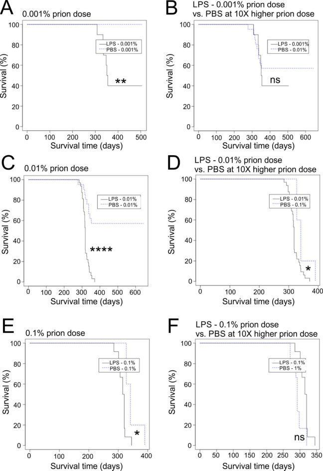
After peripheral infections, the initial accumulation of prions within secondary lymphoid tissues is essential for the transmission of disease to the brain. Macrophages are considered to sequester or destroy prions, but little was known of their impact on disease susceptibility after a peripheral infection. Inflammation in the peritoneal cavity can trigger the macrophage disappearance reaction, whereby the macrophages are temporarily contained within cellular aggregates on the mesothelium. We studied the impact of the bacterial lipopolysaccharide (LPS)-mediated macrophage disappearance reaction on susceptibility to an intraperitoneal prion infection. Intraperitoneal LPS injection significantly enhanced prion disease susceptibility approximately 100X when given 24-3 h before infection. The effects on disease susceptibility coincided with the reduced abundance of macrophages within the peritoneal cavity at the time of infection and the enhanced early accumulation of prions in the spleen. This suggests that the reduced recoverable abundance of macrophages in the peritoneal cavity following acute LPS-treatment, increased disease susceptibility by enhancing the initial propagation of the prions from site of exposure (peritoneal cavity) to the spleen from where they subsequently spread to the brain. Further studies may help identify novel macrophage-targeted treatments that can reduce susceptibility to peripherally acquired prion infections.

Keywords: Disease susceptibility; LPS; Macrophage; Peritoneal cavity; Prion diseases; Spleen; Transmissible spongiform encephalopathies.


Conflict of interest statement

Declarations. Competing interests: The authors declare no competing interests.

Figures

Fig. 1
Fig. 1
LPS treatment enhances prion disease susceptibility when given 3 h before intraperitoneal (IP) infection. Survival plots showing the impact of LPS treatment on prion disease susceptibility. Groups of mice (n = 8–10) were given a single IP injection with a limiting dose of prions (0.01%) and also injected IP with LPS (10 µg) or PBS (control) at the following intervals in relation to the timing of the prion infection: (A) untreated (prions alone); (B) −6 days; (C) −48 h; (D) −24 h; (E) −3 h; (F) + 3 h. *, P < 0.05; ***, P < 0.001; ns, not significant; Log-rank (Mantel-Cox) test. (G) Survival rate statistics for all treatment group comparisons.
Fig. 2
Fig. 2
LPS-treatment 3 h before intraperitoneal (IP) prion infection does not affect the development of the neuropathological signs of CNS prion disease. Mice were first injected IP with LPS (10 µg, n = 10) or PBS (control, n = 9), and 3 h later injected IP with a limiting dose of prions (0.01%). Brains were collected at the terminal clinical stage or the end of the experiment at ≥546 days after injection. (A), Representative images showing high levels of spongiform pathology (upper row), prion disease-specific PrPd accumulation (brown, 2nd row), reactive astrocytosis (GFAP + cells, brown, 3rd row) and microgliosis (AIF1 + cells, brown, bottom row) in the hippocampus of the brains of clinically affected mice (Clin+) at the terminal clinical stage. No histopathological signs of prion disease were detected in the brains of the clinically negative (Clin-) survivors. Sections counterstained with haematoxylin to detect cell nuclei (blue). Scale bar, 50 μm. (B) The relative abundance of AIF1 + immunostaining in the brains of the clinically affected mice (Clin+) at the terminal clinical stage, or clinically negative (Clin-) survivors. **, P < 0.01; ***, P < 0.001; ns, not significant; Student’s t-test. (C) The severity and distribution of the spongiform pathology (vacuolation) within each brain from each treatment group was scored in the following gray matter and white matter regions: G1, dorsal medulla; G2, cerebellar cortex; G3, superior colliculus; G4, hypothalamus; G5, thalamus; G6,hippocampus; G7, septum; G8, retrosplenial and adjacent motor cortex; G9, cingulate and adjacent motor cortex; W1, inferior and middle cerebellar peduncles; W2, decussation of superior cerebellar peduncles; and W3, cerebellar peduncles. Data points represent mean ± SD.
Fig. 3
Fig. 3
LPS treatment 3 h before intraperitoneal (IP) prion infection enhances disease susceptibility. Groups of mice (n = 5–32) were treated with LPS (10 µg) or PBS (control) 3 h before IP injection with a range of prion doses ranging from 1–0.001% and survival times recorded. (A, C, E) Survival plots for LPS and PBS-treated mice injected IP with a (A) 0.001% dose, (C) 0.01% dose, or (E) 0.1% dose of prions. (B, D, E) Survival plots showing LPS-treated mice injected with a (B) 0.001% dose, (D) 0.01% dose, or (F) 0.1% dose of prions, compared to PBS-treated controls injected with 10X higher doses of prions. *, P < 0.05; **, P < 0.01; ****, P < 0.0001; ns, not significant; Log-rank (Mantel-Cox) test.
Fig. 4
Fig. 4
Low dose LPS-treatment enhances prion disease susceptibility. Groups of mice (n = 6–10) were first given a single intraperitoneal (IP) injection with a range of LPS doses from 50 µg to 0.1 µg, or PBS (control), and 3 h later injected IP with a limiting dose of prions (0.01%). Survival plots for mice injected with either (A) 50 µg, (B) 10 µg, (C) 1.0 µg or (D) 0.1 µg LPS. *, P < 0.05; **, P < 0.01; Log-rank (Mantel-Cox) test.
Fig. 5
Fig. 5
LPS-treatment enhances the early accumulation of prions in the spleen. (A) Experimental design. Groups of mice (n = 5) were first given a single intraperitoneal (IP) injection of LPS (10 µg) or PBS (control), 3 h later injected IP with a low dose of prions (1.0%) and spleens were harvested 35 days-post injection. (B) Immunohistochemical analysis of follicular dendritic cell (FDC)-associated (CD21/CD35+ cells, upper panels), F4/80 + macrophages (middle panels) and disease-specific prion protein (PrPd) accumulations (lower panels) in spleens of mice from each treatment group at 35 days post-injection with prions. Sections were counterstained with hematoxylin (blue). Scale bar = 500 μm. (C) LPS treatment did not affect the density of FDC networks in the spleen compared to PBS-treated controls. Data points represent individual mice. Bars, median. Not significantly different, Student’s t-test. (D) LPS treatment did not affect the abundance of F4/80 + immunostaining in the spleen compared to PBS-treated controls. Data points represent individual mice. Bars, median. Not significantly different, Student’s t-test. (E) The relative abundance of PrPd+ FDC networks (% positive networks) was increased in the spleens of LPS-treated mice compared to PBS-treated controls. *, P < 0.05, Student’s t-test. (F) PET blot analysis of adjacent sections confirmed the presence of relatively proteinase K-resistant prion disease specific PrPSc (black, arrows). Scale bar = 200 μm. (G) The relative abundance of PrPSc+ FDC networks (% positive networks) was increased in the spleens of LPS-treated mice compared to PBS-treated controls. **, P < 0.01, Student’s t-test. (H-J) Relative prion seeding activities in spleens from each treatment group were quantified in vitro by RT-QuIC analysis. Individual traces represent spleens from individual mice. (H) Spleens from terminally infected mice and uninfected mice were used as positive and negative controls, respectively. (I) Spleens from LPS-treated mice and (J) PBS-treated mice collected 35 days after injection with prions. Data points derived from mean values from two sections/spleen.
Fig. 6
Fig. 6
LPS treatment reduces the abundance of peritoneal macrophages in the peritoneal cavity. Groups of mice (n = 5) were given a single intraperitoneal injection with LPS (10 µg) or PBS as a control, and 3 h later the abundance of macrophages, neutrophils and monocytes in peritoneal lavages was anaylsed by flow cytometry. cDC, conventional dendritic cells; LPM, large peritoneal macrophages; SPM, small peritoneal macrophages. (A) Gating strategy. (B) Total numbers of CD102 + LPM, neutrophils, B cells and T cells. (C) Total numbers of Ly6Chi monocytes, CD11c + SPM, cDC1, cDC1 and eosinophils. Data points represent individual mice. Bars, median. *, P < 0.05; **, P < 0.01, Student’s t-test.

References

    1. Legname, G. et al. Synthetic mammalian prions. Science305, 673–676. 10.1126/science.1100195 (2004). - PubMed
    1. Wang, F., Wang, X., Yuan, C. G. & Ma, J. Generating a prion with bacterially expressed Recombinant prion protein. Science327, 1132–1135. 10.1126/science.1183748 (2010). - PMC - PubMed
    1. Mabbott, N. How do PrPSc prions spread between and within host species, and within hosts? Pathogens. 10.3390/pathogens6040060 (2017). - PMC - PubMed
    1. Sigurdson, C. J. et al. Oral transmission and early lymphoid tropism of chronic wasting disease PrPres in mule deer fawns (Odocoileus hemionus). J. Gen. Virol.80, 2757–2764. 10.1099/0022-1317-80-10-2757 (1999). - PubMed
    1. Brown, K. L. et al. Scrapie replication in lymphoid tissues depends on PrP-expressing follicular dendritic cells. Nat. Med.5, 1308–1312. 10.1038/15264 (1999). - PubMed

LinkOut - more resources