. 2025 Mar 21;13(1):64.
doi: 10.1186/s40478-025-01984-3.

Curative timed NK cell-based immunochemotherapy aborts brain tumour recurrence driven by mesenchymal glioma stem cells

Affiliations

Curative timed NK cell-based immunochemotherapy aborts brain tumour recurrence driven by mesenchymal glioma stem cells

Brian Meehan et al. Acta Neuropathol Commun. .

Abstract

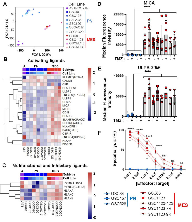
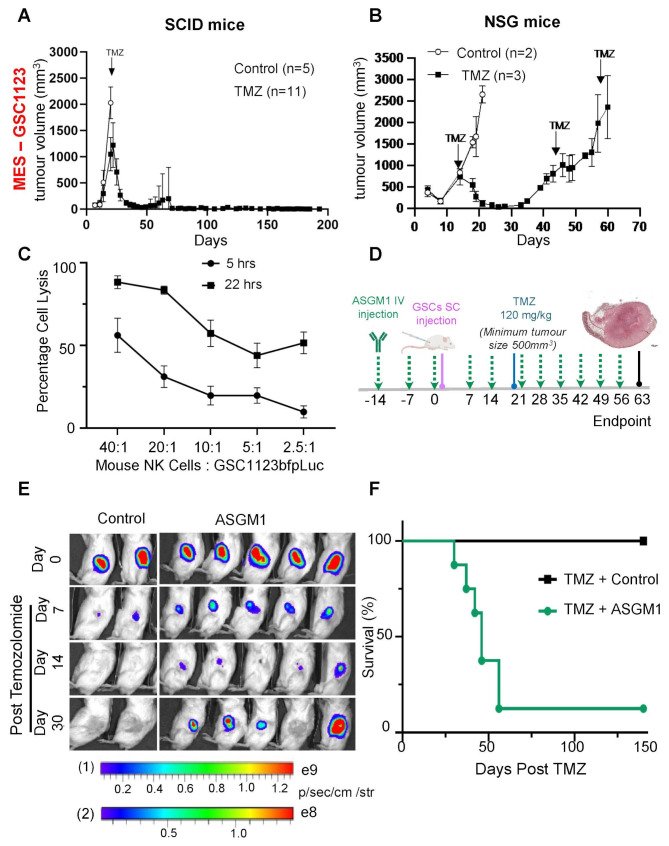
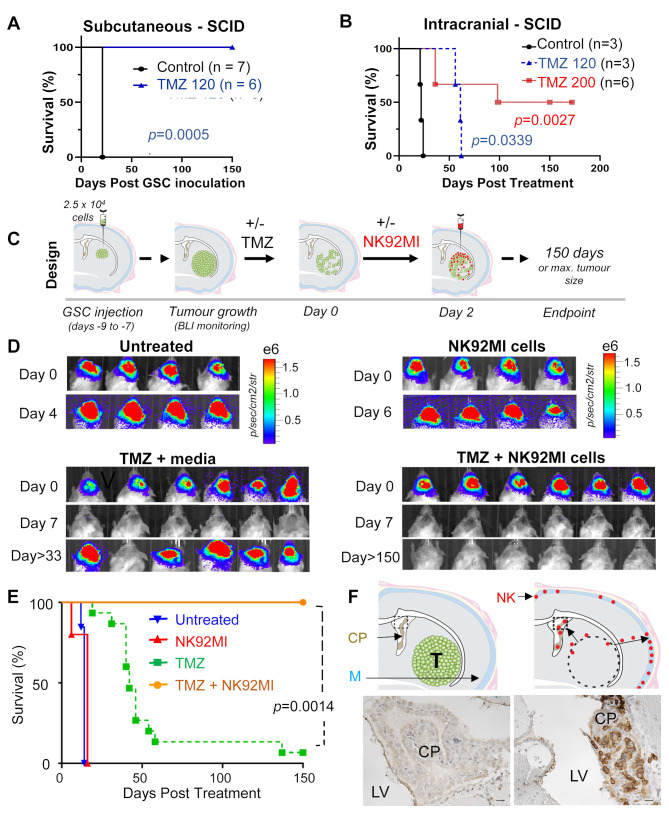
High grade gliomas (HGG) are incurable brain cancers, where inevitable disease recurrence is driven by tumour-initiating glioma stem cells (GSCs). GSCs survive and expand in the brain after surgery, radiation and temozolomide (TMZ) chemotherapy, amidst weak immune and natural killer (NK) cell surveillance. The present study was designed to understand how to enhance the contribution of innate immunity to post TMZ disease control. Strikingly, molecular subtypes of HGG impacted the repertoire of NK cell sensitivity markers across human HGG transcriptomes, and in a panel of GSCs with either proneural (PN-GSC) or mesenchymal (MES-GSC) phenotypes. Indeed, only MES-GSCs (but not PN-GSCs) were enriched for NK cell ligands and sensitive to NK-mediated cytotoxicity in vitro. While NK cells alone had no effect on HGG progression in vivo, the post-chemotherapy (TMZ) recurrence of MES-GSC-driven xenografts was aborted by timed intracranial injection of live or irradiated NK (NK92MI) cells, resulting in long term survival of animals. This curative effect declined when NK cell administration was delayed relative to TMZ exposure pointing to limits of the immune control over resurging residual tumour stem cell populations that survived chemotherapy. Overall, these results suggest that chemotherapy-dependent tumour depopulation may create a unique window of opportunity for NK-mediated intervention with curative effects restricted to a subset of HGGs driven by mesenchymal brain tumour initiating cells.

Keywords: Glioblastoma; High grade glioma; Intracranial immunotherapy; Intracranial xenografts; Mesenchymal glioma stem cells; Molecular subtypes of glioma; NK cells; Proneural glioma stem cells; Relapse; Temozolomide.


Conflict of interest statement

Declarations. Ethics approval and consent to participate: Not applicable. Consent for publication: Not applicable. Competing interests: The authors declare no competing interests.

Figures

Fig. 1
Fig. 1
Differential expression of NK cell ligands and susceptibility to NK cell-mediated cytotoxicity between patient-derived mesenchymal and proneural glioma stem cell populations. (A) Principal component analysis (PCA) of total transcriptomes of indicated cells: proneural GSCs (PN), mesenchymal GSCs (MES) and astrocytes. (B-C) In silico analysis of mRNA expression levels (microarrays) for NK cell ligands in a panel of PN and MES GSCs and Astrocytes. Activating NK ligand transcripts (B) and transcripts for multifunctional NK ligands (C). (D-E) Differential cell surface expression of NK ligands interacting with cytotoxic NKG2D by MES-GSCs and PN-GSCs (FACS); MICA (D) and ULBP-2/5/6 (E). GSCs were assayed with or without pretreatment with temozolomide (TMZ). MICA and ULBP-2/5/6 protein levels were expressed as median fluorescent intensity +/- SEM, for n = 3–5 independent repetitions, p values were determined for group comparisons by unpaired two-tailed t-test, p < 0.0001 (F) NK cell specific target cell (GSC) lysis analyzed by bioluminescence assay. GSCs were incubated with NK92MI human immortalized NK cells for 4 h at indicated ratios. Experiments were performed at least 3 times, **** p < 0.0001 as determined for group comparison between PN and MES time points by one way ANOVA with Tukey’s multiple comparison test +/- SEM
Fig. 2
Fig. 2
NK cell ligand expression by intracranial GSC xenografts in the presence or absence of temozolomide therapy. (A) MICA immunohistochemical staining of proneural (GSC528) and mesenchymal (GSC1123 and GSC83) intracranial xenografts in SCID mice (magnification = 200X (size bar = 50 μm). (B-C) FACS analysis of NK ligand expression in dissociated GSC1123 intracranial tumours. MICA (B) and ULBP-2/5/6 (C) expression by cancer cells in mice either untreated (Control) or 48 h post temozolomide (TMZ) treatment. Mean fluorescence intensity +/- SEM (right side panels in B and C) and corresponding FACS histograms; Unstained GSC123 cells (blue), MICA- or ULBP-2/5/6-stained GSC1123 control cells from culture (red), and three independent stained GSC1123 cell populations (n = 3) isolated from untreated (light green, dark green and pink) or TMZ treated tumours (light purple, dark purple and dark blue). Statistical analysis by unpaired T-test, ** p < 0.01
Fig. 3
Fig. 3
Impact of endogenous mouse NK cells on post-chemotherapy recurrence of mesenchymal glioma stem cell xenografts. (A) Endogenous NK cell proficiency of SCID mice supports eradication of mesenchymal glioma stem cell (GSC1123) - driven subcutaneous xenografts by single dose of temozolomide (TMZ; 120 mg/kg); data expressed as mean +/- SD; (B) Endogenous NK cell deficiency of NSG mice is associated with post-TMZ relapse of GSC1123 subcutaneous xenografts (mean +/- SD); (C) Purified mouse NK cells efficiently kill GSC1123 mesenchymal glioma stem cells in vitro in a time dependent manner (BLI assay with readings conducted at 5 h and 22 h post NK treatment; mean value +/- SEM); (D) Experimental design to explore the effects of pharmacological NK inhibition (Asialo-GM1) on progression of mesenchymal GSC subcutaneous xenografts post TMZ-mediated tumour depopulation; (E) Asialo-GM1 treatment aborts TMZ-mediated GSC1123 tumour eradication in NK-proficient SCID mice– bioluminescent images; (F) Inhibition of endogenous NK cell cytotoxicity (Asialo-GM1) counteracts pro-survival effects of TMZ in NK-proficient SCID mice. Error is represented by SEM
Fig. 4
Fig. 4
Intracranial delivery of exogenous NK cells blocks post-temozolomide recurrence of mesenchymal glioma xenografts. (A) Kaplan-Meier curves indicating responsiveness of subcutaneous GSC1123 xenografts to TMZ treatment (120 mg/kg, i.p) in SCID mice. (B) Kaplan-Meier curves documenting relative refractoriness of intracranial GSC1123 xenografts to TMZ therapy (120 or 200 mg/kg i.p.) in SCID mice. (C) Experimental design of intracranial NK92MI therapy. (D) BLI representative scans of SCID mice in indicated treatment groups: untreated, NK92MI, TMZ + Media or TMZ + NK92MI. (E) Kaplan-Meier curves of GSC1123 tumour bearing mice for corresponding treatment groups show efficacy of TMZ and NK92MI combination. Statistical analysis was conducted by Curve comparison using the Gehan-Breslow-Wilcoxon test. Untreated group (n = 13 mice, 2 repeats), NK92MI treated group (n = 5 mice, 1 repeat), TMZ + media group (n = 15 mice, 4 repeats), TMZ + NK92MI group (n = 6 mice, 2 repeats). (F) Distribution of exogenous NK92MI cells in the brain of mice with GSC1123 xenografts: untreated (left) and TMZ + NK92MI treated mice (right) showing infiltration of NK92MI cells (brown) into the choroid plexus (CP) of lateral ventricle (LV) and meninges (M) 150 days post treatment
Fig. 5
Fig. 5
Improved survival of brain tumour bearing SCID mice following combination therapy with temozolomide and irradiated NK92MI-IR cells. (A) Experimental protocol involving GSC1123-initiated brain tumours in SCID mice and therapy with systemic TMZ and intracranial injection of irradiated NK92MI cells (NK92MI-IR). (B-C) Examples of BLI imagery of brain tumour formation and therapeutic response to indicated agents. (D) Kaplan-Meier survival curves of mice bearing GSC1123 tumours and treated with NK92MI-IR cells alone (n = 5, 1 repeat) or combination therapy of TMZ + intracranial NK92MI-IR (n = 8, 2 repeats). These results are overlayed with previously collected GSC1123 survival data, including: untreated mice (n = 13), NK92MI alone (n = 5), TMZ + media (n = 15) and TMZ + NK92MI (n = 8). (E) Kaplan-Meier survival curves for SCID mice with GSC83 intracranial xenografts including: untreated tumour bearing mice (n = 5) and treatments with NK92MI-IR cells (n = 4), TMZ + media (n = 6, 3 repeats) or combination therapy of TMZ + NK92MI-IR (n = 5, 2 repeats) cells. Statistical analysis comparing groups: TMZ + Media vs. TMZ + NK92MI-IR, for both, GSC tumour models, was conducted by curve analysis using the Gehan-Breslow-Wilcoxon test
Fig. 6
Fig. 6
Impact of NK cell therapy timing on their ability to prevent post-temozolomide relapse of intracranial mesenchymal glioma stem cell xenografts. (A) Experimental design to examine the effects of delay in NK92MI-IR intracranial injection relative to injection of TMZ to trigger tumour cell depopulation in GSC1123 brain xenografts; (B) Kaplan-Mejer symptom-free survival curves at different intervals between TMZ and NK92MI-IR therapies. Statistical analysis comparing groups: TMZ + media vs. TMZ + NK92MI-IR, for both, GSC tumour models, was conducted by curve analysis using the Gehan-Breslow-Wilcoxon test

References

    1. Abel AM, Yang C, Thakar MS, Malarkannan S (2018) Natural killer cells: development, maturation, and clinical utilization. Front Immunol 9:1869. 10.3389/fimmu.2018.01869 - PMC - PubMed
    1. Adnani L, Kassouf J, Meehan B, Spinelli C, Tawil N, Nakano I, Rak J (2022) Angiocrine extracellular vesicles impose mesenchymal reprogramming upon proneural glioma stem cells. Nat Commun 13:5494. 10.1038/s41467-022-33235-7 - PMC - PubMed
    1. Bagley SJ, Logun M, Fraietta JA, Wang X, Desai AS, Bagley LJ, Nabavizadeh A, Jarocha D, Martins R, Maloney E al (2024) Intrathecal bivalent CAR T cells targeting EGFR and IL13Rα2 in recurrent glioblastoma: phase 1 trial interim results. Nat Med 30:1320–1329. 10.1038/s41591-024-02893-z - PubMed
    1. Bastola S, Pavlyukov MS, Yamashita D, Ghosh S, Cho H, Kagaya N, Zhang Z, Minata M, Lee Y, Sadahiro H al (2020) Glioma-initiating cells at tumor edge gain signals from tumor core cells to promote their malignancy. Nat Commun 11:4660. 10.1038/s41467-020-18189-y - PMC - PubMed
    1. Bosma GC, Custer RP, Bosma MJ (1983) A severe combined immunodeficiency mutation in the mouse. Nature 301:527–530. 10.1038/301527a0 - PubMed

Publication types

MeSH terms

Substances