. 2025 May;12(19):e2405434.
doi: 10.1002/advs.202405434. Epub 2025 Mar 22.

Inhalable Hsa-miR-30a-3p Liposomes Attenuate Pulmonary Fibrosis

Affiliations

Inhalable Hsa-miR-30a-3p Liposomes Attenuate Pulmonary Fibrosis

Shuo Liu et al. Adv Sci (Weinh). 2025 May.

Abstract

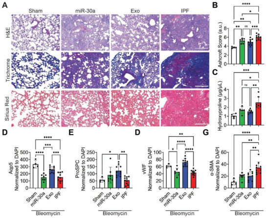
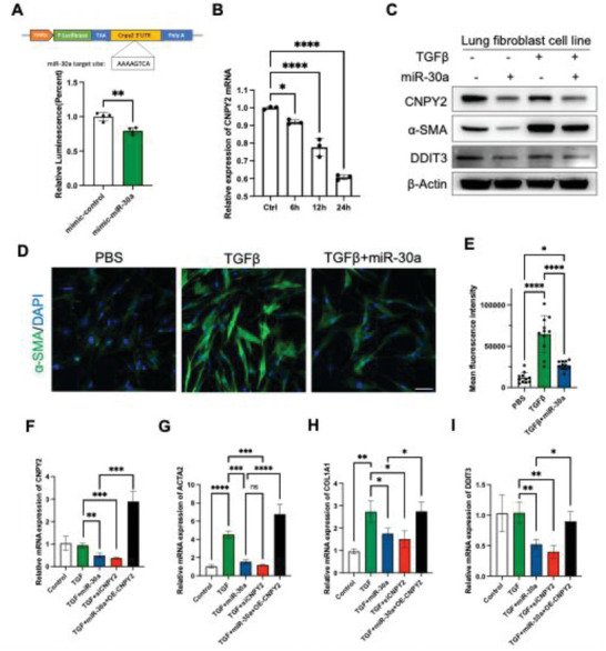
Idiopathic pulmonary fibrosis (IPF) remains an incurable form of interstitial lung disease with sub-optimal treatments that merely address adverse symptoms or slow fibrotic progression. Here, inhalable hsa-miR-30a-3p-loaded liposomes (miR-30a) for the treatment of bleomycin-induced pulmonary fibrosis in mice are presented. It was previously found that exosomes (Exo) derived from lung spheroid cells are therapeutic in multiple animal models of pulmonary fibrosis and are highly enriched for hsa-miR-30a-3p. The present study investigates this miRNA as a singular factor to treat IPF. Liposomes containing miR-30a mimic can be delivered to rodents through dry powder inhalation. Inhaled miR-30a and Exo consistently lead to improved pulmonary function across six consecutive pulmonary function tests and promote de-differentiation of profibrotic myofibroblasts. The heterogenous composure of Exo also promotes reparative alveolar type I and II cell remodeling and vascular wound healing through broad transforming growth factor-beta signaling downregulation, while miR-30a targets myofibroblast de-differentiation through CNPY2/PERK/DDIT3 signaling. Overall, inhaled miR-30a represses the epithelial-mesenchymal transition of myofibroblasts, providing fibrotic attenuation and subsequent improvements in pulmonary function.

Keywords: CNPY2; idiopathic pulmonary fibrosis; inhalation; miR‐30a‐3p.


Conflict of interest statement

The authors report no conflict of interest.

Figures

Figure 1
Figure 1
Characterization of nanoparticles. A) Schematic of hsa‐miR‐30a‐3p loading into liposomes, dry powder formulation, and dry powder inhalation. Created with BioRender.com. B) Immunoblots of CD63 and CD9 in NP lysate. C) TEM images of miR‐30a and Exo; scale bar = 50 nm. D) NTA size distribution analysis of NPs. E) Quantification of NTA size distribution analysis of the average mean ± standard error of three replicates. F) Standard curves of the normalized expression (dCt) of hsa‐miR‐30a‐3p to the LSC Control in LSC RNA co‐cultured with miR‐30a Lipo by qPCR; n = 2 per particle number. G) Relative expression (ddCt) of hsa‐miR‐30a‐3p to the RNU6 control gene and LSC Control in LSC RNA co‐cultured with Naked Mimic, miR‐30a, or Exo by qPCR; n = 2 per group. H) Relative expression (ddCt) of hsa‐miR‐30a‐3p to the RNU6 control gene and Sham in murine lung RNA that received DPI treatments of miR‐30a, Exo, or PBS (Sham and IPF) by qPCR; n = 4–10 per group. I,J), Representative ex vivo images I) and quantitative analysis J) of mouse major organs that received miR‐30a and Exo before and 24 h post‐inhalation. The control group consists of mice that did not receive any treatment. p values were determined by one‐way ANOVA using GraphPad PRISM software. * p < 0.05, ** p < 0.01, *** p < 0.001, **** p < 0.0001; ns, not significant.
Figure 2
Figure 2
Pulmonary function changes following inhalation of nanoparticles. A) Schematic of baseline pulmonary function measurements, DPI treatments, and endpoint pulmonary function measurements. Created with BioRender.com. B) Quantification of endpoint resistance normalized to baseline resistance per mouse; n = 4–10 per group. C) Quantification of endpoint compliance normalized to baseline compliance per mouse; n = 3–10 per group. D) Quantification of endpoint elastance normalized to baseline elastance per mouse; n = 4–10 per group. E) Quantification of endpoint inspiratory capacity normalized to baseline inspiratory capacity per mouse; n = 4–9 per group. F) Quantification of endpoint hysteresis area normalized to baseline hysteresis area per mouse; n = 4–10 per group. G) Quantification of endpoint FEV0.2/FVC normalized to baseline FEV0.2/FVC per mouse; n = 4–10 per group. p values were determined by one‐way ANOVA using GraphPad PRISM software. * p < 0.05, ** p < 0.01, *** p < 0.001, **** p < 0.0001; ns, not significant.
Figure 3
Figure 3
Tissue remodeling and regeneration following inhalation of nanoparticles. A) Representative H&E, Gomori's trichrome, and sirius red images of mouse lung tissue sections after DPI treatments; scale bar = 500 µm for H&E and trichrome sections and 250 µm for sirius red sections. B) Quantification of fibrosis by Ashcroft scoring; n = 4–10 per group. C) Quantification of hydroxyproline concentrations from murine lung tissue; n = 4–7 per group. D) Quantification of Aqp5 pixel intensity from mouse lung tissue images normalized to nuclei; n = 4–10 per group. E) Quantification of ProSPC pixel intensity from mouse lung tissue images normalized to nuclei; n = 4–10 per group. F) Quantification of vWF pixel intensity from mouse lung tissue images normalized to nuclei; n = 4–10 per group. G) Quantification of α‐SMA pixel intensity from mouse lung tissue images normalized to nuclei; n = 4–10 per group. p values were determined by one‐way ANOVA using GraphPad PRISM software. * p < 0.05, ** p < 0.01, *** p < 0.001, **** p < 0.0001; ns, not significant.
Figure 4
Figure 4
Inhaled hsa‐miR‐30a‐3p downregulates CNPY2/PERK/DDIT3 signaling. A) Heatmap of TGF‐β signaling genes normalized to Sham and β‐actin from lung tissue lysate. Detection of genes is in duplicates. B) Apoptosis array from lung tissue lysate. Detection of genes is in duplicates. C) Cytokine array from mouse serum. Detection of genes is in duplicates. D) Volcano plot of target genes of hsa‐miR‐30a‐3p. Red indicates significantly upregulated genes, grey indicates not significant genes and blue indicates significantly downregulated genes. Detection of genes is in duplicates. E) Immunoblots of CNPY2, PERK, DDIT3, E‐cadherin, N‐cadherin, and β‐actin in lung tissue lysate. p values were determined by one‐way ANOVA using GraphPad PRISM software. * p < 0.05, ** p < 0.01, *** p < 0.001, **** p < 0.0001; ns, not significant.
Figure 5
Figure 5
The hsa‐miR‐30a‐3p regulates fibrosis through CNPY2. A) Duel luciferase reporter assay to determine miR‐30a‐CNPY2 mRNA interactions. B) Relative CNPY2 mRNA expression after hsa‐miR‐30a‐3p transfection in lung fibroblast cells. The control group is from cells that did not receive hsa‐miR‐30a‐3p transfection. C) CNPY2, α‐SMA, and DDIT3 expression in lung fibroblast cells after TGFβ and miR‐30a treatment. D) Immunostaining of α‐SMA in TGFβ and hsa‐miR‐30a‐3p treated lung fibroblast cells. Scale bar = 30 µm. E) Quantification of D). F‐I) Relative mRNA expressions after transfections indicated. The control groups are from cells that did not receive any treatment. n = 3 per group. p values were determined by one‐way ANOVA using GraphPad PRISM software. * p < 0.05, ** p < 0.01, *** p < 0.001, **** p < 0.0001; ns, not significant.

References

    1. Richeldi L., Collard H. R., Jones M. G., Lancet 2017, 389, 1941. - PubMed
    1. Kolb M., Vašáková M., Respir. Res. 2019, 20, 57. - PMC - PubMed
    1. Raghu G., Collard H. R., Egan J. J., Martinez F. J., Behr J., Brown K. K., Colby T. V., Cordier J., Flaherty K. R., Lasky J. A., Lynch D. A., Ryu J. H., Swigris J. J., Wells A. U., Ancochea J., Bouros D., Carvalho C., Costabel U., Ebina M., Hansell D. M., Johkoh T., Kim D. S., King Jr T. E., Kondoh Y., Myers J., Müller N. L., Nicholson A. G., Richeldi L., Selman M., Dudden R. F., et al., Am. J. Respir. Crit. Care Med. 2011, 183, 788. - PMC - PubMed
    1. Richeldi L., du Bois R. M., Raghu G., Azuma A., Brown K. K., Costabel U., Cottin V., N. Engl. J. Med. 2014, 370, 2071. - PubMed
    1. Noble P. W., Albera C., Bradford W. Z., Costabel U., Glassberg M. K., Kardatzke D., King Jr T. E., Lancaster L., Sahn S. A., Szwarcberg J., Valeyre D., du Bois R. M., Lancet 2011, 377, 1760. - PubMed

LinkOut - more resources