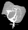
Challenges of Duplicated Portal Vein in Elective Laparoscopic Cholecystectomy: A Case Report
- PMID: 40120106
- PMCID: PMC11939121
- DOI: 10.12659/AJCR.946151
Challenges of Duplicated Portal Vein in Elective Laparoscopic Cholecystectomy: A Case Report
Abstract
BACKGROUND Anatomical variations of the portal system are not uncommon. Misidentifying structures of the hepatoduodenal ligament can precipitate tremendous adverse events during elective cholecystectomy. Preoperative radiological imaging is usually limited to ultrasound examination, which alone does not provide sufficient anatomical knowledge of the liver hilum. CASE REPORT This report presents a case of a 61-year-old woman after cholecystectomy, with iatrogenic bile duct injury and packing, due to abdominal hemorrhage derived from portal vein rupture. The patient required immediate relaparotomy and abdominal depacking, due to excessive compression of the hepatoduodenal ligament and insufficient portal blood flow. Surgery was limited to depacking and repair of the lacerated portal vein. The abdominal drainage was performed to stabilize the patient's general condition. Intraoperative ultrasound identified poor portal flow (V<10 cm/s) and intrahepatic portal thrombosis. Further treatment continued in the Intensive Care Unit, where she received anticoagulation treatment and was qualified for liver transplantation. The cavernous transformation of the portal vein was identified, along with several other anatomical variations, including a low-positioned splenomesenteric venous confluence, right-shifted pancreas, and intestinal malrotation, among other minor vascular abnormalities. During the next days, her general condition improved; following extubation, she was transferred to the Surgery Unit. A biliary fistula was managed by percutaneous transhepatic drainage and biliary stenting. Liver transplantation was not necessary. CONCLUSIONS This case highlights the extremes of vascular and biliary injury following elective cholecystectomy, partially due to lack of preoperative radiological examination, and portrays the elevated risk of mortality and burden of further medical treatment.
Conflict of interest statement
Figures


Similar articles
-
Right hepatectomy due to portal vein thrombosis in vasculobiliary injury following laparoscopic cholecystectomy: a case report.J Med Case Rep. 2014 Dec 7;8:412. doi: 10.1186/1752-1947-8-412. J Med Case Rep. 2014. PMID: 25481385 Free PMC article.
-
Portal vein thrombosis: an unusual complication of laparoscopic cholecystectomy.JSLS. 2005 Jan-Mar;9(1):87-90. JSLS. 2005. PMID: 15791978 Free PMC article.
-
From Laparoscopic Cholecystectomy to Liver Transplantation: When the Gallbladder Becomes the Pandora s Box.Chirurgia (Bucur). 2016 Sept-Oct;111(5):450-454. doi: 10.21614/chirurgia.111.5.450. Chirurgia (Bucur). 2016. PMID: 27819646
-
Emergency liver resection for combined biliary and vascular injury following laparoscopic cholecystectomy: case report and review of the literature.South Med J. 2007 Mar;100(3):317-20. doi: 10.1097/01.smj.0000242793.15923.1a. South Med J. 2007. PMID: 17396740 Review.
-
Biliary tract injuries during laparoscopic cholecystectomy: three case reports and literature review.G Chir. 2010 Jan-Feb;31(1-2):16-9. G Chir. 2010. PMID: 20298660 Review.
References
-
- Tomizawa N, Akai H, Akahane M, et al. Prepancreatic postduodenal portal vein: A new hypothesis for the development of the portal venous system. Jpn J Radiol. 2010;28(2):157–61. - PubMed
-
- Du G, Li J, Qi Y. An unusual developmental anomaly of duplicated portal vein. Folia Morphol (Warsz) 2024;83(3):737–39. - PubMed
Publication types
MeSH terms
LinkOut - more resources
Full Text Sources

