. 2025 May;301(5):108432.
doi: 10.1016/j.jbc.2025.108432. Epub 2025 Mar 20.

Structural and functional analysis of Pseudomonas aeruginosa PelA provides insight into the modification of the Pel exopolysaccharide

Affiliations

Structural and functional analysis of Pseudomonas aeruginosa PelA provides insight into the modification of the Pel exopolysaccharide

Jaime C Van Loon et al. J Biol Chem. 2025 May.

Abstract

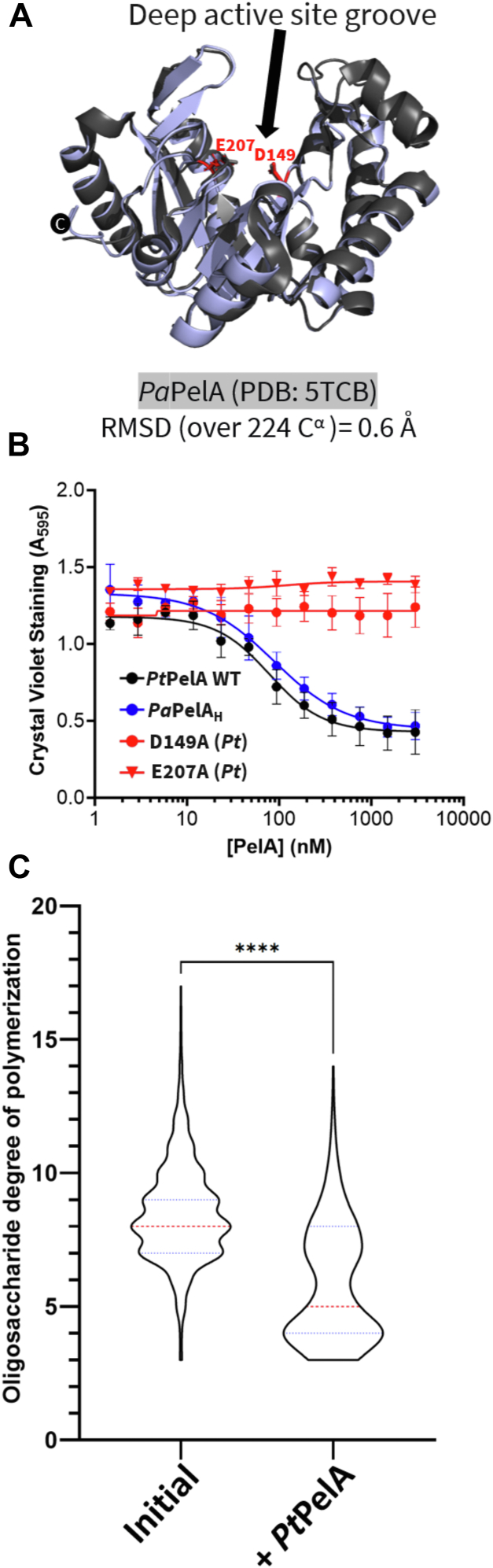
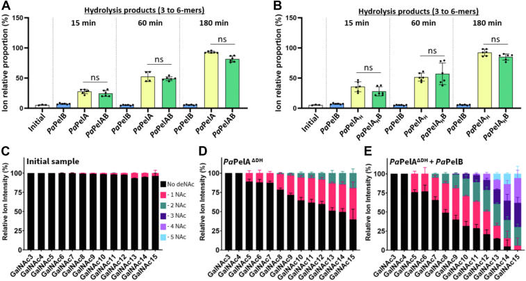
A major biofilm matrix determinant of Pseudomonas aeruginosa is the partially deacetylated α-1,4 linked N-acetylgalactosamine polymer, Pel. After synthesis and transport of the GalNAc polysaccharide across the inner membrane, PelA partially deacetylates and hydrolyzes Pel before its export out of the cell via PelB. While the Pel modification and export proteins are known to interact in the periplasm, it is unclear how the interaction of PelA and PelB coordinates these processes. To determine how PelA modifies the polymer, we determined its structure to 2.1 Å and found a unique arrangement of four distinct domains. We have shown previously that the hydrolase domain exhibits endo-α-1,4-N-acetylgalactosaminidase activity. Characterization of the deacetylase domain revealed that PelA is the founding member of a new carbohydrate esterase family, CE21. Further, we found that the PelAB interaction enhances the deacetylation of N-acetylgalactosamine oligosaccharides. Using the PelA structure in conjunction with AlphaFold2 modeling of the PelAB complex, we propose a model wherein PelB guides Pel to the deacetylase domain of PelA and subsequently to the porin domain of PelB for export. Perturbation or loss of the PelAB interaction would result in less efficient deacetylation and potentially increased Pel hydrolysis. In PelA homologs across many phyla, the predicted structure and active sites are conserved, suggesting a common modification mechanism in Gram-negative bacterial species containing a functional pel operon.

Keywords: Pseudomonas aeruginosa; biofilm; crystallography; pel polysaccharide; structure-function.


Conflict of interest statement

Conflict of interest The authors declare that they have no conflicts of interest with the contents of this article.

Figures

Figure 1
Figure 1
The full-length structure of PtPelA.A, linear representation of PtPelA domains based on the structure and available literature. The domain architecture was visualized using DOG 2.0 Illustrator of Protein Domain Structures (77). Amino acids that are required for enzymatic activity are highlighted. B, cartoon representation of the crystal structure of residues 37 to 937 of PtPelA shown in two opposing views. The domains as colored as depicted in (A). Amino acids required for the deacetylase and hydrolase activities are shown in black and red, respectively. N and C represent the N- and C-termini, respectively. The dashed lines represent regions of the protein that could not be built due to the poor quality of the electron density. C, comparison of the hydrolase domain of PelA as determined from the structure from Pseudomonas thermotolerans (blue) and the structure of PaPelAH (5TCB, gray) with the secondary structure labeled. Secondary structural elements in the AF2 model of PaPelA that are missing in the structure of PtPelA are labeled in red. D, the mixed α/β domain of PtPelA. E, the deacetylase domain of PtPelA. F, the β-rich domain of PtPelA. SS, signal sequence.
Figure 2
Figure 2
PelA is the founding member of a novel CE family.A, surface representation of the AF2 model of PaPelA from two opposing views. The polymer in the first view of PaPelA is representative of nine GalNAc residues (38). B, the PaPelAAF2 deacetylase domain (left). Inset of a structural alignment of the catalytic residues of PaPelA (pink) with representative CE4 and CE18 enzymes: Streptococcus pneumoniae PgdA (2C1G, blue) and Aspergillus fumigatus Agd3 (6NWC, green), respectively. The zinc that cocrystallized with SpPgdA is shown as a blue sphere. C, primary sequence alignment of the catalytic motifs MT1-5 and CM1-4 characteristic of CE4 and CE18 enzymes, respectively, as determined by structural alignment. The putative PaPelA catalytic base (D528), metal coordinating triad (D530, H600, and H604), and putative catalytic acid (H761) are highlighted in orange, yellow, and purple, respectively. The aspartate that coordinates the catalytic acid (E723) and the histidine that is proposed to coordinate the catalytic base (H526) is shown in cyan and pink, respectively. D, proposed metal-dependent de-N-acetylation reaction for PaPelA (first listed amino acid) and PtPelA (second listed amino acid) based on the mechanisms for CE4 and CE18 enzymes and mutagenesis data.
Figure 3
Figure 3
PtPelA has structural differences in comparison to CE4 and CE18 enzymes.A and B, cartoon and surface representations to compare the AF2 model of PaPelA (A) and the structure of SpPgdA (B). Regions where there are major structural differences are colored and labeled accordingly. Active site residues are shown in black. The black lines highlight the active site grooves. C and D, cartoon and surface representations to compare the AF2 model of PaPelA (C) and the structure of Agd3 (D).
Figure 4
Figure 4
PtPelA is a metal-dependent CE21 enzyme.A, detection of WT or mutant PtPelA esterase activity using the AMMU pseudosubstrate. The putative catalytic base and metal coordinating triad are highlighted in orange and yellow, respectively. Statistical significance was calculated using an ordinary one-way ANOVA with Dunnett's multiple comparison test between WT and mutant PtPelA. The error bars show the SEM for the four independent assays with two technical replicates. ∗∗∗∗p < 0.0001; ∗∗p < 0.0021; ∗p < 0.0332; ns, not significant. B, detection of PtPelA metal-dependent esterase activity using the AMMU substrate. Statistical significance was calculated using a one-way ANOVA with Dunnett's multiple comparison test between PtPelA as isolated and the other reaction conditions with chelator or metal chloride. AMMU, acetoxymethyl-4-methylumbelliferone.
Figure 5
Figure 5
PtPelA has α-1,4-N-acetylgalactosaminidase activity.A, structural comparison of the hydrolase domain of PtPelA (blue) with the hydrolase domain of PaPelA (5TCB, gray). Amino acids required for the hydrolase activity of PtPelA (D149 and E207) and PaPelA are shown in red and gray, respectively. B, a biofilm disruption assay. Increasing concentrations of WT PtPelA (black circle), PaPelaH (blue circle), or PtPelA hydrolase domain mutants D149A (red circle) or E207A (red triangle) were added to preformed PA14 Pel biofilms. The error bars show the SEM for the average of three technical replicates in three independent assays. C, population statistical study of the MALDI-TOF MS enzyme spectra of the oligomers released due to hydrolysis after incubating WT PtPelA. The data represent three biological replicates each with three technical replicates. Statistical significance was calculated using a one-way ANOVA with Dunnett's multiple comparison test between the indicated reaction conditions. ∗∗∗∗p < 0.0001. The median size of the oligosaccharides is shown by the dotted red line. The 25th and 75th quartiles are shown by the blue dotted lines, respectively.
Figure 6
Figure 6
Interaction with PaPelB does not affect PaPelA hydrolase activity but increases the deacetylation of α-1,4-GalNAc oligosaccharides by PaPelA.A and B, the ion relative proportion of the MALDI-TOF MS enzyme spectra of the 3 to 6-mer oligomers released due to hydrolysis after incubating WT PaPelA (A) or PaPelAH (B) in the absence or presence of PaPelB. The data represent three biological replicates each with two technical replicates. Statistical significance was calculated using Kruskal–Wallis multiple comparison tests between the indicated reaction conditions. CE, MALDI-TOF MS analysis of the deacetylation products for the oligosaccharides in isolation (C) and after incubating PaPelADH in the absence (D) or presence (E) of PaPelB. Ns, not significant.
Figure 7
Figure 7
Full-length PelA is distributed across multiple Gram-negative phyla and has conserved active site residues.A, phylogenetic distribution of PelA among bacterial taxa. The circular tree represents the approximately-maximum-likelihood distance between 96 unique PelA sequences, which was constructed using FastTree and Escherichia coli LpoB as an outgroup. PelA sequences were identified by running a FoldSeek search of PaPelA. B and C, multiple sequence alignment of PaPelA homologs identified through FoldSeek (B) or BLASTP (C) and visualized using WebLogo 3. The catalytic motifs MT1-5 and CM1-4 are characteristic of CE4 and CE18 enzymes.
Figure 8
Figure 8
Structural models of PaPelB, PaPelC, and the PaPelBC complex.A, AF2 model of full-length PaPelB complexed with PaPelA (colored by domains as in Fig. 1). PaPelB is colored by domain and aligned to the structure of PaPelB332-436 (5WFT, magenta). B, the α-rich region of PaPelB from two opposing views. The black arrow indicates the proposed trajectory of Pel through PelB. C, electrostatic surface representation of PaPelB calculated by APBS in PyMol and visualized from −5 (blue) to 5 (red) kT/e. D, electrostatic surface representation of the α-rich region of PaPelB.
Figure 9
Figure 9
Model of the PaPelABC outer membrane Pel modification and secretion complex.A, AF2 model of full-length PaPelB (light cyan) complexed with PaPelA (colored by domains as in Fig. 1). AF2 model of amino acids 600 to 1193 of PaPelB complexed with the 12-subunit ring of PaPelC (green) were aligned to the PaPelAB complex prediction. The fragment of PaPelB used for the PaPelBC prediction was then excluded from this figure. B, cartoon representation of dodecameric PaPelC and the surface representation of PaPelAB from (A) as colored by electrostatics, which were calculated by APBS in PyMol and visualized from −5 (blue) to 5 (red) kT/e. The solid black line indicates the proposed trajectory of Pel export while PaPelA and PaPelB interact. The dotted black line indicates when the polymer is enclosed by the pore formed by the PaPelAB complex. C, surface representation of (A). The active site amino acids of the deacetylase and hydrolase domains of PaPelA are shown in black.

References

    1. Flemming H.-C., Wingender J. The biofilm matrix. Nat. Rev. Microbiol. 2010;8:623–633. - PubMed
    1. Karygianni L., Ren Z., Koo H., Thurnheer T. Biofilm matrixome: extracellular components in structured microbial communities. Trends Microbiol. 2020;28:668–681. - PubMed
    1. Pang Z., Raudonis R., Glick B.R., Lin T.-J., Cheng Z. Antibiotic resistance in Pseudomonas aeruginosa: mechanisms and alternative therapeutic strategies. Biotechnol. Adv. 2019;37:177–192. - PubMed
    1. Mulcahy L.R., Isabella V.M., Lewis K. Pseudomonas aeruginosa biofilms in disease. Microb. Ecol. 2014;68:1–12. - PMC - PubMed
    1. Salerian A.J. Burn wound infections and Pseudomonas aeruginosa. Burns. 2020;46:257–258. - PubMed

MeSH terms

Substances

LinkOut - more resources