. 2025 Jul;44(7):1122-1134.
doi: 10.1016/j.healun.2025.02.1696. Epub 2025 Mar 20.

Lung rehabilitation using xenogeneic cross-circulation does not lead to hyperacute rejection in a human lung transplantation model

Affiliations

Lung rehabilitation using xenogeneic cross-circulation does not lead to hyperacute rejection in a human lung transplantation model

Kaitlyn M Tracy et al. J Heart Lung Transplant. 2025 Jul.

Abstract

Background: Access to life-saving lung transplantation remains limited by a shortage of donor organs. We have previously described rehabilitation of discarded human donor lungs to a quality suitable for transplantation using cross-circulation of whole blood between xeno-support swine and human lungs. However, the immunologic implications of transplanting rehabilitated lungs remain unknown.

Methods: Human donor lungs declined for clinical transplantation (N = 5) and underwent xenogeneic cross-circulation (XC) for up to 12 hours. To model subsequent human transplantation, lungs were re-exposed to autologous human whole blood via normothermic ex vivo machine perfusion for up to 6 hours. Upon human blood re-exposure (HBR), lungs were evaluated for evidence of hyperacute rejection (HAR) through physiologic assessments and tissue analyses including histology, immunostaining, and flow cytometry.

Results: Upon HBR, lungs showed no significant change in physiologic function relative to the end of cross-circulation (PaO2/FiO2: p = 0.41; vascular resistance: p = 0.27; dynamic compliance: p = 0.24) and histologic features of HAR were absent in all lungs. Despite pulmonary deposition of porcine IgG during cross-circulation, HBR resulted in decreased complement deposition (p = 0.019) with no change in membrane attack complex formation (p = 0.65) or apoptotic signaling (p = 0.93). Endothelial integrity was maintained after HBR with preservation of microvascular tight junctions, decreasing endothelial injury marker p-selectin (p = 0.34), and intact vascular response to alpha-adrenergic stimulation.

Conclusions: Our findings indicate that transient exposure of human donor lungs to XC does not result in HAR upon simulated human transplantation, representing an important step toward clinical translation of this donor organ rehabilitation platform.

Keywords: human lung transplantation model; lung rehabilitation; lung transplantation; transplant immunology; xenogeneic cross-circulation.


Conflict of interest statement

Disclosures

MB is an inventor on a patent related to this work filed by Columbia University (No. WO2018013849A1) filed July 13, 2017 and published January 18, 2018. WKW, RU, and MB are inventors on a patent application related to this work filed by Vanderbilt University (No. 63/165,773) filed March 25, 2021. All other authors declare they have no competing interests.

Figures

Fig. 1.
Fig. 1.. Experimental overview.
(A) Human donor lungs declined for transplantation are recovered on a human-xenogeneic cross-circulation platform and subsequently re-exposed to autologous human whole blood. Immunosuppression is administered for each experimental phase. (B-D) Gross photographs: (B) Left atrial reconstruction with donor pericardium for pulmonary vein (PV) cannula placement. (C) Venovenous cannulation of bilateral internal jugular veins of xeno-support swine. (D) Ex vivo machine perfusion platform for autologous human blood re-exposure. CVF: cobra venom factor; PA: pulmonary artery; RIJ: right internal jugular vein; LIJ: left internal jugular vein. Schematic created using BioRender.com.
Fig. 2.
Fig. 2.. Functional evaluation of human donor lungs during xenogeneic cross-circulation and human blood re-exposure.
(A) PaO2/FiO2. (B) Pulmonary vascular resistance. (C) Dynamic compliance. For (A to C): N=5 independent experiments. (D) Peak and mean airway pressures of lungs ventilated using volume control mode with tidal volume of 6 mL/kg donor body weight (N=2 independent experiments). Data are presented as mean (SD). Mixed-effects model for repeated measures used for all comparisons. Functional parameters of Lungs 1–5 are in Fig. S4. HBR: Human blood re-exposure; PIP: peak inspiratory pressure; Pmean: mean airway pressure; XC: cross-circulation.
Fig. 3.
Fig. 3.. Multiscale evaluation of human donor lungs during xenogeneic cross-circulation and human blood re-exposure.
Representative gross photography (A), thermography (B), and hematoxylin and eosin (H&E) staining of lung parenchyma (C) before XC (Pre XC), after XC (Post XC), and after HBR (Post HBR). (D) Histopathologic injury score (N=5 independent experiments). Data are presented as mean (SD). Mixed-effects model for repeated measures used for all comparisons with Tukey’s multiple comparisons test. Representative images of Lungs 1–5 are in Figs. S5–S9. HBR: Human blood re-exposure; XC: cross-circulation.
Fig. 4.
Fig. 4.. Immunoglobulin deposition in human donor lungs during xenogeneic cross-circulation and human blood re-exposure.
(A) Representative immunofluorescence staining of human and porcine immunoglobulin G (IgG) in human lungs before XC (Pre XC), after XC (Post XC), and after HBR (Post HBR). White arrowhead indicates colocalization of porcine IgG and human IgG on a single cell during XC and HBR. (B and C) Quantification of immunofluorescence staining for porcine (B) and human (C) IgG+ cells in human lungs. (D) Quantification of cells bound with both porcine and human IgG. For (A to D), N=5 independent experiments. (E) Flow cytometric evaluation of porcine IgG and porcine immunoglobulin M (IgM) binding specificity to human immune, epithelial, or endothelial cells. Data presented as a percentage of porcine IgG+ (or IgM+) live, singlet cells (Pre XC: N=4, Post XC: N=5, and Post HBR: N=3 independent experiments, due to insufficient cell count in remaining samples). Data are presented as mean (SD). Mixed-effects model for repeated measures used for all comparisons with Tukey’s multiple comparisons test. *P<0.05, **P<0.01. Representative immunofluorescence staining images of Lungs 1–5 are in Figs. S5–S9; negative controls are in Fig. S14. The flow cytometry gating strategy can be found in Reference . HBR: Human blood re-exposure; XC: cross-circulation.
Fig. 5.
Fig. 5.. Characterization of complement activation and apoptosis in human donor lungs during xenogeneic cross-circulation and human blood re-exposure.
(A-D) Representative immunohistochemical staining for C4d (A) membrane attack complex (MAC) (B) cleaved Caspase-3 (C) and TUNEL (D) in human lungs before XC (Pre XC), after XC (Post XC), and after HBR (Post HBR). (E) Quantification of immunohistochemical staining for markers of complement activation and apoptosis. For (A to E), N=5 independent experiments for each stain. (F and G) Quantification of plasma complement activity (CH50) during XC (F) and HBR (G) as a percentage of normal complement activity (N=5 independent experiments for XC and HBR). Data are presented as mean (SD). Mixed-effects model for repeated measures used for all comparisons with Tukey’s multiple comparisons test. *P < 0.05. Representative immunohistochemical staining images of Lungs 1–5 are in Figs. S5–S9; positive controls are in Fig. S15. CH50 values for Lungs 1–5 are in Fig. S3. C4d staining score as previously defined. CVF: cobra venom factor; HBR: human blood re-exposure; XC: cross-circulation.
Fig. 6.
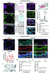
Fig. 6.. Evaluation of endothelial and epithelial integrity after autologous human blood re-exposure.
(A) Representative immunofluorescence staining for endothelial marker (CD31) and tight junction protein zonula occludens-1 (ZO-1) in alveoli of human lungs. (B) Quantification of CD31+ cells, ZO-1+ cells, and CD31+ ZO-1+ cells as a percentage of total cells. (C and D) Representative immunofluorescence staining for CD31, ZO-1 (C), and α-smooth muscle actin (α-SMA) (D) in pulmonary arterioles in human lungs. For (A to D), N=5 independent experiments. (E) Transmission electron microscopy (TEM) demonstrating intact alveolar-capillary barrier (pseudocolored): red blood cells (red), endothelial cell nucleus (orange) and membrane (blue), type I pneumocyte membrane (arrow heads), alveolar space (*) (N=1 independent experiment, Lung 5). (F) P-selectin in plasma Pre XC, Post XC, Post HBR, and from autologous human whole blood perfusate (WB) (N=5 independent experiments). (G) Pulmonary arterial (PA) pressure after administration of α−1 agonist (N=2 independent experiments). (H and I) Representative immunofluorescence staining for epithelial cell marker (EpCAM) (H) with quantification of EpCAM+ cells as a percentage of total cells (I) (N=5 independent experiments). (J) TEM image of a type II pneumocyte (*) with preserved lamellar bodies containing surfactant (arrow) (N=1 independent experiment, Lung 5). (K and L) Representative immunofluorescence staining for α-SMA, EpCAM (K) and CD31, ZO-1 (L) in bronchioles of human lungs (N=5 independent experiments). (M and N) Peak and mean airway pressures after administration of cholinergic agonist (M) and β−2 agonist (N) (N=1 independent experiment, Lung 5). Data are presented as mean (SD). Mixed-effects model for repeated measures used for all comparisons with Tukey’s multiple comparisons test. *P < 0.05. Representative immunofluorescence staining images of Lungs 1–5 are in Figs S5–S9; negative controls are in Fig. S14. HBR: Human blood re-exposure; PIP: peak inspiratory pressure; Pmean: mean airway pressure; XC: cross-circulation.

References

    1. Valapour M, Lehr CJ, Schladt DP, et al. OPTN/SRTR 2022 Annual Data Report: Lung. Am J Transplant. 2024;24(2S1):S394–S456. doi: 10.1016/j.ajt.2024.01.017 - DOI - PubMed
    1. Cypel M, Yeung JC, Hirayama S, et al. Technique for Prolonged Normothermic Ex Vivo Lung Perfusion. J Heart Lung Transplant. 2008;27(12):1319–1325. doi: 10.1016/j.healun.2008.09.003 - DOI - PubMed
    1. Moreno P, González-García J, Ruíz-López E, Alvarez A. Lung Transplantation in Controlled Donation after Circulatory-Determination-of-Death Using Normothermic Abdominal Perfusion. Transpl Int. 2024;37:12659. doi: 10.3389/ti.2024.12659 - DOI - PMC - PubMed
    1. Gouchoe DA, Sanchez PG, D’Cunha J, et al. Ex vivo lung perfusion in donation after circulatory death: A post hoc analysis of the Normothermic Ex Vivo Lung Perfusion as an Assessment of Extended/Marginal Donors Lungs trial. J Thorac Cardiovasc Surg. 2024;168(3):724–734.e7. doi: 10.1016/j.jtcvs.2024.03.011 - DOI - PubMed
    1. Hozain AE, O’Neill JD, Pinezich MR, et al. Xenogeneic cross-circulation for extracorporeal recovery of injured human lungs. Nat Med. 2020;26(7):1102–1113. doi: 10.1038/s41591-020-0971-8 - DOI - PMC - PubMed