. 2025 Mar 22;8(1):477.
doi: 10.1038/s42003-025-07897-0.

ZDHHC9-mediated CD38 palmitoylation stabilizes CD38 expression and promotes pancreatic cancer growth

Affiliations

ZDHHC9-mediated CD38 palmitoylation stabilizes CD38 expression and promotes pancreatic cancer growth

Hui Guo et al. Commun Biol. .

Abstract

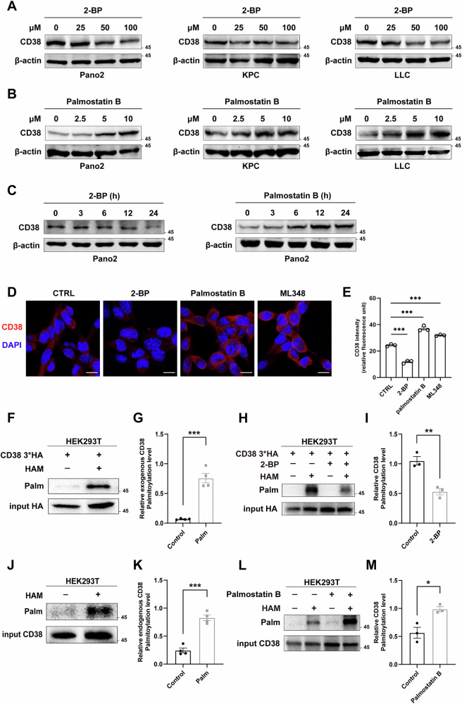
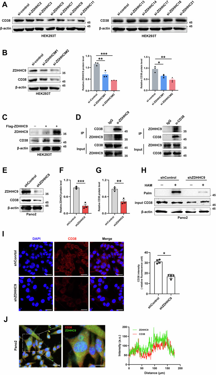
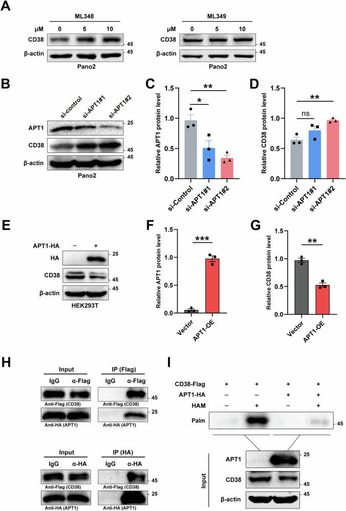
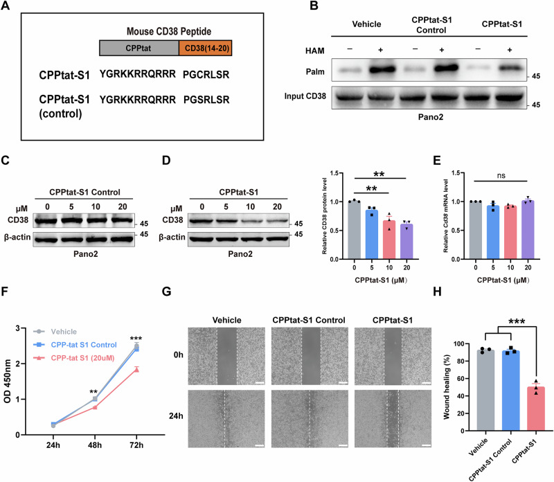
The cluster of differentiation 38 (CD38) is a multifunctional transmembrane protein involved in numerous physiological and pathological processes including aging, neurodegenerative diseases, and tumorigenesis, hence is an attractive drug target. However, the mechanisms underlying the regulation of CD38 expression remain enigmatic. Herein, we report for the first time that CD38 is palmitoylated at Cys16, and that S-palmitoylation is required to maintain CD38 protein expression in tumor cells. Furthermore, we identify DHHC9 as the palmitoyl transferase and APT1 as the acylprotein thioesterase responsible for this crucial post-translational modification. Finally, we designed a competitive peptide of CD38 palmitoylation that decreases CD38 expression in tumor cells and suppresses tumor progression in vivo. These findings provide novel insight into CD38 regulation and highlight potential therapeutic strategies targeting CD38 palmitoylation for cancer treatment.


Conflict of interest statement

Competing interests: The authors declare no competing interests.

Figures

Fig. 1
Fig. 1. CD38 palmitoylation dynamically modulates the protein expression of CD38.
A, B Pano2, KPC, and LLC cells were incubated with indicated concentrations of 2-BP (A) and palmostatin B (B) for 24 h and expression of CD38 was evaluated by western blotting analysis. C Western blotting showing the protein expression of CD38 in Pano2 cells treated with 2-BP (100 μM) and palmostatin B (10 μM) at the indicated time points. D Immunofluorescence assay detecting CD38 expression in Pano2 cells treated with 100 µM 2-BP, 10 µM palmostatin B, and 10 µM ML348 for 24 h. CD38 is stained red and the cell nucleus is stained with 4′,6-diamidino-2-phenylindole (DAPI) and appears blue. Scale bars, 10 μm. E Quantification of the intensity of the immunofluorescence of CD38 using ImageJ software. F Exogenous CD38 palmitoylation was detected in HEK293T cells using ABE assay and quantification (G). H Analysis of the ABE assay showing the level of CD38 palmitoylation in HEK293T cells with or without 2-BP (100 μM) treatment and quantification (I). J Palmitoylation of endogenous CD38 in HEK293T cells was detected using ABE assay and quantification (K). L ABE assay analysis demonstrated that the palmitoylation level of CD38 increased following treatment with palmostatin B and quantification (M). All results are presented as mean ± SEM. E Statistical analyses were calculated using a one-way ANOVA with a Tukey post hoc test (n = 3). G, I, K, M Statistical analyses were calculated using unpaired Student’s t-test (n = 3 or 4) (ns not significant, *p < 0.05, **p < 0.01, and ***p < 0.001).
Fig. 2
Fig. 2. CD38 is palmitoylated at Cys16.
A Sequences of amino acids of CD38 in various species showing conserved cysteine at position 16. B Schematic diagram of the structure of the CD38 molecule. The CD38 domains contained intracellular (20 amino acids), transmembrane (23 amino acids), and extracellular (257 amino acids). C Topology model of CD38 palmitoylation. D Western blotting analysis showing the level of S-palmitoylation of CD38 WT and C16S in HEK293T cells. E Western blotting analysis showing decreased expression of the CD38 protein in Pano2 cells treated with 2-BP and CHX at the indicated time compared to DMSO treatment. F Western blotting analysis of CD38 stability in CHX pretreated CD38-WT and CD38-C16S B16F10 cells and qualification (H) and (G). The CD38-C16S B16F10 cells were treated with MG132 and the western blotting assay was used to detect CD38 protein levels and qualification (I). J CD38 ubiquitination assay in B16F10 cells. The CD38-WT and CD38-C16S mutant cells were treated with MG132 followed by the western blotting assay to detect CD38 and ubiquitin. All results are presented as mean ± SEM. D Statistical analyses were calculated using unpaired Student’s t-test (n = 3) (ns not significant, *p < 0.05, **p < 0.01, and ***p < 0.001).
Fig. 3
Fig. 3. CD38 is palmitoylated by ZDHHC9.
A Western blotting analysis showing the expression of CD38 after transfected with different siRNA from ZDHHC enzymes in HEK293T cells. B Western blotting assay detecting the protein level of CD38 in HEK293T cells that were transfected with two independent siRNAs targeting ZDHHC9 and quantification. C Expression of CD38 in HEK293T cells transfected with different concentration flag-tagged ZDHHC9 vector. D A reciprocal co-IP assay was performed to detect the interaction between endogenously expressed ZDHHC9 and CD38 in HEK293T cells. E Western blotting was used to detect the endogenous expression of the CD38 protein in Pano2 cells with ZDHHC9 stably knocked down. F Quantification of ZDHHC9 protein levels. G Quantification of CD38 protein levels. H Endogenous CD38 protein was immunoprecipitated from ZDHHC9 knockdown cells and subjected for ABE analysis. I Left, representative immunofluorescence images of CD38 staining (red) of shZDHHC9 cells and shControl cells (blue, DAPI-labelled cell nuclei). (Scale bars, 20 µm). Right, corresponding quantification analysis. J Immunofluorescence assay showing the colocalization of CD38 and ZDHHC9 in Pano2 cells and statistical analysis. (Scale bars, 50 µm). All results are presented as mean ± SEM. B Statistical analyses were calculated using a one-way ANOVA with a Tukey post hoc test (n = 3). F, G, I Statistical analyses were calculated using unpaired Student’s t-test (n = 3 or 4) (ns not significant, *p < 0.05, **p < 0.01, and ***p < 0.001).
Fig. 4
Fig. 4. APT1 is the primary acylprotein thioesterase for CD38.
A Pano2 cells were incubated with indicated concentrations of ML348 and ML349 for 24 h and the expression of CD38 was evaluated by western blotting analysis. B Western blotting assay detecting the protein level of CD38 in Pano2 cells that were transfected with two independent siRNAs targeting APT1 and quantification (C) and (D). E Expression of CD38 in HEK293T cells overexpressing APT1 and quantification (F) and (G). H Co-IP analysis of the interaction of CD38 and APT1 in HEK293T cells. I CD38 palmitoylation in HEK293T cells with ectopic expression of APT1 detected by ABE assay. All results are presented as mean ± SEM. C, D Statistical analyses were calculated using a one-way ANOVA with a Tukey post hoc test (n = 3). F, G Statistical analyses were calculated using unpaired Student’s t-test (n = 3) (ns not significant, *p < 0.05, **p < 0.01, and ***p < 0.001).
Fig. 5
Fig. 5. A peptide designed to inhibit CD38 palmitoylation and protein expression.
A Sequence diagram of mouse CD38 peptide. B Pano2 cells were treated with CPPtat-S1 Control peptide or the CPPtat-S1 peptide and subjected to the ABE assay. C Western blot analysis showing the effect of CPPtat-S1 Control peptide incubation on CD38 protein expression in Pano2 cells. D Pano2 cells were treated with CPPtat-S1 peptide and subjected to western blotting assay and quantification analysis. E The mRNA level of Cd38 analyzed by qPCR after CPPtat-S1 peptide treatment. F Pano2 cells were pretreated with peptide, then cell viability was analyzed using the CCK8 cell proliferation assay. G The effect of peptide treatment on Pano2 cell migration was detected by the scratch test and quantification (H). (Scale bars, 100 μm). All results are presented as mean ± SEM. DH Statistical analyses were calculated using a one-way ANOVA with a Tukey post hoc test (n = 3) (ns not significant, **p < 0.01, and ***p < 0.001).
Fig. 6
Fig. 6. Inhibition of CD38 palmitoylation by peptides slows Pano2 pancreatic cancer growth.
AD C57BL/6 mice were subcutaneously injected with Pano2 cells and treated with DMSO (Vehicle group), CPPtat-S1 Control peptide, or CPPtat-S1 peptide (10 mg/kg). A Schematic diagram showing the treatment plan. BD Tumor volumes were measured every 2 days. Tumor size was weighed and photographed at the end of treatment. E After peptide treatment, a part of the tumor tissue was removed and paraffin embedded, and representative images of HE staining and IHC staining (Ki67, Tunel, CD38) were obtained. The scale is 50 μm. F, G The number of positive cells in Ki67 and Tunel under 40× mirror field was quantified by IHC staining. All results are presented as mean ± SEM. BG Statistical analyses were calculated using a one-way ANOVA with a Tukey post hoc test (n = 5) (ns not significant, *p < 0.05, **p < 0.01, and ***p < 0.001).

References

    1. Camacho-Pereira, J. et al. CD38 Dictates Age-Related NAD Decline and Mitochondrial Dysfunction through an SIRT3-Dependent Mechanism. Cell Metab.23, 1127–1139 (2016). - PMC - PubMed
    1. Chini, C. C. S. et al. CD38 ecto-enzyme in immune cells is induced during aging and regulates NAD(+) and NMN levels. Nat. Metab.2, 1284–1304 (2020). - PMC - PubMed
    1. Guerreiro, S., Privat, A. L., Bressac, L. & Toulorge, D. CD38 in Neurodegeneration and Neuroinflammation. Cells10.3390/cells9020471 (2020). - PMC - PubMed
    1. Jin, D. et al. CD38 is critical for social behaviour by regulating oxytocin secretion. Nature446, 41–45 (2007). - PubMed
    1. Chini, E. N., Chini, C. C. S., Espindola Netto, J. M., de Oliveira, G. C. & van Schooten, W. The Pharmacology of CD38/NADase: an emerging target in cancer and diseases of aging. Trends Pharm. Sci.39, 424–436 (2018). - PMC - PubMed