. 2025 Mar 22;8(1):89.
doi: 10.1038/s42004-025-01472-8.

A quantitative analysis of ligand binding at the protein-lipid bilayer interface

Affiliations

A quantitative analysis of ligand binding at the protein-lipid bilayer interface

Allison Pearl Barkdull et al. Commun Chem. .

Abstract

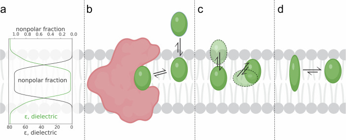
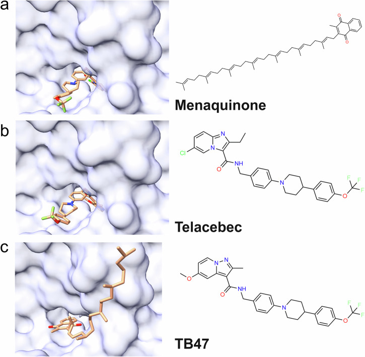
The majority of drugs target membrane proteins, and many of these proteins contain ligand binding sites embedded within the lipid bilayer. However, targeting these therapeutically relevant sites is hindered by limited characterization of both the sites and the molecules that bind to them. Here, we introduce the Lipid-Interacting LigAnd Complexes Database (LILAC-DB), a curated dataset of 413 structures of ligands bound at the protein-bilayer interface. Analysis of these structures reveals that ligands binding to lipid-exposed sites exhibit distinct chemical properties, such as higher calculated partition coefficient (clogP), molecular weight, and a greater number of halogen atoms, compared to ligands that bind to soluble proteins. Additionally, we demonstrate that the atomic properties of these ligands vary significantly depending on their depth within and exposure to the lipid bilayer. We also find that ligand binding sites exposed to the bilayer have distinct amino acid compositions compared to other protein regions, which may aid in the identification of lipid-exposed binding sites. This analysis provides valuable guidelines for researchers pursuing structure-based drug discovery targeting underexploited ligand binding sites at the protein-lipid bilayer interface.


Conflict of interest statement

Competing interests: The authors declare no competing interests.

Figures

Fig. 1
Fig. 1. Thermodynamic considerations for ligand binding at the protein-lipid interface.
a Dielectric and nonpolar fraction with respect to depth in a lipid bilayer. Qualitative reproduction of data from. b Ligand partitioning into the bilayer localizing near its target. c Ligand oriented at a preferred depth and orientation in the bilayer. d Ligand preferentially adopting a specific conformation in the lipid environment.
Fig. 2
Fig. 2. Dataset of ligands bound at the protein-lipid interface.
a Pie chart describing the functional classes of proteins represented in the dataset. b Membrane associated ligands (blue) bound to GPCRs (representative protein structure PDB ID: 4PHU). c Membrane associated ligands (purple) bound to cytochrome complexes (representative protein structure PDB ID: 6XVF). d Membrane associated ligands (pink) bound to transient receptor potential (TRP) channels (representative protein structure PDB ID: 5IRX).
Fig. 3
Fig. 3. Atomic properties of ligands with respect to depth in the bilayer and solvent exposure.
a Binned average of atomic Crippen contribution of exposed and buried ligand atoms with respect to the position in the lipid bilayer. The shaded region represents standard error. b Violin plots of atomic Crippen contribution of ligand atoms separated by exposure and membrane region. Means of distribution are displayed as dotted black lines. c Binned average of the magnitude of espaloma partial charge of exposed and buried ligand atoms with respect to the position in the lipid bilayer. The shaded region represents standard error. d Violin plots of magnitude of espaloma partial charge of ligand atoms separated by exposure and membrane region. Means of distribution are displayed as dotted black lines.
Fig. 4
Fig. 4. Comparison of amino acid composition of binding sites and other protein regions with respect to solvent.
Comparison of population residue types within binding sites (hatch pattern) and outside binding sites (solid fill) across different protein regions (Tail Group, Head Group, and Soluble Proteins). Residues are classified as either buried (solvent-exposed surface area (SASA) <15% in the absence of ligand, left) or solvent-exposed (SASA >15% in the absence of ligand, right). Error bars were generated using bootstrap sampling (2000 iterations) to calculate standard deviations of residue proportions.
Fig. 5
Fig. 5. Poses of ligands bound to CaSR.
a Cinacalcet-bound (blue, PDB ID 7M3F, chain A) and evocalcet-bound (purple, PDB ID 7M3G, chain A) CaSR with ligands in the bent conformation. b Cinacalcet-bound (blue, PDB ID 7M3F, chain B) and evocalcet-bound (purple, PDB ID 7M3G, chain B) CaSR with ligands in the extended conformation. c Tecalcet-bound (green, PDB ID 7SIL, chain A) CaSR. d NPS-2143-bound (sand, PDB ID 7M3E) CaSR. e Chemical structures of the ligands cinacalcet, evocalcet, tecalcet, and NPS-2143.
Fig. 6
Fig. 6. Drug and lipid binding to T-type calcium channel Cav3.3.
a Cav3.3 (white) and lipid (black) (PDB ID: 7WLI). b Mibefradil (orange) bound to Cav3.3 (white) and lipid (black) (PDB ID: 7WLJ). c Otilonium bromide (pink) bound to Cav3.3 (white) and lipid (black) (PDB ID: 7WLK). d Pimozide (green) bound to Cav3.3 (white) and lipid (black) (PDB ID: 7WLL).
Fig. 7
Fig. 7. Ligands bound to the Mycobacterium tuberculosis cytochrome bcc complex.
a Menaquinone (teal) bound to M. tuberculosis cytochrome bcc (white) (PDB ID: 7E1V). b Telacebec (orange) bound to M. tuberculosis cytochrome bcc (white) (PDB ID: 7E1W). c TB47 (pink) bound to M. tuberculosis cytochrome bcc (white) (PDB ID: 7E1X).

References

    1. Yin, H. & Flynn, A. D. Drugging membrane protein interactions. Annu. Rev. Biomed. Eng.18, 51–76 (2016). - PMC - PubMed
    1. Santos, R. et al. A comprehensive map of molecular drug targets. Nat. Rev. Drug Discov.16, 19–34 (2017). - PMC - PubMed
    1. Payandeh, J. & Volgraf, M. Ligand binding at the protein–lipid interface: strategic considerations for drug design. Nat. Rev. Drug Discov.20, 710–722 (2021). - PubMed
    1. Wang, Y., Yu, Z., Xiao, W., Lu, S. & Zhang, J. Allosteric binding sites at the receptor–lipid bilayer interface: novel targets for GPCR drug discovery. Drug Discov. Today26, 690–703 (2021). - PubMed
    1. Szlenk, C. T., Gc, J. B. & Natesan, S. Does the lipid bilayer orchestrate access and binding of ligands to transmembrane orthosteric/allosteric sites of G protein-coupled receptors? Mol. Pharmacol.96, 527–541 (2019). - PMC - PubMed

LinkOut - more resources