. 2025 Mar 22;15(1):9992.
doi: 10.1038/s41598-025-94165-0.

Viral mediated α-synuclein overexpression results in greater transgene levels and α-synuclein overload in mice bearing kinase dead mutation of LRRK2

Affiliations

Viral mediated α-synuclein overexpression results in greater transgene levels and α-synuclein overload in mice bearing kinase dead mutation of LRRK2

Federica Albanese et al. Sci Rep. .

Abstract

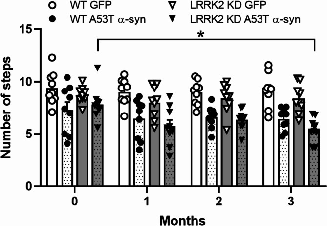
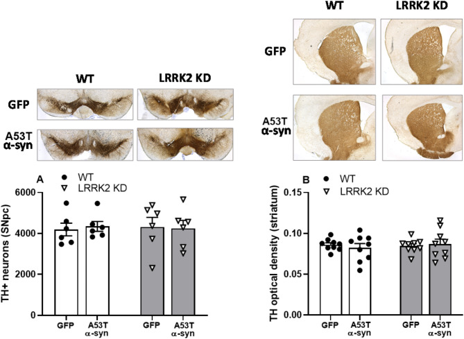
The relationship between LRRK2 mutations and susceptibility to synuclein pathology in Parkinson's disease (PD) is still unclear. We here investigate whether the mice carrying the D1994S kinase-dead (KD) mutation of LRRK2 show enhanced susceptibility to synucleinopathy. Twelve-month-old LRRK2 KD and WT mice were injected with AAV2/9 carrying human A53T α-synuclein (AAV-h-A53Tα-syn) or AAV2/9-GFP as a control. Three months after injection, α-synuclein pathology and nigrostriatal dopaminergic neuron degeneration were assessed along with motor behaviour. AAV-h-A53Tα-syn-injected LRRK2 KD mice showed a decline in stepping activity in the drag test compared to baseline levels and AAV-GFP-injected controls. This was associated with higher transgene levels and Serine129 α-syn phosphorylation in striatum and substantia nigra measured by immunohistochemistry. Total α-synuclein levels were also elevated in the substantia nigra but not striatum of AAV-h-A53Tα-syn LRRK2 KD mice compared to AAV-h-A53Tα-syn controls. Stereological counting of nigral dopaminergic neurons and densitometric analysis of striatal dopaminergic terminals did not reveal overt nigrostriatal degeneration. We conclude that silencing of kinase activity results in greater α-syn load due to greater viral transduction and/or defective α-syn clearance, possibly related to autophagy-lysosomal pathway impairment, however, with no consequence upon dopaminergic neuron survival in the mouse.

Keywords: AAV2/9; D1994S LRRK2; Kinase-dead; Parkinson’s disease; Viral vectors; Α-synuclein.


Conflict of interest statement

Declarations. Competing interests: The authors declare no competing interests.

Figures

Fig. 1
Fig. 1
LRRK2 KD mice injected with AAV-h-A53Tα-syn show mild motor deficits. Stepping activity was evaluated in 12-month-old WT and LRRK2 KD mice injected with AAV-h-A53Tα-syn or AAV-GFP using the drag test. Motor tests were carried out before (baseline, time 0) and 1, 2, 3 months after surgery. Data are expressed as number of steps and are mean ± SEM of 9 mice per group. *p < 0.05 (three-way ANOVA followed by Tukey’s test for multiple comparisons).
Fig. 2
Fig. 2
AAV-h-A53Tα-syn injection results in greater transgene levels in LRRK2 KD mice compared to WT mice. h-A53Tα-syn transgene expression was evaluated by immunohistochemistry in 12-month-old WT and LRRK2 KD mice 3 months after AAV-h-A53Tα-syn or AAV-GFP injection. Representative images and quantification of h-A53Tα-syn in SNpc (A) and striatum (B). Data are expressed as mean percentage ± SEM of: panel (A), n = 9 mice (KD AAV-h-A53Tα-syn, KD AAV-GFP, WT AAV-h-A53Tα-syn) or 10 mice (WT AAV-GFP) per group; panel (B), n = 9 mice per group. **p < 0.01 (ART ANOVA followed by the Bonferroni’s test for multiple comparisons).
Fig. 3
Fig. 3
AAV-h-A53Tα-syn injection results in greater pSer129 α-syn levels in the SN of LRRK2 KD mice compared to WT mice. Immunohistochemistry analysis of pSer129 α-syn and total α-syn staining in the SNpc of 12-month-old WT and LRRK2 KD mice 3 months after AAV-h-A53Tα-syn or AAV-GFP injection. Representative images and quantification of nigral pSer129 α-syn (A) and total α-syn (B) staining. Data are expressed as immunopositive surface of the structure of interest ± SEM of: panel (A), n = 8 mice per group; panel (B), n = 9 (WT AAV-h-A53Tα-syn, KD AAV-GFP) or n = 10 (WT AAV-GFP, KD AAV-h-A53Tα-syn) mice per group. **p < 0.01 (Student’s t test two-tailed for unpaired data or two-way ANOVA followed by the Tukey’s test for multiple comparisons).
Fig. 4
Fig. 4
AAV-h-A53Tα-syn injection results in greater pSer129 α-syn levels in the striatum of LRRK2 KD mice compared to WT mice. Immunohistochemistry analysis of pSer129 α-syn and total α-syn staining in the striatum of 12-month-old WT and LRRK2 KD mice 3 months after AAV-h-A53Tα-syn or AAV-GFP injection. Representative images and quantification of striatal pSer129 α-syn (A) and total α-syn (B) staining. Data are expressed as immunopositive surface of the structure of interest ± SEM of: panel (A), n = 9 (WT AAV-h-A53Tα-syn) or n = 10 (KD AAV-h-A53Tα-syn) mice per group; panel (B), n = 9 (WT AAV-h-A53Tα-syn, KD AAV-h-A53Tα-syn, KD AAV-GFP) or n = 10 (WT AAV-GFP) mice per group. *p < 0.05, **p < 0.01 (Student’s t test two-tailed for unpaired data or two-way ANOVA followed by the Tukey’s test for multiple comparisons).
Fig. 5
Fig. 5
AAV-h-A53Tα-syn injection does not result in degeneration of the nigrostriatal dopaminergic pathway. Stereological count of nigral neurons and optical density of striatal TH + terminals were carried out in 12-month-old WT and LRRK2 KD mice 3 months after AAV-h-A53Tα-syn or AAV-GFP injection. Representative images and quantification of TH+ neurons in SNpc (A) and terminals in striatum (in grey scale arbitrary units; B). Data are expressed as mean percentage ± SEM of n = 6 mice per group (A) or n = 9 mice per group (B).

Similar articles

References

    1. Paisan-Ruiz, C. et al. Cloning of the gene containing mutations that cause PARK8-linked Parkinson’s disease. Neuron44, 595–600. 10.1016/j.neuron.2004.10.023 (2004). - PubMed
    1. Zimprich, A. et al. Mutations in LRRK2 cause autosomal-dominant parkinsonism with pleomorphic pathology. Neuron44, 601–607. 10.1016/j.neuron.2004.11.005 (2004). - PubMed
    1. Nalls, M. A. et al. Large-scale meta-analysis of genome-wide association data identifies six new risk loci for Parkinson’s disease. Nat. Genet.46, 989–993. 10.1038/ng.3043 (2014). - PMC - PubMed
    1. Cookson, M. R. The role of leucine-rich repeat kinase 2 (LRRK2) in Parkinson’s disease. Nat. Rev. Neurosci.11, 791–797. 10.1038/nrn2935 (2010). - PMC - PubMed
    1. West, A. B. et al. Parkinson’s disease-associated mutations in leucine-rich repeat kinase 2 augment kinase activity. Proc. Natl. Acad. Sci. U. S. A.. 102, 16842–16847. 10.1073/pnas.0507360102 (2005). - PMC - PubMed

MeSH terms

Substances

LinkOut - more resources