. 2025 Jun;107(6):1051-1063.
doi: 10.1016/j.kint.2025.02.024. Epub 2025 Mar 22.

Life-supporting functional kidney replacement by integration of embryonic metanephros-bladder composite tissue transplants

Affiliations

Life-supporting functional kidney replacement by integration of embryonic metanephros-bladder composite tissue transplants

Yoshitaka Kinoshita et al. Kidney Int. 2025 Jun.

Abstract

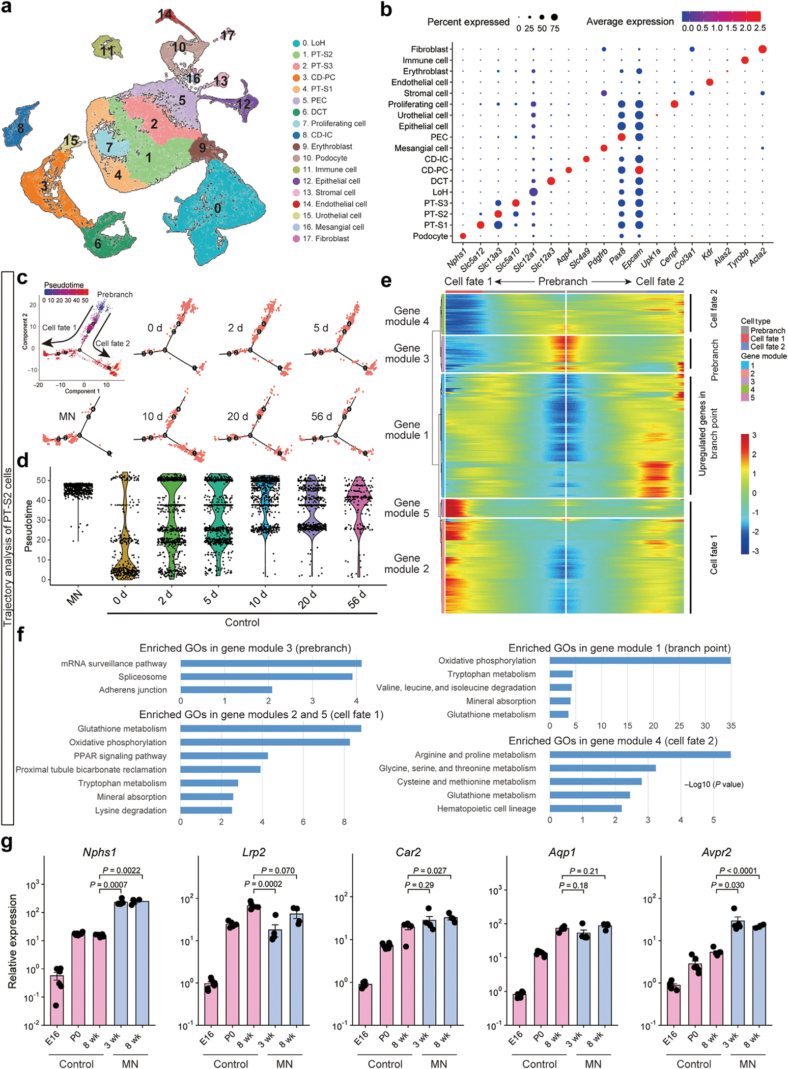
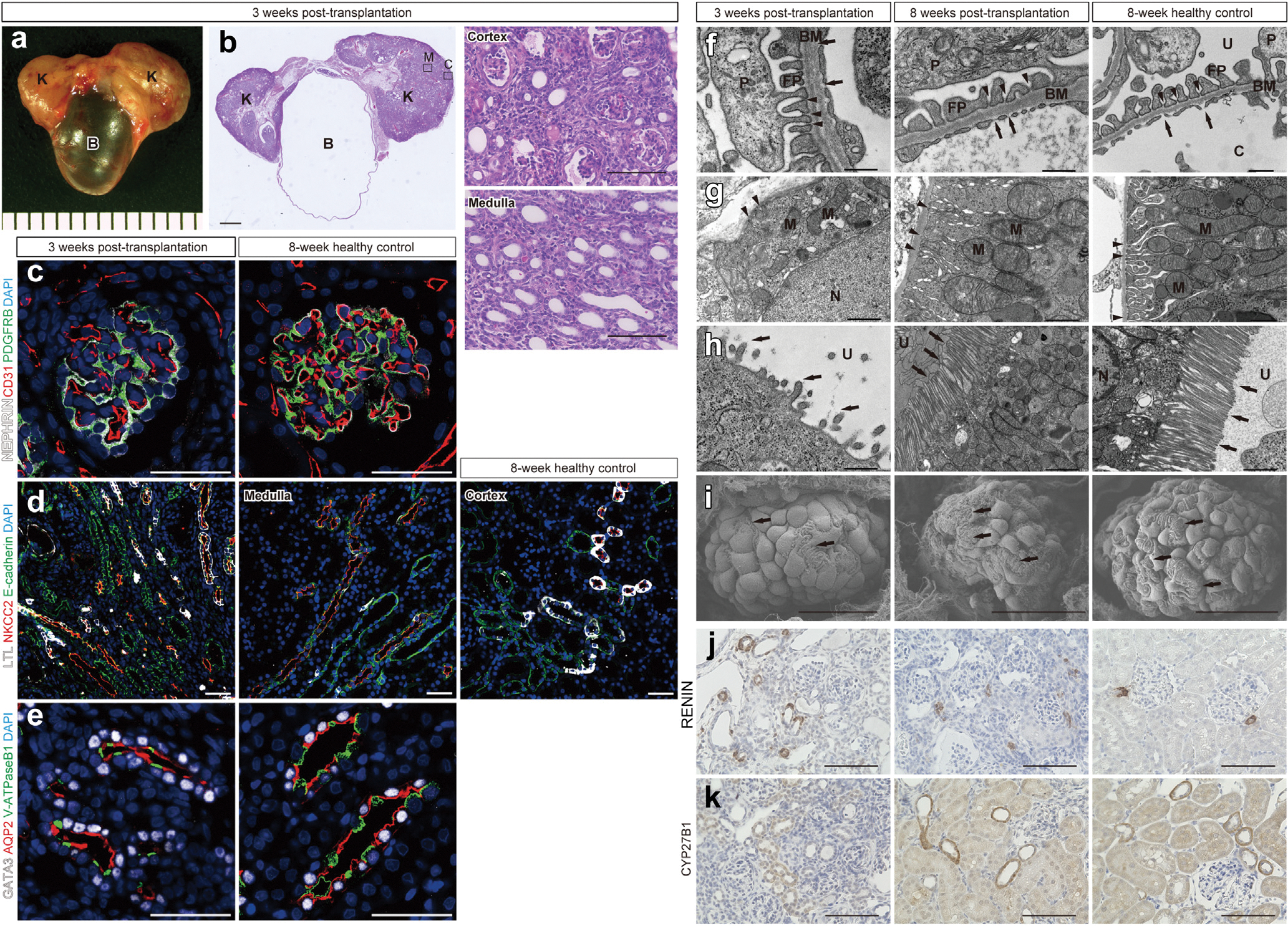
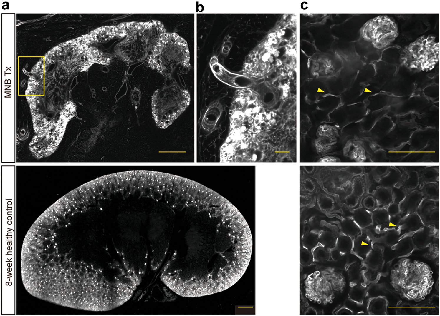
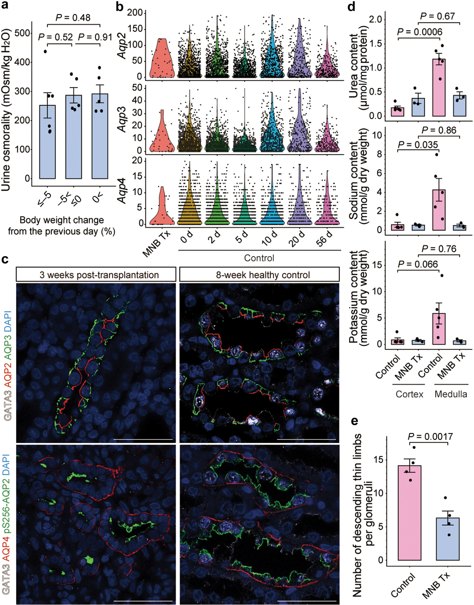
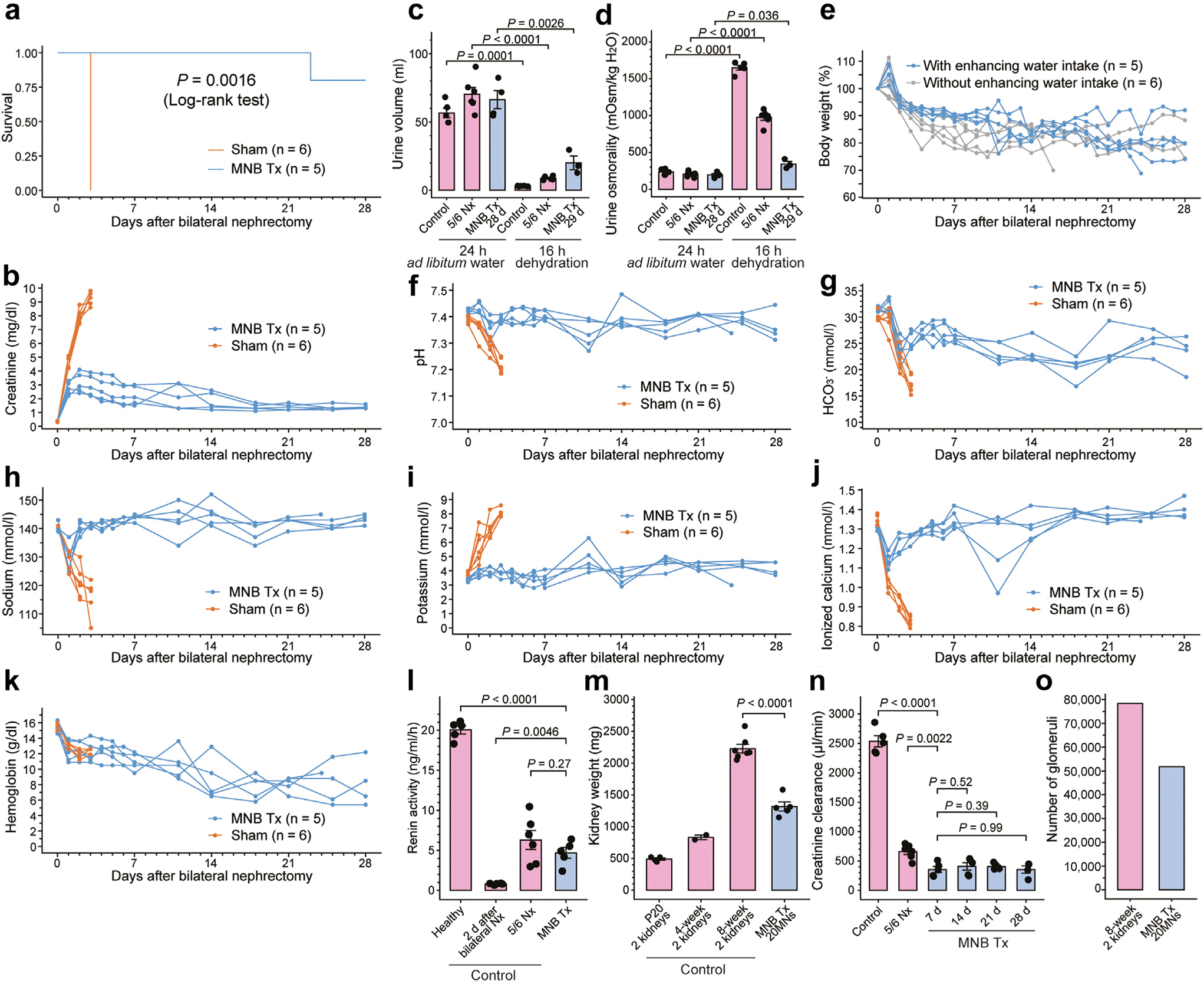
Novel transplantable organs need to be developed to address the global organ shortage. Transplantation of embryonic kidney tissue, or metanephros, facilitates glomerular and tubular maturation and offers partial organ functional support. However, adult environments do not permit exponential growth in size, limiting the life-supporting functionality and organ replacement effect of this approach. Here, we developed a novel strategy that combines the fusion of embryonic bladders with multiple anastomoses to the host ureter, enabling a significant increase in metanephros transplantation and urinary tract integration. By surgically anastomosing divided bladder segments, we reconstructed the excretory pathways by merging four metanephroi into each bladder and integrating them with the host ureter. Following the transplantation and integration of 20 metanephroi at the para-aortic region, anephric rats survived for over a month and generated approximately 50,000 nephrons in vivo. Ultrastructural and single-cell-transcriptomic analyses revealed that the maturity of the transplanted metanephroi was comparable to that of adult kidneys, although their small size likely contributed to their decreased urine concentration ability. Postoperative support helped normalize physiological homeostasis, including solute clearance, acid-base balance, electrolyte levels, and kidney hormone levels, within vital ranges. Our findings underscore the functional maturation capacity and dose-dependent therapeutic efficacy of embryonic kidney tissue, suggesting its potential as a transplantable organ system.

Keywords: embryonic kidney; homeostasis; life-support; maturation; metanephros; transplantation.


Conflict of interest statement

DISCLOSURE

A patent application for the technology involved in creating an integrated MNB graft was filed on January 20, 2023 (Japanese Patent Application Number 2023–007159): the applicant was Bios Co., Ltd.; the inventors were YK, TY, and EK. TY and EK are members of Bios Co., Ltd. All the other authors declared no competing interests.

Figures

Figure 1 |
Figure 1 |. Dosage effect of integrated metanephros–bladder composite (MNB) transplantation.
(a) Schematic (left) and a representative image (right) of an integrated MNB graft. The bladder segment of the embryonic day 16 (E16) MNB is divided, and the anterior and posterior walls are sutured. Each tick on the background represents 1 mm. (b) Immunofluorescence of the fused bladder of integrated MNB grafts transplanted (Tx) into immunocompromised mice at 3 weeks post-transplantation, derived from green fluorescent protein (GFP)–positive and –negative rat MNBs. GFP-positive and -negative cells form a contiguous urothelial barrier stained with uroplakin III and share the bladder lumen. Bar = 100 μm. (c) Hematoxylin and eosin staining of integrated MNB grafts 5 weeks after anastomosing to the host ureter at 3 weeks post-transplantation into syngeneic rats. Four transplanted kidneys (K) with no hydronephrosis surround the fused bladder (B). Bar = 1 mm. (d) Three-dimensional reconstructed images (left [L]), schematic representation (right [R]), and axial images (bottom) of contrast-enhanced computed tomography to visualize the urinary tract. Urine from 4 transplanted kidneys converges into the fused bladder via the transplanted ureters (arrowhead), with excretion observed from the host ureters (arrow). The axial images highlight the junction where the host ureter (left) and the 4 transplanted embryonic ureters (second, third, and fourth from the left) connect to the fused bladder. Bars = 5 mm. (e) Experimental workflow of multiple integrated MNB graft transplantation, urinary tract reconstruction, host kidney removal, and survival analysis in a syngeneic rat model. (f) Kaplan–Meier curve showing the survival of rats transplanted with 12, 16, and 20 metanephros (MNs). Euthanasia for sample collection and deaths attributed to technical errors during anesthesia were censored. To optimize viewing of this image, please see the online version of this article at www.kidney-international.org.
Figure 2 |
Figure 2 |. Transcriptomic profiling of transplanted metanephros (MN) grafts and adult kidneys.
Single-cell RNA-sequencing (scRNA-seq) data from transplanted MNs sampled 3 weeks after bilateral nephrectomy (16 weeks after integrated MNB transplantation) were analyzed after integration with scRNA-seq data obtained from kidney samples at postnatal days 0 (P0), 2, 5, 10, 20, and 56. (a) Uniform manifold approximation and projection displaying unsupervised clustering of all 53,994 cells into 18 cell types with annotations based on the expression of established marker genes. (b) Dot plot showing the expression levels of representative marker genes in the 18 clusters. (c) t-Distributed stochastic neighbor embedding (t-SNE) plot showing Monocle 2-based trajectory analysis of proximal tubule segment 2 (PT-S2) cells, the cluster with the highest cell count in the MN sample. Cells are colored based on the predicted pseudotime. (d) Violin plot showing pseudotime distribution of PT-S2 cells from MN sample and healthy controls at various ages (P0–P56). Most cells derived from the MN samples show advanced pseudotime. (e) Heatmap depicting branch-dependent genes during the transition from pseudotime-young to pseudotime-old cells along the trajectory timeline. Five gene modules associated with branches in t-SNE were identified. Cells from the MN samples were predominantly observed in the cell fate 1 branch, showing upregulation in gene modules 2 and 5. (f) Gene ontologies (GOs) enriched in gene modules corresponding to each state along the trajectory: module 3 to prebranch state, module 1 to branch point, modules 2 and 5 to cell fate 1, and module 4 to cell fate 2, respectively. (g) Quantitative real-time polymerase chain reaction analysis of maturation-related markers in various nephron segments. Gene expression was compared among embryonic day 16 (E16), P0, and 8-week control kidneys, and MNs at 3 weeks post-transplantation (without urinary tract reconstruction) and 8 weeks post-transplantation (5 weeks after urinary tract reconstruction). Expression values are normalized to Gapdh and shown relative to one of the E16 control samples. Data are mean ± SEM. Points represent biological replicates from at least 4 independent experiments. Statistical significance was determined by 2-tailed Student’s unpaired t tests. CD-IC, intercalated cell of the collecting duct; CD-PC, principal cell of the collecting duct; DCT, distal convoluted tubule; LoH, Loop of Henle; PEC, glomerular parietal epithelial cell.
Figure 3 |
Figure 3 |. Longitudinal structural and functional maturation of metanephros (MN) grafts.
(a) Metanephros-bladder composite (MNB) 3 weeks post-transplantation (before urinary reconstruction). Each tick on the background represents 1 mm. (b) Hematoxylin and eosin staining of MNB 3 weeks post-transplantation into syngeneic rats. Two kidneys (K) have developed adjacent to the bladder, filled with urine (B). Higher magnification images of cortex and medulla are shown on the right (corresponding to the regions marked C and M in the left panel). Bars = 1 mm (left) and 100 μm (right). (c–e) Immunofluorescence of MNs (left) and 8-week healthy control kidneys (right). Bars = 50 μm. (c) Glomerular structures supported by PDGFRB-positive mesangial cells, nephrin-positive podocytes, and cluster of differentiation 31 (CD31)–positive endothelial cells. (d) Tubular segments, including lotus tetragonolobus lectin (LTL)–positive proximal tubules, sodium-potassium-chloride cotransporter 2 (NKCC2)–positive loops of Henle, and E-cadherin-positive distal tubules. (e) Collecting ducts formed by AQP2-positive principal cells and vacuolar-type ATPase B1 (V-ATPaseB1)–positive intercalated cells. Nuclei of the collecting ducts stained with GATA3. (f–k) Ultrastructural observation and immunohistochemistry of MN samples at 3 weeks post-transplantation (before urinary reconstruction), 8 weeks post-transplantation (5 weeks after urinary reconstruction), and 8-week healthy control kidneys. (f–h) Transmission electron microscopy images of glomeruli (f) and the basolateral (g) and luminal (h) sides of proximal tubular cells. (f) Endothelial cells supported by the 3-layered glomerular basement membrane (BM), along with podocytes (P) and foot processes (FPs). Slit diaphragms (arrowheads) are visible between FPs. Arrows represent the fenestrae of endothelial cells. Bar = 500 nm. (g,h) At 3 weeks post-transplantation, microvilli (arrows) are sparse and short, with mitochondria (M) present but showing limited basolateral infoldings (arrowheads). By 8 weeks post-transplantation, microvilli (arrows) are extended, and mitochondria are aligned along the developed basolateral infoldings (arrowheads). Bars =− 1 μm (g), 500 nm (h, left) and 2 μm (h, middle and right). (i) Scanning electron microscopy images of glomeruli. At 3 weeks post-transplantation, interdigitation of FPs (arrows) extending from podocytes can be observed; however, it is segmental and not well-developed in some areas. By 8 weeks post-transplantation, it developed prominently in the glomerulus. Bars = 20 nm (left), 30 nm (middle), and 50 nm (right). (j,k) Immunohistochemistry of renin and CYP27B1. Bars = 100 μm. (j) Renin-producing cells were present in the arterial wall apart from the glomerulus at 3 weeks post-transplantation but gradually assemble in the juxtaglomerular apparatus by 8 weeks post-transplantation. (k) CYP27B1 expression has gradually strengthened and reached a comparable level to that in adults by 8 weeks post-transplantation. C, capillary; DAPI, 4′,6-diamidino-2-phenylindole; N, nucleus; U, urinary space (Bowman’s space). To optimize viewing of this image, please see the online version of this article at www.kidney-international.org.
Figure 4 |
Figure 4 |. Confirmation of blood flow and visualization of vasculature within metanephros (MN) grafts.
Visualization of blood vessels in MN grafts at 13 weeks post-transplantation and 8-week healthy control kidneys using fluorescently labeled lectin perfusion and tissue clearing. (a) Global vascular networks of MN graft and control kidney. Distinct spots of lectin accumulation represent glomeruli. Yellow box indicates the region shown in (b). Bars = 1 mm. (b) Blood vessels infiltrating from the capsular region of MN grafts. Bar = 100 μm. (c) Higher magnification images showing well-developed glomerular capillaries and peritubular capillaries (arrowheads). Bars = 100 μm. MNB, metanephros–bladder composite; Tx, transplanted grafts. To optimize viewing of this image, please see the online version of this article at www.kidney-international.org.
Figure 5 |
Figure 5 |. Small kidney size impairs urine-concentrating ability.
(a) Osmolality of urine collected overnight using metabolic cages during ad libitum water and food intake. Despite the decrease in body weight from the previous day, the rise in urine osmolality is minimal. (b) Violin plots showing the expression of Aqp2, Aqp3, and Aqp4 genes in the principal cells of the collecting ducts in transplanted kidneys (metanephros–bladder composite [MNB] Tx) and healthy rat kidneys at postnatal days 0, 2, 5, 10, 20, and 56 (control) obtained from single-cell RNA-sequencing data. Comparable expression levels are observed in transplanted kidneys and healthy controls. (c) Immunofluorescence showing localization of each aquaporin protein in collecting duct principal cells of MNs at 3 weeks post-transplantation (left) and 8-week healthy control kidneys (right). Bars = 50 μm. (d) Tissue concentrations of urea, sodium, and potassium in the cortex and medulla of the embryonic kidneys at least 8 weeks post-transplantation (MNB Tx) and in the kidneys of healthy 8-week-old rats (controls). Medullary osmolyte concentration has increased compared with that in the cortex in the controls; however, this increase is limited in the transplanted MNs. (e) Number of AQP1-positive thin descending limb cross-sections per glomerulus, as quantified via tissue immunostaining. To estimate the length of AQP1-positive segments, AQP1-positive tubular and PDGFRB-positive glomerular cross-sections were counted in the maximum cross-sectional slice and subsequently divided. (a,d,e) Data are mean ± SEM. Points are biological replicates examined over at least 3 independent experiments. Statistical analyses were performed using 2-tailed Student’s unpaired t tests. DAPI, 4′,6-diamidino-2-phenylindole. To optimize viewing of this image, please see the online version of this article at www.kidney-international.org.
Figure 6|
Figure 6|. Renal functional assessment after refined postoperative management.
(a,b) Kaplan–Meier curve showing the survival (a) and changes in serum creatinine levels (b) in 5-integrated metanephros-bladder composite graft-transplanted rats (MNB Tx) and sham-operated (sham) rats. (c,d) Urine volume (c) and osmolality (d) of 5-integrated MNB Tx rats and healthy 8-week-old and 5/6 nephrectomy (5/6 Nx) rats (control). Urine during a 24-hour period with ad libitum access to food and water and a 16-hour fasting period was collected using metabolic cages. (e) Changes in body weight of 5-integrated MNB Tx rats with or without water intake enhancement. Data were normalized to the body weight on the day of host kidney removal. Early postoperative weight loss was alleviated through water intake enhancement. (f–k) Changes in blood levels of pH (f), bicarbonate (HCO3) (g), sodium (h), potassium (i), ionized calcium (j), and hemoglobin (k) in 5-integrated MNB Tx rats and sham rats. (l) Renin activity of 5-integrated MNB Tx rats and 8-week-old healthy, anephric (2 days after kidney removal), and 5/6 Nx rats (control). (m) Weights of 20 transplanted embryonic kidneys (MNB Tx) and bilateral kidneys of 20-day-, 4-week-, and 8-week-old healthy rats (control). (n) The 24-hour creatinine clearance levels of 5-integrated MNB Tx rats at 7, 14, 21, and 28 days after host kidney removal (MNB Tx) and those of healthy 8-week-old and 5/6 Nx rats (control). (o) Total number of glomeruli counted as distinct spherical structures, attributed to lectin accumulation in cleared tissues. Twenty metanephros (MNs) contain approximately 50,000 glomeruli, which is 60% of the glomeruli of bilateral healthy kidneys. (c,d,l–n) Data are mean ± SEM. Points are biological replicates examined over at least 3 independent experiments. Statistical analyses were performed using 2-tailed Student’s unpaired t tests. P20, postnatal day 20.

References

    1. Griffith BP, Goerlich CE, Singh AK, et al. Genetically modified porcine-to-human cardiac xenotransplantation. N Engl J Med. 2022;387:35–44. - PMC - PubMed
    1. Anand RP, Layer JV, Heja D, et al. Design and testing of a humanized porcine donor for xenotransplantation. Nature. 2023;622:393–401. - PMC - PubMed
    1. Takebe T, Sekine K, Enomura M, et al. Vascularized and functional human liver from an iPSC-derived organ bud transplant. Nature. 2013;499:481–484. - PubMed
    1. Sugimoto S, Kobayashi E, Fujii M, et al. An organoid-based organ-repurposing approach to treat short bowel syndrome. Nature. 2021;592:99–104. - PubMed
    1. Yoshihara E, O’Connor C, Gasser E, et al. Immune-evasive human islet-like organoids ameliorate diabetes. Nature. 2020;586:606–611. - PMC - PubMed

Supplementary concepts

LinkOut - more resources