. 2025 Aug 7;74(9):1500-1513.
doi: 10.1136/gutjnl-2024-334094.

Longitudinal paired liver biopsies and transcriptome profiling in alcohol-associated hepatitis reveal dynamic changes in cellular senescence

Affiliations

Longitudinal paired liver biopsies and transcriptome profiling in alcohol-associated hepatitis reveal dynamic changes in cellular senescence

Daniel Rodrigo-Torres et al. Gut. .

Abstract

Background and aims: Alcohol-associated hepatitis (AH) is an acute form of alcohol-related liver disease (ALD) with high mortality rate. AH is histologically characterised by cellular processes, including steatosis, inflammation and cell death. Apoptosis is the most studied form of cell death in AH; however, the role of cellular senescence, another response to cellular injury, in AH is unknown. Here, we explore the mechanisms of ALD pathophysiology and describe the role of senescence in AH.

Methods: We performed RNA sequencing and bioinformatics analysis of 0- and 28-day transjugular liver biopsies (n=65) from patients with AH participating in the IL-1 Signal Inhibition In Alcoholic Hepatitis (ISAIAH) clinical trial. Additional bioinformatics reanalysis of existing AH transcriptomic datasets was conducted to confirm our findings. We also performed multiomic analysis of an in vitro model of AH with ethanol-treated hepatocytes overexpressing ethanol-metabolising enzymes.

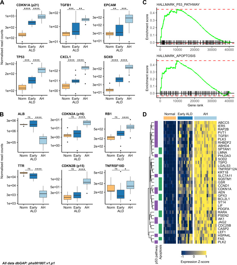
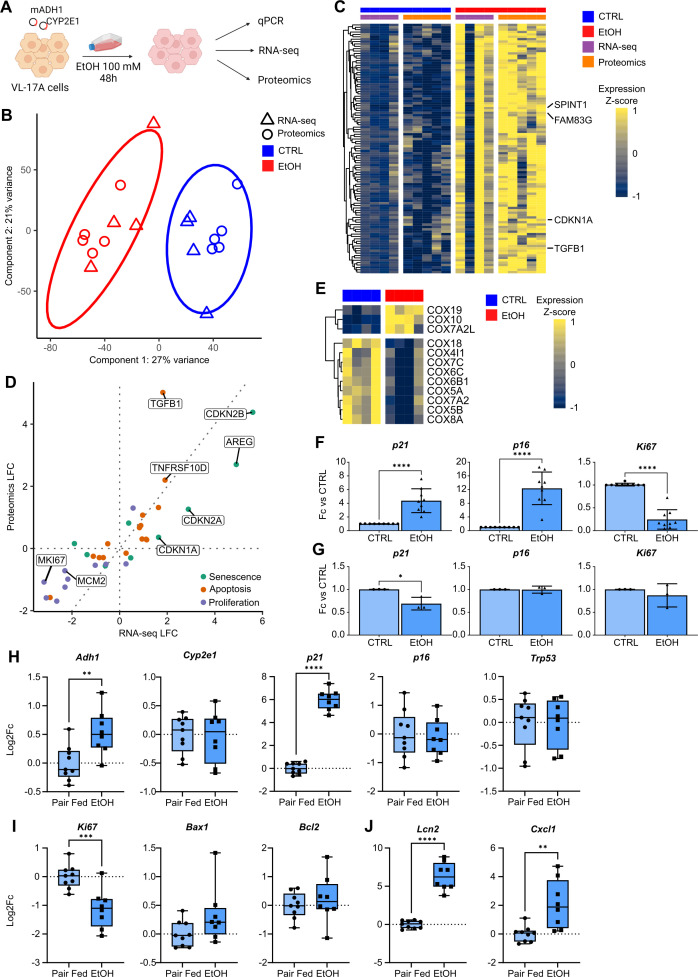
Results: Our longitudinal analysis revealed that senescence and inflammation were reduced at transcriptomic level following AH resolution; the expression of hepatocyte markers was increased. We identified two senescence-associated protein complexes, cytochrome c oxidase and the proteasome, which may act as senescence-induction mechanisms. We confirmed that senescence markers and pathways were increasingly expressed in hepatocytes as ALD progressed towards AH; this was partially reversed following AH resolution. Our in vitro model revealed that ethanol directly induces senescence and was dependent on ethanol metabolism.

Conclusions: Our results suggest a possible pathogenic role for senescence in AH and indicate cellular senescence as a potential therapeutic target in early ALD to limit AH severity.

Keywords: ALCOHOLIC LIVER DISEASE; ETHANOL; INFLAMMATION; LIVER REGENERATION.


Conflict of interest statement

Competing interests: AMK is a consultant for Resolution Therapeutics. SF-G is founder of SensiBile. MRT is a consultant for Surrozen, Hepatx, Resolution Therapeutics and Durect, and advisor for Intercept. SJF is founder of Resolution Therapeutics, SensiBile and an advisor of Cytotheryx.

Figures

Figure 1
Figure 1. Senescence marker expression decreases in patients with sAH following AH resolution. Analysis of ISAIAH patients at d0 and d28. Points represent patients; lines connect matched measurements from the same patient, where available. (A) MELD measurements. (B) mDF measurements. (C) Heatmap of expression for significantly DE genes; at both timepoints, patients are ordered by increasing d0 MELD. (D) Representative staining of p21 and p53 in ISAIAH liver biopsies. (E) Quantifications of the percentage of p21+ and p53+ hepatocytes at d0 and d28 in paired biopsies (p21, n=12 paired biopsies and p53, n=7 paired biopsies). (F) CLR transformed read counts for hepatocyte markers. (G) Volcano plot of gene coefficient and significance between d0 and d28; points represent genes. Scale bar: 50 µm. *FDR<0.05; ***FDR<0.001; ****FDR<0.0001. AH, alcohol-associated hepatitis; DE, differentially expressed; FDR, false discovery rate; mDF, Maddrey’s discriminant function; MELD, model for end-stage liver disease; sAH, severe AH; SASP, senescence-associated secretory phenotype.
Figure 2
Figure 2. Senescence-associated protein complexes are dysregulated with AH severity. (A) CLR transformed read counts for ALB versus MELD, in ISAIAH patients, (d0, d28). Points represent patients; arrows connect matched measurements from the same patient, where available. (B) Correlation of CLR transformed read counts for senescence and SASP markers versus MELD, in ISAIAH patients, (d0, d28). Heatmaps of significantly DE (C) COX subunit and (D) proteasome subunit gene expression in ISAIAH patients. Patients are ordered by decreasing MELD; point colours represent timepoint. (E) Volcano plot of gene coefficient and significance with respect to MELD. Points represent genes; significantly DE COX and proteasomal subunit genes are highlighted. AH, Alcohol-associated hepatitis; ALB, albumin; COX, cytochrome c oxidase; DE, differentially expressed; MELD, model for end-stage liver disease; PSM, Proteasome; SASP, senescence-associated secretory phenotype.
Figure 3
Figure 3. ALD progression is characterised by transcriptomic dysregulation of the proliferation/senescence axis. Boxplots of normalised read counts for (A) senescence, SASP and biliary markers; (B) hepatic and senescence markers in normal liver (n=10), early ALD (n=12) and AH (n=18). (C) GSEA for the MSigDB Hallmark P53 pathway and apoptosis gene sets, comparing AH to normal liver. (D) Heatmap of genes associated with the P53 pathway and apoptosis in normal liver, early ALD and AH. AH, alcohol-associated hepatitis; ALD, alcohol-related liver disease; GSEA, gene set enrichment analysis; SASP, senescence-associated secretory phenotype.
Figure 4
Figure 4. AH is characterised histologically by increased presence of p21-positive cells. (A) Representative staining of p21, Ki67 and γH2A.X in normal, cirrhotic and AH livers, including digital magnification of p21, Ki67 and γH2A.X stainings in AH. Far right, quantification of percentage of p21+ and Ki67+ hepatocytes in normal (n=5–6), cirrhotic (n=5) and (n=4) patients with AH (mean±SD). (B) Representative staining of p21 in AH liver biopsies. Top far right, digital magnification of area showing p21+BECs (red arrows), bottom far right, digital magnification of area showing p21+ hepatocytes. Dual immunofluorescence staining of p21 (red) and (C) hepatocyte marker HNF4α (green) and (D) p21 (red) and proliferative marker Ki67 (green) from patients with AH, counterstained with DAPI (blue). Scale bar: 100 µm. *p<0.05, ns=non-significant; **FDR<0.01; ***FDR<0.001; ****FDR<0.0001. AH, alcohol-associated hepatitis; BEC, biliary epithelial cell; DAPI, 4′,6-diamidino-2-phenylindole; FDR, false discovery rate.
Figure 5
Figure 5. Ethanol-induced toxicity in the hepatocyte VL-17A cell line directly induces senescence marker expression in vitro. (A) Schematic of in vitro experimental design. (B) Scatterplot of RNA-seq and proteomics samples in dimensionally reduced space. (C) Heatmap of progressive ALD markers also significantly upregulated in VL-17A cells. (D) Fold change in RNA-seq and proteomic analysis for significantly DE senescence, apoptosis and proliferation markers. (E) Heatmap of COX subunits significantly DE in RNA-seq of VL-17A cells. (F) Gene expression changes in ethanol-treated (EtOH; n=9) versus untreated VL-17A cells (CTRL; n=9) (mean±SD). (G) Differences in expression in senescence and proliferation markers between CTRL (n=3) and EtOH (n=3) HepG2 cells (mean±SD). Gene expression changes in (H) ethanol-metabolising enzyme Adh1, senescence markers (p21, p16 and Trp53), (I) proliferation marker Ki67 and (J) SASP/inflammation markers (Cxcl1 and Lcn2) between pair-fed (n=9) and ethanol-fed (n=8) mice. *p<0.05; **p<0.01; ***p<0.001 ****p<0.0001. ALD, alcohol-related liver disease; COX, cytochrome c oxidase; DE, differentially expressed; Fc, fold change; SASP, senescence-associated secretory phenotype.

References

    1. Seitz HK, Bataller R, Cortez-Pinto H, et al. Alcoholic liver disease. Nat Rev Dis Primers. 2018;4:16. doi: 10.1038/s41572-018-0014-7. - DOI - PubMed
    1. Thursz M, Morgan TR. Treatment of Severe Alcoholic Hepatitis. Gastroenterology. 2016;150:1823–34. doi: 10.1053/j.gastro.2016.02.074. - DOI - PMC - PubMed
    1. Vergis N, Atkinson SR, Knapp S, et al. In Patients With Severe Alcoholic Hepatitis, Prednisolone Increases Susceptibility to Infection and Infection-Related Mortality, and Is Associated With High Circulating Levels of Bacterial DNA. Gastroenterology. 2017;152:1068–77. doi: 10.1053/j.gastro.2016.12.019. - DOI - PMC - PubMed
    1. Yoon EL, Kim W. Current and future treatment for alcoholic-related liver diseases. J Gastroenterol Hepatol. 2023;38:1218–26. doi: 10.1111/jgh.16257. - DOI - PubMed
    1. Vergis N, Patel V, Bogdanowicz K, et al. IL-1 Signal Inhibition in Alcohol-Related Hepatitis: A Randomized, Double-Blind, Placebo-Controlled Trial of Canakinumab. Clin Gastroenterol Hepatol. 2024 doi: 10.1016/j.cgh.2024.07.025. - DOI - PubMed

LinkOut - more resources