. 2025 Mar 24;44(1):104.
doi: 10.1186/s13046-025-03346-2.

AMP-activated protein kinase mediates adaptation of glioblastoma cells to conditions of the tumor microenvironment

Affiliations

AMP-activated protein kinase mediates adaptation of glioblastoma cells to conditions of the tumor microenvironment

Nadja I Lorenz et al. J Exp Clin Cancer Res. .

Abstract

AMP-activated protein kinase (AMPK) is an energy sensor that regulates cellular metabolic activity. We hypothesized that in glioblastoma (GB), AMPK plays a pivotal role in balancing metabolism under conditions of the tumor microenvironment with fluctuating and often low nutrient and oxygen availability. Impairment of this network could thus interfere with tumor progression. AMPK activity was modulated genetically by CRISPR/Cas9-based double knockout (DKO) of the catalytic α1 and α2 subunits in human GB cells and effects were confirmed by pharmacological AMPK inhibition using BAY3827 and an inactive control compound in primary GB cell cultures. We found that metabolic adaptation of GB cells under energy stress conditions (hypoxia, glucose deprivation) was dependent on AMPK and accordingly that AMPK DKO cells were more vulnerable to glucose deprivation or inhibition of glycolysis and sensitized to hypoxia-induced cell death. This effect was rescued by reexpression of the AMPK α2 subunit. Similar results were observed using the selective pharmacological AMPK inhibitor BAY3827. Mitochondrial biogenesis was regulated AMPK-dependently with a reduced mitochondrial mass and mitochondrial membrane potential in AMPK DKO GB cells. In vivo, AMPK DKO GB cells showed impaired tumor growth and tumor formation in CAM assays as well as in an orthotopic glioma mouse model. Our study highlights the importance of AMPK for GB cell adaptation towards energy depletion and emphasizes the role of AMPK for tumor formation in vivo. Moreover, we identified mitochondria as central downstream effectors of AMPK signaling. The development of AMPK inhibitors could open opportunities for the treatment of hypoxic tumors.

Keywords: AMP-activated protein kinase; AMPK; Glioblastoma; Hypoxia; Metabolic adaptation.


Conflict of interest statement

Declarations. Ethics approval and consent to participate: Not applicable. Consent for publication: Not applicable. Competing interests: JPS has received honoraria for consulting or advisory board membership as well as travel or accommodation support from Abbvie, Medac, Novocure, Roche, Servier and UCB. MWR has received research funding from UCB as well as honoraria for advisory board participation from Alexion and Servier. All other authors declare that they have no competing interests.

Figures
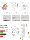
Fig. 1
Fig. 1
Knockout of AMPK catalytic subunits prevents AMPK activation and deregulates metabolic adaptation under hypoxic conditions in human GB cells (A) Dimensional reduction (UMAP) of the single cell reference GBMap dataset including neurons from the Allen Institute database. Celltypes are colored based on their transcriptional subtypes. (B, C) UMAP representation of AMPK gene expression (B) and for the AMPK pathway activation (C). (D) LNT-229, LN-308 and G55T2 wildtype (wt) or AMPK catalytic subunits double knockout (DKO) cells were incubated in serum-free DMEM supplemented with 2 mM glucose for 8 h in normoxia (21% O2) or hypoxia (0.1% O2). Immunoblots with antibodies for P-ACC, P-AMPK, AMPK α1/2 were performed. (E) Protein network analysis of LNT-229 wildtype and AMPK DKO cells using WGCNA. Representation of the modules is indicated in the Dendrogram in the upper panel. The differentially enriched modules across the conditions are demonstrated in the bottom panel. (F) UMAP representation of the protein expression modules. (G) Geneset Enrichment analysis of the modules which indicate significance between LNT-229 wildtype and AMPK DKO
Fig. 2
Fig. 2
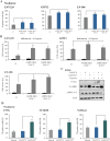
Inhibition of AMPK sensitizes GB cells to nutrient starvation and hypoxia (A) LNT-229, LN-308 and G55T2 wildtype (wt) and AMPK catalytic subunits double knockout (DKO) cells were treated in serum- and glucose-free DMEM for 24 h. Cell death was analyzed by PI staining and quantified by flow cytometry (n = 3, mean ± SD, **p < 0.01, Student’s t-test). (B) Cell death of LNT-229, LN-308 and G55T2 wildtype and AMPK DKO was analyzed by an LDH release assay after incubation of the cells in serum-free DMEM containing 2 mM glucose in normoxia or hypoxia (0.1% O2) (n = 4, mean ± SD, *p < 0.05, **p < 0.01, Student’s t-test). (C) Primary GB cells (P3NS) were treated with vehicle (DMSO), 1 µM BAY974 or 1 µM BAY3827 in serum-free medium containing 2 mM glucose for 8 h in normoxia or hypoxia (0.1% O2). Cellular lysates were analyzed by immunoblot with antibodies for P-ACC, P-AMPK, AMPKα1/2 and actin. (D) Human primary glioblastoma cells P3NS, NCH60 and NCH644 cells were treated with vehicle (DMSO), 1 µM BAY974 or 1 µM BAY3827 in serum-free medium without glucose. Cell death was analyzed by PI staining and flow cytometry (n = 3, mean ± SD, **p < 0.01, Student’s t-test)
Fig. 3
Fig. 3
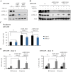
Retransfection of the catalytic subunit of AMPK in knockout cells restores adaptation to starvation conditions (A) LNT-229 wildtype (wt) and AMPK catalytic subunits double knockout (DKO) cells were stably transfected with empty vector (control) or PRKAA2. Cellular lysates were analyzed by immunoblot with antibodies for AMPK α1, AMPK α2 and actin. (B) Immunoblot analysis of LNT-229 wildtype and AMPK DKO PRKAA2 lysates treated in serum-free medium with 2 mM glucose for 8 h in normoxia or hypoxia (0.1% O2) was performed with antibodies for P-ACC, AMPK α2 and actin. (C) LNT-229 wildtype and AMPK DKO PRKAA2 cells were treated in serum-free medium without glucose. Cell death was analyzed by PI uptake and flow cytometry (n = 3, mean ± SD, n.s. not significant, **p < 0.01, Student’s t-test). (D) Cell death of LNT-229 wildtype and AMPK DKO cells was analyzed by LDH release assay after incubation in serum-free medium supplemented with 2 mM glucose in normoxia and hypoxia (0.1% O2) (n = 4, mean ± SD, *p < 0.05, **p < 0.01, Student’s t-test)
Fig. 4
Fig. 4
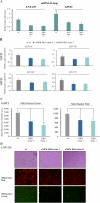
Mitochondrial mass and activity are impaired by AMPK catalytic subunits knockout in human GB cell lines (A) LNT-229 and G55T2 wildtype (wt) and AMPK catalytic subunits double knockout (DKO) cells were analyzed for the mRNA expression of mtDNA D-loop by qPCR. 18 S and SDHA were used for normalization (n = 3, mean ± SD). (B) mRNA expression of mitochondrial encoded as well as mitochondrial associated genes (ATP5G1, MT-CYB, MT-ND1 and MT-CO1) of G55T2 wildtype and AMPK DKO cells was determined by qPCR. 18 S and SDHA were used as housekeeping genes for normalization (n = 3, mean ± SD). (C) G55T2 wildtype and AMPK DKO cells were incubated in serum-free DMEM for 24 h. Cells were stained with 100 nM MitoTracker Green or 100 nM MitoTracker Red for 20 min and analyzed by flow cytometry. Mean fluorescence intensities are shown (n = 3, mean ± SD, *p < 0.05, **p < 0.01, Student’s t-test). (D) LNT-229 wildtype and AMPK DKO cells were treated as described in (C). Bright-field (upper panel) and fluorescence microscopy (RFP channel: middle panel, GFP channel: lower panel) were used for analysis (48x magnification). Representative images are shown
Fig. 5
Fig. 5
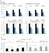
AMPK catalytic subunits double knockout cells are sensitized to 2-deoxyglucose treatment (A) CV staining of LNT-229 wildtype and AMPK catalytic subunits double knockout (DKO) cells was performed after treatment with vehicle (DMSO), 2 mM (left panel) or 5 mM (right panel) 2-DG in serum-free medium supplemented with 5 mM glucose for 72 h. Results are shown as absorption at 595 nm and relative to T0, reflecting the respective cell density at the beginning of the experiment. (B) LNT-229 wildtype and AMPK DKO cells were treated with vehicle (DMSO) or 2 mM 2-DG in serum-free DMEM with 2 mM glucose in normoxia or hypoxia (0.1% O2). LDH release assay was used for cell death analysis (n = 4, mean ± SD, n.s. not significant, *p < 0.05, **p < 0.01, Student’s t-test)
Fig. 6
Fig. 6
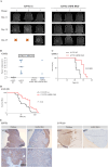
Knockout of AMPK catalytic subunits impairs tumor growth in vivo (A) Athymic nude mice were intracranially injected with 1 × 105 G55T2 wildtype (wt) or AMPK catalytic subunits double knockout (DKO) cells (n = 10 mice per group). MRI measurements were performed on day 11, 18 and 25 after tumor cell injection. Images of 3 mice per group are shown exemplarily. (B) Tumor volumes were calculated based on MRI measurements on day 11 and 18 using the ITK Snap software. (C) Survival of mice (n = 10 per group) injected with G55T2 wildtype or AMPK DKO cells was analyzed by Kaplan-Meier Plot. Significance was tested using Log-Rank test. (D) Survival of mice (n = 20 per group) injected with LNT-229 wildtype or AMPK DKO cells was analyzed by Kaplan-Meier Plot. Significance was tested using Log-Rank test. Data of two independent experiments were pooled. (E) G55T2 wildtype and AMPK DKO tumors (left panel) were analyzed immunohistochemically with antibody for P-ACC. Scale bar represents 200 μm (10x magnification, bottom panel) or 1 mm (2x magnification, upper panel). LNT-229 wildtype and AMPK DKO tumors (right panel) were also analyzed with antibody for P-ACC

References

    1. Monteiro AR, Hill R, Pilkington GJ, Madureira PA. The role of hypoxia in glioblastoma invasion. Cells 2017; 6(4). - PMC - PubMed
    1. Stupp R, Mason WP, van den Bent MJ, Weller M, Fisher B, Taphoorn MJB, et al. Radiotherapy plus concomitant and adjuvant Temozolomide for glioblastoma. N Engl J Med. 2005;352(10):987–96. - PubMed
    1. Hanahan D, Weinberg RA. Hallmarks of cancer: the next generation. Cell. 2011;144(5):646–74. - PubMed
    1. Weyandt JD, Thompson CB, Giaccia AJ, Rathmell WK. Metabolic alterations in Cancer and their potential as therapeutic targets. Am Soc Clin Oncol Educ Book. 2017;37:825–32. - PMC - PubMed
    1. Hardie DG, Schaffer BE, Brunet A. AMPK: an Energy-Sensing pathway with multiple inputs and outputs. Trends Cell Biol. 2016;26(3):190–201. - PMC - PubMed

Substances

LinkOut - more resources