

Regionally resolved cardiac metabolism using a dipole-loop array coil for 7 T 31P-MRSI
- PMID: 40123193
- PMCID: PMC12137756
- DOI: 10.1002/mrm.30492
Regionally resolved cardiac metabolism using a dipole-loop array coil for 7 T 31P-MRSI
Abstract
Purpose: We introduce a novel commercial phosphorus-31 (31P) dipole-loop array coil, describing the coil hardware and testing its performance on phantoms. We used this coil to assess cardiac metabolism per region in healthy volunteers.
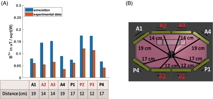
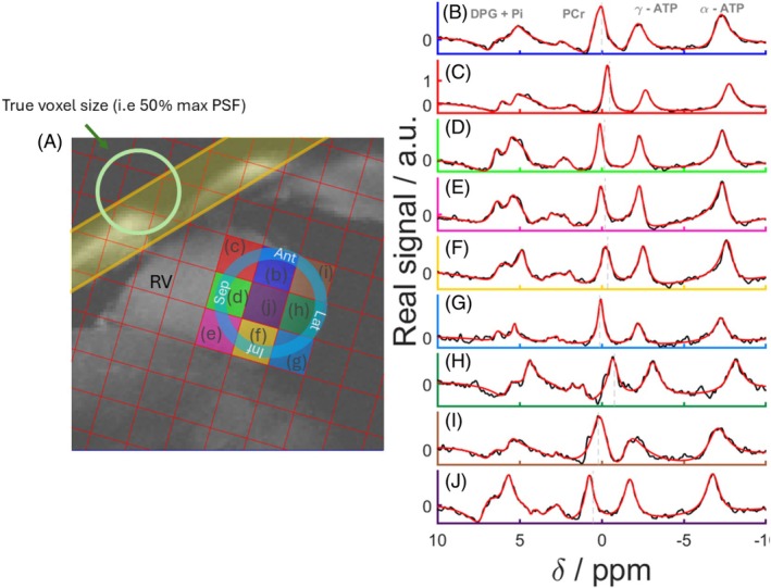
Methods: B1 + field maps were simulated and compared to maps measured with a set of CSI sequences with varying voltages. Seventeen volunteers were scanned with 7 T phosphorus-31 magnetic resonance spectroscopic imaging (31P-MRSI). Reproducibility was assessed in nine of these volunteers. Strain was measured for six of these volunteers at 3 T.
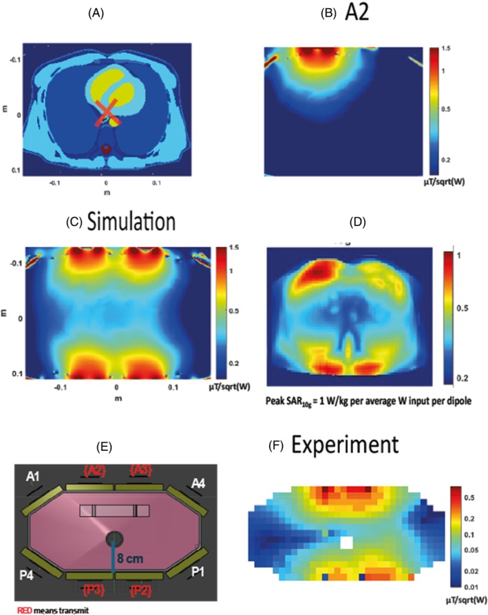
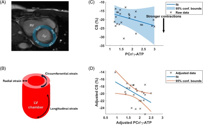
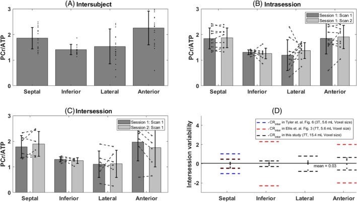
Results: Blood- and saturation-corrected Phosphocreatine/γ-adenosine triphosphate (PCr/ATP) ratios were measured for four regions of the left ventricle: 1.86 in septum, 2.25 in anterior wall, 1.41 in inferior wall, and 1.53 in lateral wall, respectively. These are in the expected range compared to previous studies. B1 + maps show good signal uniformity around the position of the heart (0.13 ± 0.06 μT/sqrt(W)). Intrasession and intersession coefficients of reproducibility were 0.22-0.88 and 0.29-0.79, respectively. Linear modeling shows that regional PCr/γATP correlates with circumferential strain but not radial strain. This requires corroboration by a larger study including patients with impaired function and energetics.
Conclusion: Dipole-loop array coils present a promising new approach for human cardiac 31P-MRSI at 7 T. Their favorable B1 + uniformity at depth and specific absorption rate over loop arrays and improved SNR when combined with loops for reception could be beneficial for further clinical studies measuring energetics by 31P-MRSI at 7 T. The new capability to assess PCr/γATP ratios across the whole left ventricle could enable clinical studies to investigate regional changes in cardiac energetics for the first time.
Keywords: 31P‐MRSI; array coils; heart energetics; heart failure; metabolic imaging; spectroscopic imaging.
© 2025 The Author(s). Magnetic Resonance in Medicine published by Wiley Periodicals LLC on behalf of International Society for Magnetic Resonance in Medicine.
Conflict of interest statement
Christopher Rodgers receives research support from Siemens for another project.
Figures
Similar articles
-
A novel multifrequency-tuned transceiver array for human-brain 31P-MRSI at 7 T.Magn Reson Med. 2025 Jun;93(6):2640-2654. doi: 10.1002/mrm.30449. Epub 2025 Feb 4. Magn Reson Med. 2025. PMID: 39902517 Free PMC article.
-
Flip-angle mapping of 31P coils by steady-state MR spectroscopic imaging.J Magn Reson Imaging. 2014 Aug;40(2):391-7. doi: 10.1002/jmri.24401. Epub 2013 Nov 4. J Magn Reson Imaging. 2014. PMID: 24925600
-
Bloch-Siegert B1+-mapping for human cardiac (31) P-MRS at 7 Tesla.Magn Reson Med. 2016 Oct;76(4):1047-58. doi: 10.1002/mrm.26005. Epub 2015 Oct 28. Magn Reson Med. 2016. PMID: 26509652 Free PMC article.
-
Measuring inorganic phosphate and intracellular pH in the healthy and hypertrophic cardiomyopathy hearts by in vivo 7T 31P-cardiovascular magnetic resonance spectroscopy.J Cardiovasc Magn Reson. 2019 Mar 14;21(1):19. doi: 10.1186/s12968-019-0529-4. J Cardiovasc Magn Reson. 2019. PMID: 30871562 Free PMC article.
-
Phosphorus liver MRSI at 3 T using a novel dual-tuned eight-channel ³¹P/¹H H coil.Magn Reson Med. 2012 Nov;68(5):1346-56. doi: 10.1002/mrm.24164. Epub 2012 Jan 27. Magn Reson Med. 2012. PMID: 22287206
References
-
- Neubauer S, Horn M, Cramer M, et al. Myocardial phosphocreatine‐to‐ATP ratio is a predictor of mortality in patients with dilated cardiomyopathy. Circulation. 1997;96:2190‐2196. - PubMed
-
- Neubauer S, Horn M, Pabst T, et al. Contributions of 31P‐magnetic resonance spectroscopy to the understanding of dilated heart muscle disease. Eur Heart J. 1995;16:115‐118. - PubMed
-
- Neubauer S. The failing heart—an engine out of fuel. N Engl J Med. 2007;356:1140‐1151. - PubMed
MeSH terms
Substances
Grants and funding
LinkOut - more resources
Full Text Sources
