. 2025 Mar 7:15:1536549.
doi: 10.3389/fonc.2025.1536549. eCollection 2025.

Iron content of glioblastoma tumours and role of ferrous iron in the hypoxic response in vitro

Affiliations

Iron content of glioblastoma tumours and role of ferrous iron in the hypoxic response in vitro

Citra Praditi et al. Front Oncol. .

Abstract

Introduction: Glioblastomas are an aggressive primary brain cancer, characterised by hypoxia and poor patient survival. Iron is the most abundant transition metal in the brain, yet data on the iron content of brain cancers is sparse. Ferrous iron is an essential cofactor for a super-family of enzymes, the iron- and 2-oxoglutarate-dependent dioxygenase enzymes (2-OGDD). These enzymes control the response to hypoxia via hydroxylation of the hypoxia-inducible factor-1α (HIF-1α), and DNA demethylation via hydroxylation of 5-methyl cytosines (5hmC).

Methods: This study used clinical glioblastoma samples from 40 patients to determine the relationship between 2-OGDD activity and iron. Elemental iron was measured using inductively coupled plasma mass spectrometry (ICP-MS) and ferrous iron was measured using the colorimetric ferrozine assay. Iron measurements were compared against patient survival and clinicopathological data, and 2-OGDD-dependent activity of HIF-1 activation and 5hmC.

Results and discussion: Elemental and ferrous iron levels were weakly related. Higher ferrous iron content of clinical glioblastoma tissue was associated with longer overall survival compared to lower ferrous iron content, but elemental iron showed no such relationship. Neither form of iron was related to clinicopathological data or markers of 2-OGDD activity. The impact of iron supplementation on the hypoxic response was assessed in three glioblastoma cell lines in vitro, similarly showing only a limited influence of iron on these 2-OGDD enzymes. Our data, together with prior studies in anaemic patients, highlight the importance of healthy iron levels in patients with glioblastoma, but further mechanistic studies are needed to elucidate the molecular pathways involved.

Keywords: brain cancer; elemental iron; ferrous iron; glioblastoma; hypoxia; survival.


Conflict of interest statement

The authors declare that the research was conducted in the absence of any commercial or financial relationships that could be construed as a potential conflict of interest. The author(s) declared that they were an editorial board member of Frontiers, at the time of submission. This had no impact on the peer review process and the final decision.

Figures

Figure 1
Figure 1
Iron content of glioblastoma samples. Correlation between ferrous iron measured by ferrozine assay and elemental iron measured by ICP-MS (Spearman’s correlation r=0.26, p=0.1, n=40).
Figure 2
Figure 2
Associations between elemental iron and clinicopathological data. Associations between iron measured by ICP-Q-MS in glioblastoma samples and (A) IDH mutation status of glioblastoma (Mann Whitney p=0.63, n=40), (B) location of tumour in the brain (Kruskal-Wallis p=0.36, n=34), (C) sex of patient (Mann Whitney p=0.31, female n=10, male n=24), and (D) age of patient (Spearman’s correlation r-0.15, n=34). Median is shown as red line.
Figure 3
Figure 3
Associations between ferrous iron and clinicopathological data. Associations between iron measured by the ferrozine assay in glioblastoma samples and (A) IDH mutation status of glioblastoma (Mann Whitney p=0.97, n=39), (B) location of tumour in the brain (Kruskal-Wallis p=0.16, n=31), (C) sex of patient (Mann Whitney p=0.83, female n=10, male n=23), and (D) age of patient (Spearman’s correlation r 0.03, n=29). Median shown as red line.
Figure 4
Figure 4
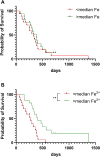
Patient survival according to iron content of glioblastoma tumours. Overall survival of patients with glioblastoma according to (A) elemental iron content of tumours (Log-rank test, p=0.50, n=34), and (B) ferrous iron of tumours (Log-rank p=0.005, n=33). Samples were divided into two groups of below or above median elemental iron (61.5 mg/kg) or median ferrous iron (112 mg/kg). ** p < 0.01.
Figure 5
Figure 5
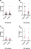
Association between iron and activity of 2-OGDDs in glioblastoma samples. Association between elemental iron and (A) above or below median relative HIF pathway (p=0.50, n=40), and (C) above or below median TET pathway (p=0.54, n=23). Association between ferrous iron and (B) above or below median relative HIF pathway (p=0.08, n=39), and (D) above or below median TET pathway (p=0.44, n=22). Associations were tested using Mann Whitney test. The median relative HIF pathway score was 2, and the median TET pathway score was 0.18% 5hmC. Median shown as red line.
Figure 6
Figure 6
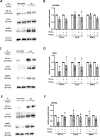
Hypoxic pathway response of glioblastoma cells exposed to iron supplementation. Cells were exposed to FeSO4 (100 μM) in normoxia, or exposed to FeSO4 in normoxia, then incubated in mild hypoxia (5% O2). Lysates of (A) U251MG, (C) T98G and (E) U87MG were collected for western blotting. Relative levels of HIF-1α, CA-IX and BNIP3 were used to analyse the hypoxic pathway, with β-actin used as loading control. Protein bands on western blots were quantified for relative protein levels in (B) U251MG, (D) T98G and (F) U87MG cells (with highest expression set as 1). Paired t-test; mean ± SD; n=3; * p<0.05.

References

    1. Ostrom QT, Gittleman H, Liao P, Vecchione-Koval T, Wolinsky Y, Kruchko C, et al. . CBTRUS Statistical Report: Primary brain and other central nervous system tumors diagnosed in the United States in 2010-2014. Neuro Oncol. (2017) 19:v1–v88. doi: 10.1093/neuonc/nox158 - DOI - PMC - PubMed
    1. Lapointe S, Perry A, Butowski NA. Primary brain tumours in adults. Lancet. (2018) 392:432–46. doi: 10.1016/S0140-6736(18)30990-5 - DOI - PubMed
    1. Weller M, van den Bent M, Preusser M, Le Rhun E, Tonn JC, Minniti G, et al. . EANO guidelines on the diagnosis and treatment of diffuse gliomas of adulthood. Nat Rev Clin Oncol. (2021) 18:170–86. doi: 10.1038/s41571-020-00447-z - DOI - PMC - PubMed
    1. Louis DN, Perry A, Reifenberger G, von Deimling A, Figarella-Branger D, Cavenee WK, et al. . The 2016 world health organization classification of tumors of the central nervous system: a summary. Acta Neuropathol. (2016) 131:803–20. doi: 10.1007/s00401-016-1545-1 - DOI - PubMed
    1. Wesseling P, Capper D. WHO 2016 classification of gliomas. Neuropathol Appl Neurobiol. (2018) 44:139–50. doi: 10.1111/nan.2018.44.issue-2 - DOI - PubMed

LinkOut - more resources