. 2025 Feb 25:18:389-399.
doi: 10.1016/j.ibneur.2025.02.012. eCollection 2025 Jun.

Exploring synergistic effects: Atorvastatin and electrical stimulation in spinal cord injury therapy

Affiliations

Exploring synergistic effects: Atorvastatin and electrical stimulation in spinal cord injury therapy

Martina Magurova et al. IBRO Neurosci Rep. .

Abstract

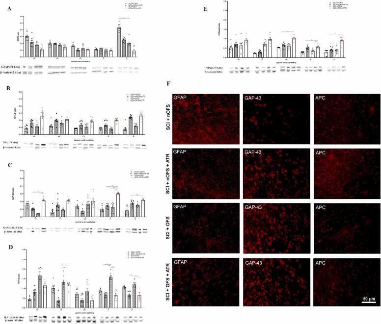
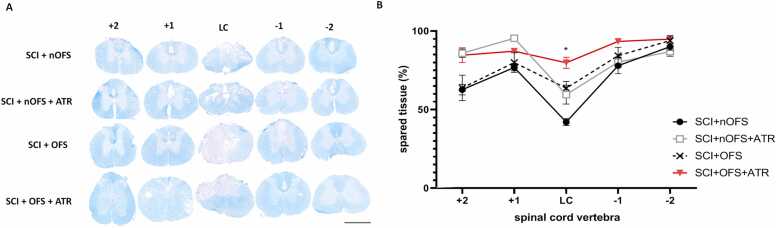
Spinal cord trauma represents a significant clinical challenge, and improving patient outcomes is a main priority for many scientific teams globally. Despite advances in the understanding its pathogenesis, the overall mechanisms occurring in the spinal cord after traumatic injury remain unclear. This study explores the possible synergistic effects of a regenerative therapy that combines electrical stimulation with the anti-inflammatory drug Atorvastatin (ATR) after spinal cord injury (SCI). SCI was induced at the T9 segment under isoflurane anesthesia and applying a compression force of 40 g for 15 minutes. An oscillating field stimulator (OFS) was implanted subcutaneously, delivering a weak electric current (50 µA) that changed polarity every 15 minutes for six weeks to promote axonal growth at the injury site. Female Wistar albino rats were divided into four groups: SCI with non-functional stimulator (SCI + nOFS), SCI with functional stimulator (SCI+OFS), and two groups that received ATR together with stimulator for 7 days after injury (SCI+OFS+ATR, SCI+nOFS+ATR). Behavioral tests (hot-plate test and BBB scale) showed improvement in sensory and motor performance in animals treated with the combination therapy. The protein levels of astrocytes (GFAP), neurofilaments (NF-L), newly sprouting axons (GAP-43), and oligodendrocytes (PLP -1, CNPase) were analysed by Western blot. The results showed increased neurofilaments, newly sprouting axons and oligodendrocytes in groups receiving both individual and combination therapies, with a decrease in their concentrations in the following order: SCI+OFS+ATR, SCI+nOFS+ATR, SCI+OFS, SCI+nOFS. In addition, astrocyte protein levels were lower in the SCI+OFS+ATR group compared with others. Histological analysis showed a significant reduction in white and gray matter after SCI, but less white and gray matter volume loss was found in the groups receiving therapies (SCI+OFS+ATR, SCI+nOFS+ATR, SCI+OFS). These results suggest that the combination of Atorvastatin with OFS stimulation promotes neural recovery after SCI, highlighting the potential of combination therapies in enhancing regenerative outcomes.

Keywords: Atorvastatin; Axonal regeneration; Behavioral assessment; Functional recovery; Oscillating field stimulation; Spinal cord injury.


Conflict of interest statement

The authors declare that they have no known competing financial interests or personal relationships that could have appeared to influence the work reported in this paper.

Figures

Fig. 1
Fig. 1
Health status of animals after T9 spinal cord compression in the following groups: rats with non-functional stimulator (SCI+nOFS), with functional stimulator (SCI+OFS), with non-functional stimulator and administered ATR (SCI+nOFS+ATR), and with functional stimulator and administered ATR (SCI+OFS+ATR). (A) - Body weight changes in adult female Wistar rats after SCI. The animals were weighed every 7 days for 6 weeks. Data are presented as percentage values. (B) – Voiding score assessment in all experimental groups after SCI. Voiding score 0 – rats unable to urinate spontaneously; Voiding score 1 – rats with the ability to urinate spontaneously. SCI – T9 spinal compression injury; OFS – oscillating field stimulator; nOFS – non-functional oscillating field stimulator; ATR – Atorvastatin. Data are presented as MEAN ± SEM. Results for weight changes were statistically evaluated using a two-way Anova followed by a post-hoc Tukey´s test. Results for urination were statistically evaluated using chi-square followed by Yates correction. The data were considered statistically significant at P < 0,05 (*).
Fig. 2
Fig. 2
Demonstration of functional improvement in motor and sensory functions after SCI in all experimental groups. (A) Assessment of hind-limb locomotion improvement in rats after SCI by BBB score. A visible improvement was observed after 3 weeks of evaluation in the combination therapy group (SCI+OFS+ATR). Slight improvement was in the individual therapy groups (SCI+OFS and SCI+nOFS+ATR) compared with the no – treatment group (SCI+nOFS). (B) Hot-plate test performed to observe recovery in sensory functions after SCI. The test was repeated every 2 weeks during the 6-week survival period. The most obvious changes were noticed in the SCI+OFS+ATR group compared to the other groups – SCI+OFS, SCI+nOFS+ATR and SCI+nOFS. SCI – T9 spinal cord compression injury; OFS – oscillating field stimulation; nOFS – non-functional electrical stimulation; ATR – Atorvastatin. Data are presented as MEAN ± SEM. Results were statistically evaluated using a two-way Anova followed by a post-hoc Tukey´s test. No statistical significance was detected.
Fig. 3
Fig. 3
Monitoring of changes in the levels of selected proteins after SCI and applied treatment by Western blot method. (A-E) – Graphs represent the protein level of accessed proteins: glial fibrillary acidic protein (GFAP), neurofilaments (NF-L), newly sprouting nerve fibers (GAP-43) and oligodendrocytes (PLP-1 and CNPase) relative to the β-actin 6 weeks after SCI. (A) GFAP protein level 6 weeks after SCI. Western blot analysis showed that the lowest GFAP protein level was in the group with applied combination therapy (SCI + OFS + ATR) compared to the group without treatment (SCI+nOFS) where the protein level was up to 2-fold higher. The GFAP protein level increased gradually in the following order: SCI+OFS+ATR, SCI+OFS, SCI+nOFS+ATR, SCI+nOFS. (B) NF-L protein level 6 weeks after SCI. The difference in NF-L protein levels was mainly observed in the spinal cord segments of ATR-treated animals. This was the case for both functional and non-functional electrical stimulator groups (SCI+OFS+ATR and SCI+nOFS+ATR). The optical density decreased in the order: SCI+OFS+ATR, SCI+nOFS+ATR, SCI+OFS and SCI+nOFS. (C) GAP-43 protein level 6 weeks after SCI. Data showed that the application of combination therapy had the highest effect – the high optical density level was in the SCI+OFS+ATR group, and then decreased in order: SCI+nOFS+ATR, SCI+OFS and SCI+nOFS. (D) PLP-1 protein level 6 weeks after SCI. For this protein, the highest levels were observed in the SCI+OFS group. In the groups where ATR was part of the treatment there was a decrease in the level of this protein (SCI+OFS+ATR and SCI+nOFS+ATR). (E) CNPase protein level 6 weeks after SCI. Western blot analysis revealed changes in CNPase protein in treated groups (SCI+OFS+ATR, SCI+OFS and SCI+nOFS+ATR) versus untreated (SCI+nOFS) group. Representative blots show protein levels for five distinct target proteins, with one band corresponding to each protein in each group. Band intensity reflects the abundance of the proteins, with dark bands representing higher exposure (and consequently higher protein concentration), and the lighter bands representing lower exposure (and lower protein concentration). All experiments were repeated independently with consistent results. (F) Representative images showing immunohistochemical staining of GFAP, GAP-43 and APC in spinal segments + 2 and −2. Scale bar – 50 µm. SCI – T9 spinal cord compression injury; OFS – oscillating field stimulation; nOFS – non-functional electrical stimulation; ATR – Atorvastatin; LC – lesion center (T9 compression). Data are presented as MEAN ± SEM. Results were statistically evaluated using a one-way Anova followed by a post-hoc Tukey´s test (*P value < 0.05; **P < 0.01; ***P < 0.001).
Fig. 4
Fig. 4
Spinal cord tissue preservation. (A) – Representative transverse spinal cord sections stained with Luxol Fast Blue / Cresyl Violet at 6 weeks post-injury taken from 1.5 cm cranial (+2, +1) and caudal (-1,−2) segments at the lesion center (T9). Scale bar – 700 µm. (B) Quantitative assessment of preserved spinal cord tissue revealed that amount of spared tissue was considerably larger in the treated groups (SCI+OFS+ATR, SCI+OFS and SCI+nOFS+ATR) in caudal segments ((-1,−2) SCI – T9 spinal cord compression injury; OFS – oscillating field stimulation; nOFS – non-functional electrical stimulation; ATR – Atorvastatin; LC – lesion center. Data are presented as MEAN ± SEM. Results were statistically evaluated using a one-way Anova followed by a post-hoc Tukey´s test (*P value < 0.05).

References

    1. Aghazadeh J., Motlagh P.S., Salehpour F., Meshkini A., Fatehi M., Mirzaei F., Alavi S.A.N. Effects of atorvastatin in patients with acute spinal cord injury. Asian Spine J. 2017;11(6):903–907. doi: 10.4184/asj.2017.11.6.903. - DOI - PMC - PubMed
    1. Ahadi R., Khodagholi F., Daneshi A., Vafaei A., Mafi A.A., Jorjani M. Diagnostic value of serum levels of GFAP, pNF-H, and NSE compared with clinical findings in severity assessment of human traumatic spinal cord injury. Spine (Philos. Pa 1976) 2015;40(14):823–830. doi: 10.1097/BRS.0000000000000654. - DOI - PubMed
    1. Ahmed R.U., Alam M., Zheng Y.P. Experimental spinal cord injury and behavioral tests in laboratory rats. Heliyon. 2019;5(3) doi: 10.1016/j.heliyon.2019.e01324. - DOI - PMC - PubMed
    1. Aigner L., Arber S., Kapfhammer J.P., Laux T., Schneider C., Botteri F., Brenner H.R., Caroni P. Overexpression of the neural growth-associated protein GAP-43 induces nerve sprouting in the adult nervous system of transgenic mice. Cell. 1995;83(2):269–278. doi: 10.1016/0092-8674(9(95)168-X. - DOI - PubMed
    1. Almad A., Sahinkaya F.R., McTigue D.M. Oligodendrocyte fate after spinal cord injury. Neurotherapeutics. 2011;8(2):262–273. doi: 10.1007/s13311-011-0033-5. - DOI - PMC - PubMed

LinkOut - more resources