. 2025 Feb 21;28(3):112080.
doi: 10.1016/j.isci.2025.112080. eCollection 2025 Mar 21.

Genomic characteristics of emerging human pathogen Rickettsia aeschlimannii isolated from two Hyalomma tick species

Affiliations

Genomic characteristics of emerging human pathogen Rickettsia aeschlimannii isolated from two Hyalomma tick species

Ning Wang et al. iScience. .

Abstract

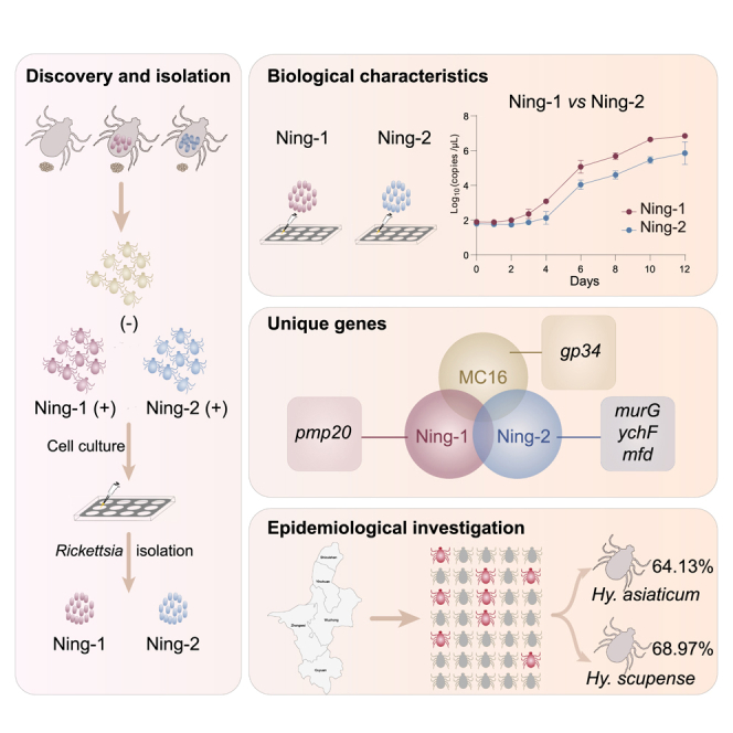
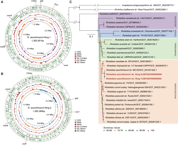
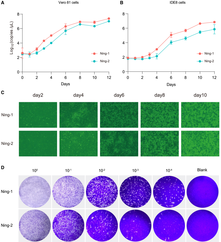
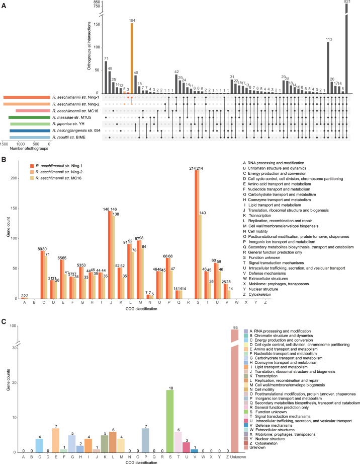
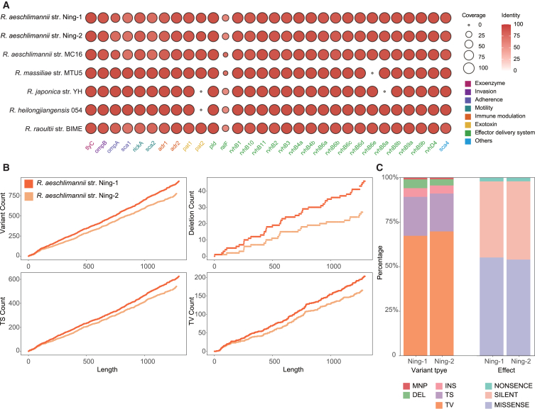
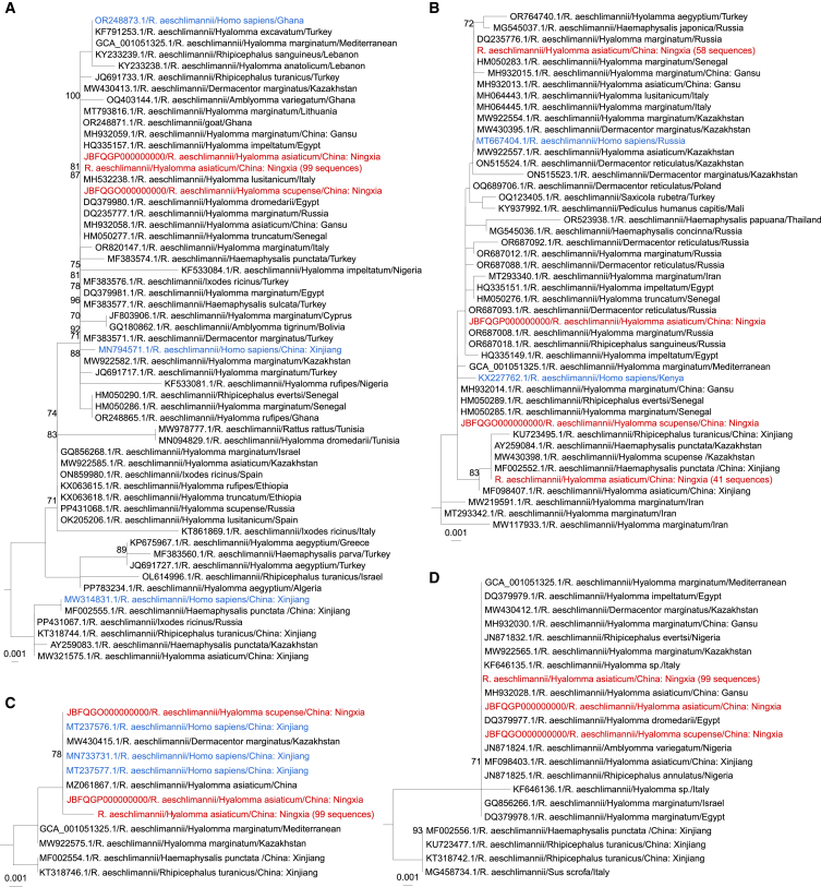
Rickettsia aeschlimannii, which emerged in Morocco in 1997, causes the Mediterranean spotted fever-like rickettsiosis in various Mediterranean countries and recently in Russia and China. Despite its increasing distribution, no available genome has been reported outside Morocco to date. Here, we isolated two strains of R. aeschlimannii from Hyalomma asiaticum (Ning-1 strain) and Hyalomma scupense (Ning-2 strain) ticks in northwestern China and assembled their complete genomes. The genomes of the two strains were smaller than the Mediterranean MC16 strain, containing fewer pseudogenes, higher ralF virulence factor coverage, and 154 unique orthogroups. The Ning-1 strain overwhelmed the Ning-2 strain with more obvious cytopathic effects, quicker growth, and faster plaque formation in cell culture, likely due to its unique pmp20 gene, higher frequency of single nucleotide polymorphisms, and missense/silent ratio. The prevalence of R. aeschlimannii was high among Hyalomma ticks in northwestern China. These findings highlight the genomic characteristics of R. aeschlimannii and the necessity for enhanced surveillance of the emerging Rickettsia in the human population.

Keywords: Genomics; Pathogenic organism.


Conflict of interest statement

The authors declare no competing interests.

Figures

None
Graphical abstract
Figure 1
Figure 1
Isolation of R. aeschlimannii from two Hyalomma species (A) Giemsa staining of R. aeschlimannii isolated from a pool of Hy. asiaticum larvae in Vero 81 cells. (B) Giemsa staining of R. aeschlimannii isolated from a pool of Hy. scupense larvae in Vero 81 cells. Scale bar represents 10 μm (A and B). (C) Transmission electron micrographs of Vero 81 cells infected with R. aeschlimannii isolated from Hy. asiaticum. (D) Transmission electron micrographs of Vero 81 cells infected with R. aeschlimannii isolated from Hy. scupense. Photomicrographs were captured with an HT7800 transmission electron microscope camera. Scale bar represents 5 μm (magnification ×2,500) and 2 μm (magnification ×6,000) (C and D).
Figure 2
Figure 2
Circular map and phylogenetic tree of two R. aeschlimannii strains (A) Bird’s eye view of the assembled genome of R. aeschlimannii str. Ning-1. (B) Bird’s eye view of the assembled genome of R. aeschlimannii str. Ning-2. From inner circle to outer circle (A and B), the map represents GC skew, GC content, proteins of - strand, contig, and proteins of + strand. The locations of tRNA, rRNA, ompA, ompB, gltA, 17 kDa, sca1, and sca4 genes within the genome are indicated. (C) Phylogenetic tree constructed using the maximum likelihood method with 1,000 replications, based on the whole genomes of 26 other publicly available established or proposed Rickettsiales species. Anaplasma phagocytophilum and Ehrlichia chaffeensis were used as outgroup species to help root the tree. Scale bar indicates 0.1 nucleotide substitutions per site. See also Figure S1.
Figure 3
Figure 3
Comparison of growth characteristics between two R. aeschlimannii strains (A) Growth curves of R. aeschlimannii str. Ning-1 and R. aeschlimannii str. Ning-2 in Vero 81 cells over 288 h. (B) Growth curves of R. aeschlimannii str. Ning-1 and R. aeschlimannii str. Ning-2 in IDE8 tick cells over 288 h. Quantitative data from three independent experiments are presented as mean ± SD (shown as error bars) (A and B). (C) Cytopathic effect in Vero 81 cells induced by the two R. aeschlimannii strains. (D) Plaque formation in Vero 81 cells by the two R. aeschlimannii strains with multiple dilutions at 8 days post-infection (dpi).
Figure 4
Figure 4
Functional annotation of R. aeschlimannii genomes (A) UpsetR plot showing the number of orthogroups in the R. aeschlimannii genomes compared with other closely related SFGR representatives. Connected circles indicate shared orthogroups among these SFGR species. (B) COG annotation of all genes from the three R. aeschlimannii strains. (C) COG annotation of shared homologous genes of Ning-1 and Ning-2 strains. See also Tables S2 and S3.
Figure 5
Figure 5
Virulence factors determination and single nucleotide polymorphism analysis of R. aeschlimannii (A) Presence of virulence factors in R. aeschlimannii and other representative SFGR genomes. The vertical axis represents various SFGRs, the horizontal axis indicates the names of the virulence factors, the size of the bubbles represents the coverage of the virulence factors, and the color intensity indicates their identity. (B) Number of variants, deletions, transitions, and transversions of R. aeschlimannii strains Ning-1 and Ning-2 with the genome of R. aeschlimannii strain MC16 as the reference. (C) Proportion of total SNPs accounted for by various types and effects of SNPs. See also Tables S4–S6.
Figure 6
Figure 6
Phylogenetic analysis of R. aeschlimannii based on nucleotide sequences of four genes (A) Phylogenetic tree based on ompA gene. (B) Phylogenetic tree based on gltA gene. (C) Phylogenetic tree based on sca1 gene. (D) Phylogenetic tree based on 17 kDa gene. (A–D), Bootstrap analysis with 1,000 replicates was conducted to evaluate phylogenetic robustness. Scale bar indicates the number of nucleotide substitutions per site. GenBank accession numbers are provided. Sequences obtained in this study are highlighted in red, while sequences identified from humans are in blue. See also Table S7.

Similar articles

Cited by

References

    1. Beati L., Meskini M., Thiers B., Raoult D. Rickettsia aeschlimannii sp. nov., a new spotted fever group rickettsia associated with Hyalomma marginatum ticks. Int. J. Syst. Bacteriol. 1997;47:548–554. doi: 10.1099/00207713-47-2-548. - DOI - PubMed
    1. Raoult D., Fournier P.E., Abboud P., Caron F. First documented human Rickettsia aeschlimannii infection. Emerg. Infect. Dis. 2002;8:748–749. doi: 10.3201/eid0807.010480. - DOI - PMC - PubMed
    1. Oteo J.A., Portillo A. Tick-borne rickettsioses in Europe. Ticks Tick. Borne. Dis. 2012;3:271–278. doi: 10.1016/j.ttbdis.2012.10.035. - DOI - PubMed
    1. Germanakis A., Chochlakis D., Angelakis E., Tselentis Y., Psaroulaki A. Rickettsia aeschlimannii infection in a man, Greece. Emerg. Infect. Dis. 2013;19:1176–1177. doi: 10.3201/eid1907.130232. - DOI - PMC - PubMed
    1. Mokrani N., Parola P., Tebbal S., Dalichaouche M., Aouati A., Raoult D. Rickettsia aeschlimannii infection, Algeria. Emerg. Infect. Dis. 2008;14:1814–1815. doi: 10.3201/eid1411.071221. - DOI - PMC - PubMed

LinkOut - more resources