Review
. 2025 Feb 13;28(3):112012.
doi: 10.1016/j.isci.2025.112012. eCollection 2025 Mar 21.

Evolving concepts of the protein universe

Affiliations
Review

Evolving concepts of the protein universe

Prakash Kulkarni et al. iScience. .

Abstract

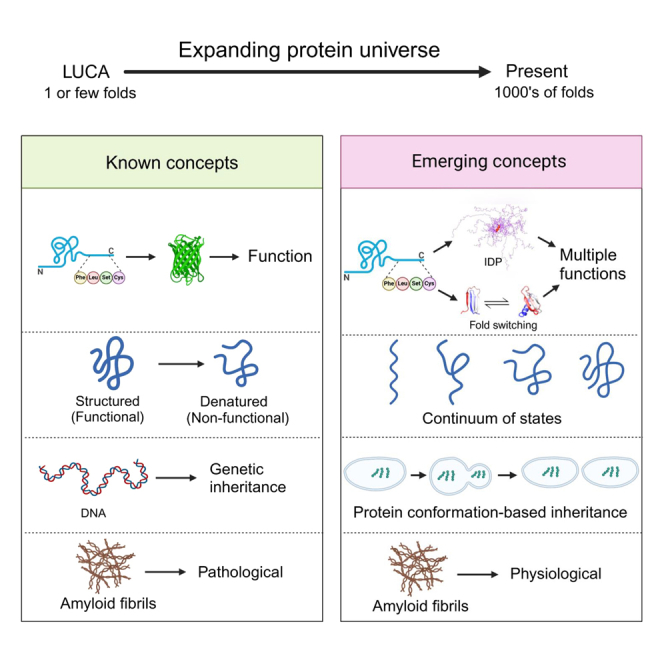
The protein universe is the collection of all proteins on earth from all organisms both extant and extinct. Classical studies on protein folding suggested that proteins exist as a unique three-dimensional conformation that is dictated by the genetic code and is critical for function. In this perspective, we discuss ideas and developments that emerged over the past three decades regarding the protein structure-function paradigm. It is now clear that ordered (active/functional) and disordered/denatured (and hence inactive/non-functional) represent a continuum of states rather than binary states. Some proteins can switch folds without sequence change. Others exist as conformational ensembles lacking defined structure yet play critical roles in many biological processes, including forming membrane-less organelles driven by liquid-liquid phase separation. Numerous diverse proteins harbor segments with the potential to form amyloid fibrils, many of which are functional, and some possess prion-like properties enabling conformation-based transfer of heritable information. Taken together, these developments reveal the remarkable complexity of the protein universe.

Keywords: Biochemistry; Biological sciences; Protein; Structural biology.


Conflict of interest statement

The authors declare no competing interests.

Figures

None
Graphical abstract
Figure 1
Figure 1
Proteins on the brink of stability can undergo a continuum of order/disorder transitions (A) Examples of transitions from top left to bottom right: transition between the extended and collapsed disordered states of prostate associated gene 4 (PAGE4), modulated by phosphorylation; 111 disorder-to-order transition of 4E-BP2 induced by phosphorylation; 23 order-to-order fold switching between GA98 and GB98, triggered by single amino acid changes or ligand binding. In contrast, stable proteins such as subtilisin (shown in dark blue) do not undergo such changes. (B) Approximate energy well diagrams for each protein from PAGE4 (top) to subtilisin (bottom) (from the study by Kulkarni P. et al. with permission).
Figure 2
Figure 2
Fold-switching proteins (A) Summary of designed switches between 3α, αβ-plait, and β-grasp fold topologies that have been characterized structurally. Color-coding indicates how the corresponding regions change in the alternative fold. The dashed lines show regions that are disordered in one of the states but not the other. (B) Top, a NusG N-terminal (NGN) fold (light gray) and a C-terminal β-roll fold (lavender) are predicted from a deep input MSA (region corresponding to the CTD shown) generated from the sequence of a NusG protein with ≤29% aligned identity to its homologs with experimentally determined structures. Predicted β sheets in the C-terminal domain that agree closely with the β sheets predicted from nuclear magnetic resonance experiments are shown with black boxes surrounding lavender bars. Bottom, a NusG N-terminal (NGN) fold (light gray) and a C-terminal α helical hairpin fold (teal) are predicted from a modified input MSA of the NusG homolog in which columns predicted to form only β-roll contacts are changed to alanine. Predicted α helices in the C-terminal domain that agree with the α helices predicted from nuclear magnetic resonance experiments are shown with black boxes surrounding teal bars. Protein structures were generated with PyMOL43.
Figure 3
Figure 3
Amyloid-forming proteins (A) Amyloid-like fibrils formed from an SH3 domain. Readapted with permission from the study by Watson J. L. et al. (B) Energy funnel illustrating the protein folding (left) and the protein aggregation (right) components. Readapted with permission from the study by Parui S. et al. (C) Curli of bacteria visible as filamentous structures as an example of functional amyloids. Readapted with permission from the study by Olsen A. et al. (D) Comparison between predicted protein aggregation profile (blue) and regions of the sequence found experimentally to form β-strands in amyloid fibrils (colored bars). Readapted with permission from the study by Westermark P. et al. (E) The four structures on the right are cross-sectional structures of amyloid fibrils along with their PDB entries and references, as extracted from the amyloid atlas. Readapted with permission from the study by Fowler D.M. et al.
Figure 4
Figure 4
Three types of protein-based inheritance described in the yeast Saccharomyces cerevisiae (A) Prions that can be generated de novo by overexpression of the prion-forming protein. The resulting amyloid fibrils (red) are fragmented by Hsp104 into smaller elements called propagons that can be transferred to daughter cells at mitosis. (B) Mnemons are generated in cells in response to a chemical signal. The only example described in detail so far is the Whi3-based mnemons (ref.90), high-molecular-weight forms of the protein (in dark blue) that are retained by the mother cell and not passed on to daughter cells. These forms are not amyloid but require Hsp70 or Hsp104 for their maintenance. (C) The prion-like proteins described by Chakrabortee et al., which are induced de novo by overexpression of a typically low-abundance protein, although the nature of the resulting altered forms of the protein (yellow) remains to be defined but are not amyloid. Depending on the protein, they either require Hsp104, Hsp90, or Hsp70 to maintain the associated phenotype. In all three cases, the cells in green show the altered phenotype. Reproduced with permission from Tuite M.F. et al.
Figure 5
Figure 5
Prions: Proteins mediating transgenerational inheritance (A) Certain proteins can refold to take up an amyloid form, triggered either by changes in the cellular environment or because of a mutational change in the protein’s sequence. As illustrated, the resulting amyloid can take up one of several different structures (amyloid polymorphs) which in turn can give rise to distinct prion-mediated phenotypes. (B) The transmissible form of the prion amyloid is generated by fragmentation of the amyloid fibrillar form of the protein. This fragmentation is mediated by the coordinated action of at least three different molecular chaperones and the resulting transmissible fragments are referred to as prion seeds or propagons. The atomic force microscopic (AFM) images refer to different mutant forms of the yeast Sup35 prion-forming domain (A) or the wild-type form of the same protein (B). AFM images kindly provided by Drs Wei-feng Xue and Ricardo Marchante, University of Kent.

References

    1. Ladunga I. Phylogenetic continuum indicates "galaxies" in the protein universe: preliminary results on the natural group structures of proteins. J. Mol. Evol. 1992;34:358–375. - PubMed
    1. Povolotskaya I.S., Kondrashov F.A. Sequence space and the ongoing expansion of the protein universe. Nature. 2010;465:922–926. - PubMed
    1. Dokholyan N.V., Shakhnovich B., Shakhnovich E.I. Expanding protein universe and its origin from the biological Big Bang. Proc. Natl. Acad. Sci. USA. 2002;99:14132–14136. - PMC - PubMed
    1. Matveev V.V. Cell theory, intrinsically disordered proteins, and the physics of the origin of life. Prog. Biophys. Mol. Biol. 2019;149:114–130. - PubMed
    1. Kauzmann W.J. Some factors in the interpretation of protein denaturation. Adv. Protein Chem. 1959;14:1–63. - PubMed

LinkOut - more resources