. 2025 Jan;5(1):4.
doi: 10.20517/jca.2024.17. Epub 2025 Feb 18.

Aging triggers mitochondrial, endoplasmic reticulum, and metabolic stress responses in the heart

Affiliations

Aging triggers mitochondrial, endoplasmic reticulum, and metabolic stress responses in the heart

Sakthijothi Muthu et al. J Cardiovasc Aging. 2025 Jan.

Abstract

Introduction: Aging is a multifaceted biological process characterized by a progressive decline in cellular and tissue function. It significantly impacts the cardiovascular system and contributes to the onset of cardiovascular diseases. The mitochondria (mt) and the endoplasmic reticulum (ER) play synergistic roles in maintaining cellular homeostasis and energy production in the heart. Nevertheless, their response to cardiac aging is not well known.

Aim: This study explores mt and ER stress responses and their associated factors, such as metabolic, cellular, and autophagic stress, in cardiac aging.

Methods and results: We utilized 10- and 25-month-old CBA/CaJ mice to evaluate mt, ER, and their associated factors, such as metabolic, cellular, and autophagic stress responses. We studied the gene expression for mitochondrial biogenesis, mt and ER stress response, autophagy and metabolic markers, and activating transcription factors that mediate cellular stress responses. We found no significant difference in mtDNA content and the mRNA expression of the mt transcription factor, Tfam; however, selective mtDNA genes, such as mt-Cytb and mt-Co2, showed significant induction in 25-month-aged compared to 10-month-young hearts. Interestingly, genes of several mitochondrial stress response proteases and their components, including Lonp1, Yme1l1, Afg3l2, and Spg7, were significantly induced, with a substantial induction of Clpp and Clpx. However, age-associated differences were not observed in the induction of mt chaperones (Hspa9 and Hspd1), but significant induction of Dnaja2, a mitochondrial co-chaperone, was observed. The ER stress transcription factors Xbp1 and Atf6 were markedly induced in aged hearts, accompanied by decreased expression of ER stress chaperone Hsp90b with no change in Hspa5 and Dnajb9 chaperones. However, induction of Dnm1l was significant, whereas Mfn1 and Fis1 were downregulated in contrast to Mfn2, suggesting dysregulated mitochondrial dynamics in the aged heart with no change in autophagy and metabolic stress regulators observed. Furthermore, aged hearts showed significantly increased oxidative damage as evidenced by elevated lipid peroxidation (4-HNE) levels.

Conclusion: These findings demonstrate that aging triggers mt, ER, and oxidative stress in the heart, which over time leads to the accumulation of oxidative damage, causing cellular impairment, highlighting these pathways as potential therapeutic targets for mitigating age-related cardiac dysfunction.

Keywords: Aging; endoplasmic reticulum stress; heart; mitochondrial stress; oxidative stress.


Conflict of interest statement

Conflicts of interest All authors declared that there are no conflicts of interest.

Figures

Figure 1.
Figure 1.
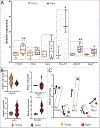
Cardiac aging alters mitochondrial biogenesis. (A) Schematic representation of the study comparing young (10-month) vs. old (25-month) CBA/CaJ mice hearts subjected to genomic DNA, mRNA, and protein isolation and investigation of mt biogenesis, mt and ER stress response, autophagy, and metabolic alterations. This schematic figure was created using BioRender (www.app.biorender.com). (B) Relative mRNA expression levels of mtDNA-encoded (Nd1, Nd4, mt-Cytb, mt-Co2, mt-Atp6) and nDNA-encoded mitochondrial Ndufs4. (C) Relative mtDNA content (top) normalized to Tert, and mRNA expression levels of Tfam normalized to Gapdh in young vs. old hearts. Experiments were conducted in triplicate, and relative expression levels were plotted. Values are presented as mean ± SEM (n = 3–10). *P < 0.05, **P < 0.01, by an unpaired Student’s t-test, using GraphPad Prism software.
Figure 2.
Figure 2.
Aging induces mitochondrial stress response and changes mt dynamics in the heart. (A) Relative mRNA expression levels of mt ATP-dependent proteases and its components in aged (25-month) vs. young (10-month) CBA/CaJ mice hearts, normalized to Gapdh. (B) Relative mRNA expression levels of mt chaperones and Atf5 in aged vs. young hearts normalized to Gapdh. (C) Relative mRNA expression levels of mt dynamic regulators in aged vs. young hearts normalized to Gapdh. Relative expression values were represented as mean ± SEM (n = 3–10). *P < 0.05, **P < 0.01 are considered significant, calculated by an unpaired Student’s t-test using GraphPad Prism software.
Figure 3.
Figure 3.
Impaired metabolic and autophagy regulation in cardiac aging. (A) Reduced mRNA expression levels of autophagy regulators in aged (25-month) vs. young (10-month) CBA/CaJ mice hearts, normalized to Gapdh. (B) Top relative mRNA expression levels of essential metabolic regulators (Mtor, Nrf1, and Prkaa1) in aged (25- month) vs. young (10-month) CBA/CaJ mice hearts, normalized to Gapdh. Relative expression values were represented as mean ± SEM (n = 3–10). Significant difference was calculated by an unpaired Student’s t-test using GraphPad Prism software.
Figure 4.
Figure 4.
Aging induces selective endoplasmic reticulum stress response in the heart. (A) Relative mRNA expression levels of ER stress response factors in aged (25-month) vs. young (10-month) CBA/CaJ mice hearts, normalized to Gapdh. (B) Relative mRNA expression levels of ER stress response factors in aged (25-month) vs. young (10-month) CBA/CaJ mice hearts, normalized to Gapdh. Relative expression values were represented as mean ± SEM (n = 3–10). *P < 0.05, **P < 0.01 are considered significant, calculated by an unpaired Student’s t-test using GraphPad Prism software.
Figure 5.
Figure 5.
Increased oxidative stress in cardiac aging. Left: Immunoblot showing the expression of 4-HNE in aged (25-month) vs. young (10-month) CBA/CaJ mice hearts and corresponding Ponceau stained loading control blot. Right: Densitometry quantification of the 4-HNE levels normalized to ponceau staining. Relative expression values were represented as mean ± SEM (n = 5). **P < 0.01 is considered significant, calculated by an unpaired Student’s t-test using GraphPad Prism software.
Figure 6.
Figure 6.
Aging induces mt and ER stress responses in the heart. During cardiac aging, mitochondria (mt) and the endoplasmic reticulum (ER) are two important organelles exposed to high energy demand. Initially, they adapt by upregulating the stress response, but as aging progresses, oxidative stress and damage increase, thereby predisposing the heart to dysfunction. Created using BioRender (www.app.biorender.com).

References

    1. Sack MN, Finkel T. Mitochondrial metabolism, sirtuins, and aging. Cold Spring Harb Perspect Biol. 2012;4:a013102. - PMC - PubMed
    1. Lesnefsky EJ, Chen Q, Hoppel CL. Mitochondrial metabolism in aging heart. Circ Res. 2016;118:1593–611. - PMC - PubMed
    1. Wallace KB, Sardão VA, Oliveira PJ. Mitochondrial determinants of doxorubicin-induced cardiomyopathy. Circ Res. 2020;126:926–41. - PMC - PubMed
    1. Tabas I, Ron D. Integrating the mechanisms of apoptosis induced by endoplasmic reticulum stress. Nat Cell Biol. 2011;13:184–90. - PMC - PubMed
    1. Boengler K, Kosiol M, Mayr M, Schulz R, Rohrbach S. Mitochondria and ageing: role in heart, skeletal muscle and adipose tissue. J Cachexia Sarcopenia Muscle. 2017;8:349–69. - PMC - PubMed

LinkOut - more resources