Review
. 2025 Dec;32(1):2476140.
doi: 10.1080/10717544.2025.2476140. Epub 2025 Mar 24.

External stimuli-responsive drug delivery to the posterior segment of the eye

Affiliations
Review

External stimuli-responsive drug delivery to the posterior segment of the eye

Shuting Xu et al. Drug Deliv. 2025 Dec.

Abstract

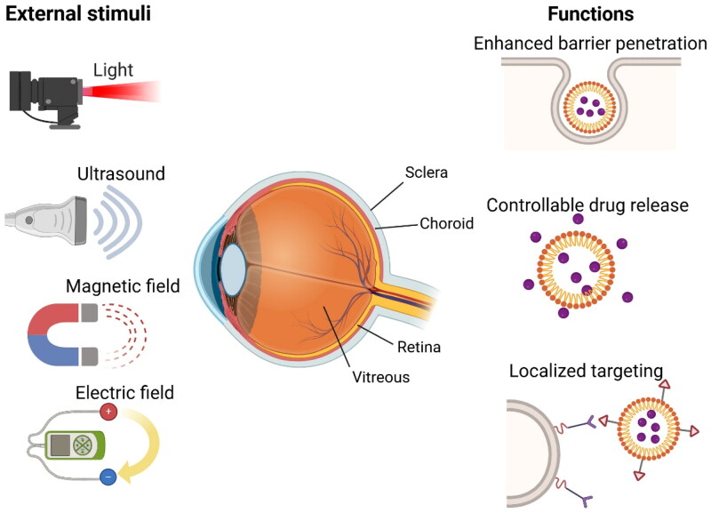
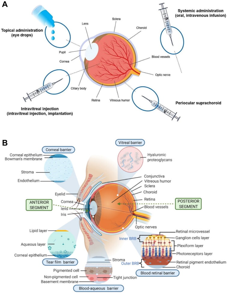
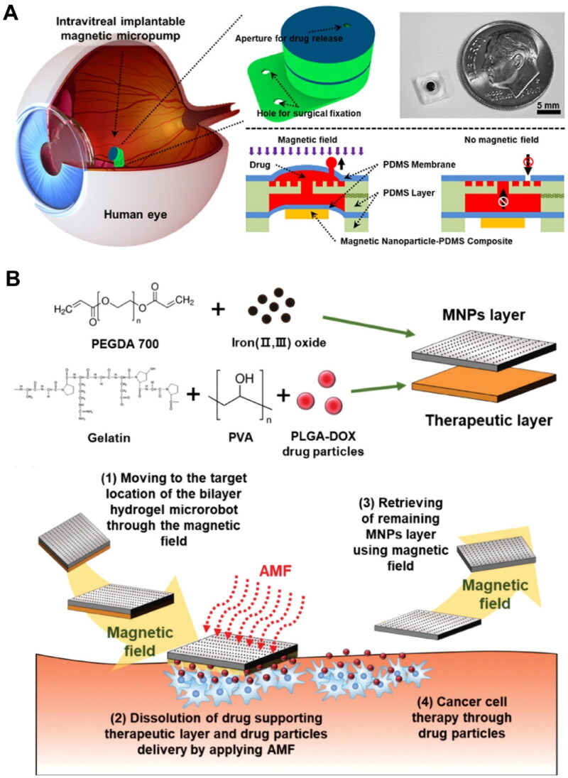
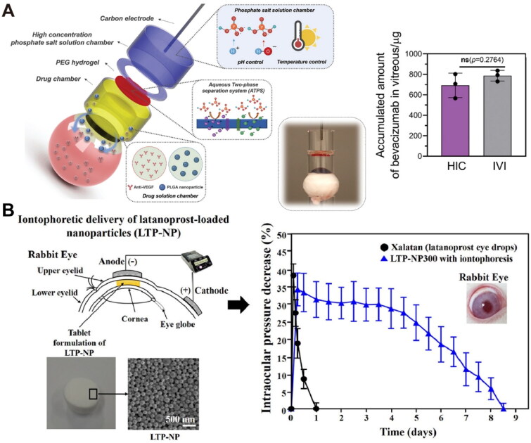
Posterior segment eye diseases represent the leading causes of vision impairment and blindness globally. Current therapies still have notable drawbacks, including the need for frequent invasive injections and the associated risks of severe ocular complications. Recently, the utility of external stimuli, such as light, ultrasound, magnetic field, and electric field, has been noted as a promising strategy to enhance drug delivery to the posterior segment of the eye. In this review, we briefly summarize the main physiological barriers against ocular drug delivery, focusing primarily on the recent advancements that utilize external stimuli to improve treatment outcomes for posterior segment eye diseases. The advantages of these external stimuli-responsive drug delivery strategies are discussed, with illustrative examples highlighting improved tissue penetration, enhanced control over drug release, and targeted drug delivery to ocular lesions through minimally invasive routes. Finally, we discuss the challenges and future perspectives in the translational research of external stimuli-responsive drug delivery platforms, aiming to bridge existing gaps toward clinical use.

Keywords: Drug delivery; external stimuli; nanomedicines; photoresponsive nanoparticles; posterior segment eye diseases.


Conflict of interest statement

No potential conflict of interest was reported by the author(s).

Figures

Figure 1.
Figure 1.
Schematic illustration of external stimuli-responsive drug delivery strategies for the treatment of posterior segment eye diseases. Commonly used external stimuli, such as light, ultrasound, magnetic field, and electric field, have demonstrated significant potential in enhancing the penetration of administered drugs across biological barriers, enabling precise control over drug release or localized targeting of nanocarriers. The image was created with BioRender.com.
Figure 2.
Figure 2.
(A) Schematic illustration of common administration routes for ocular drug delivery. Reproduced from Liu et al. (2024) with permission. Copyright: 2024 the authors. Journal of Nanobiotechnology published by Springer Nature. (B) Schematic representation of physiological barriers of the eye. Reproduced from Adrianto et al. (2022) with permission. Copyright: 2021 the authors. Drug Delivery and Translational Research published by Springer Nature.
Figure 3.
Figure 3.
Representative examples of light-responsive drug delivery based on the photocleavage mechanisms for the treatment of posterior segment eye diseases. (A) Schematic representation of light-controlled intraocular drug release from a photoresponsive nanosystem for retinoblastoma therapy. (B) Schematic illustration and representative images showing in vivo biodistribution of the photoresponsive nanoparticles 1 h after intravenous administration, with or without light irradiation to the mouse eyes. (A-B) Reproduced from Long et al. (2021) with permission. Copyright: 2021 the authors. Advanced Science published by Wiley-VCH GmbH. (C) Schematic representation of light-triggered targeting of intravenously injected nanoparticles to the eye. (D) Representative confocal images and quantitative analysis of choroid-RPE flat-mounts showing light-triggered accumulation of fluorescein (AMF)-loaded nanoparticles (NP-AMF-[CPP]) in CNV lesions. Data were presented as mean ± standard deviation (n = 8). (C-D) Reproduced from Wang et al. (2019) with permission. Copyright: 2019 the authors. Nature Communications published by Springer Nature.
Figure 4.
Figure 4.
Representative examples of light-responsive drug delivery based on photothermal and photodynamic effects for the treatment of posterior segment eye diseases. (A) Schematic illustration of a photoresponsive hydrogel system containing gold nanorods as the photothermal agent and an ATOX1 inhibitor for the combination treatment of uveal melanoma. Reproduced from Wang et al. (2021) with permission. Copyright: 2021 the authors. Advanced Science published by Wiley-VCH GmbH. (B) Schematic representation of a photoresponsive prodrug nanosystem composed of a ROS-sensitive anti-angiogenic prodrug and a photosensitizer for the combination treatment of AMD. Reproduced from Xu et al. (2023) with permission. Copyright: 2023 the authors. Advanced Science published by Wiley-VCH GmbH.
Figure 5.
Figure 5.
Representative examples of magnetic field-responsive drug delivery for the treatment of posterior segment eye diseases. (A) The working mechanism and drug release profile of an intravitreal implantable magnetic micropump for controlled release of vascular endothelial growth factor receptor (VEGFR) inhibitor. The open check valve enables the upward release of the encapsulated anti-Flt1 gold nanoparticles under a magnetic field, while the closed check valve prevents the payload diffusion. PDMS, polydimethylsiloxane. Reproduced from Wang et al. (2018) with permission. Copyright 2018 Elsevier B.V. (B) Schematic illustration of the composition of an intravitreal bilayer hydrogel microrobot and its treatment process. DOX, doxorubicin; AMF, alternating magnetic fields. Reproduced from Kim et al. (2020) with permission. Copyright 2020 WILEY-VCH verlag GmbH & Co. KGaA.
Figure 6.
Figure 6.
Representative examples of electric field-responsive drug delivery for the treatment of posterior segment eye diseases. (A) Schematic illustration of a hydrogel ionic circuit (HIC)-based iontophoresis device for high-intensity transscleral iontophoresis and quantitative analysis of the accumulated bevacizumab in ocular tissues after high-intensity transscleral iontophoresis (100 mA, 20 min) (n = 3). Reproduced from Zhao et al. (2022) with permission. Copyright 2021 Wiley-VCH GmbH. (B) Schematic representation of an iontophoretic device for transcorneal delivery of latanoprost-loaded nanoparticles and profiles of intraocular pressure of rabbit eyes after the treatments with various latanoprost formulations with or without iontophoresis (n = 4). Data were presented as mean ± standard deviation. Reproduced from Kim et al. (2022) with permission. Copyright 2022 Elsevier B.V.

Similar articles

References

    1. Abdelmohsen, H.A.M., Copeland, N.A., and Hardy, J.G., 2023. Light-responsive biomaterials for ocular drug delivery. Drug delivery and translational research, 13 (8), 2159–2182. doi: 10.1007/s13346-022-01196-5. - DOI - PMC - PubMed
    1. Adrianto, M.F., et al. , 2022. In vitro dissolution testing models of ocular implants for posterior segment drug delivery. Drug delivery and translational research, 12 (6), 1355–1375. doi: 10.1007/s13346-021-01043-z. - DOI - PMC - PubMed
    1. Afarid, M., Mahmoodi, S., and Baghban, R., 2022. Recent achievements in nano-based technologies for ocular disease diagnosis and treatment, review and update. Journal of nanobiotechnology, 20 (1), 361. doi: 10.1186/s12951-022-01567-7. - DOI - PMC - PubMed
    1. Agban, Y., et al. , 2019. Depot formulations to sustain periocular drug ­delivery to the posterior eye segment. Drug discovery today, 24 (8), 1458–1469. doi: 10.1016/j.drudis.2019.03.023. - DOI - PubMed
    1. Ahmed, S., Amin, M.M., and Sayed, S., 2023. Ocular drug delivery: a comprehensive review. AAPS PharmSciTech, 24 (2), 66. doi: 10.1208/s12249-023-02516-9. - DOI - PubMed