. 2025 Mar 24;20(3):e0314252.
doi: 10.1371/journal.pone.0314252. eCollection 2025.

Eye features and retinal photoreceptors of the nocturnal aardvark (Orycteropus afer, Tubulidentata)

Affiliations

Eye features and retinal photoreceptors of the nocturnal aardvark (Orycteropus afer, Tubulidentata)

Leo Peichl et al. PLoS One. .

Abstract

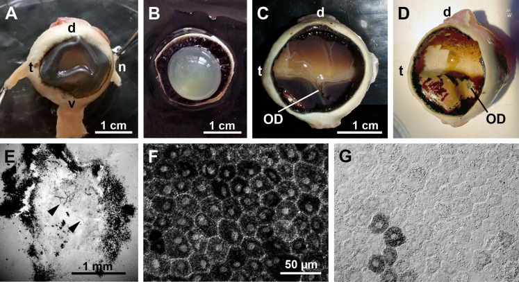
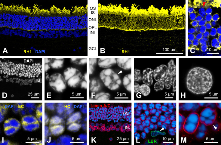
The nocturnal aardvark Orycteropus afer is the only extant species in the mammalian order Tubulidentata. Previous studies have claimed that it has an all-rod retina. In the retina of one aardvark, we found rod densities ranging from 124,000/mm² in peripheral retina to 214,000/mm² in central retina; the retina of another aardvark had 163,000 - 245,000 rods/mm². This is moderate in comparison to other nocturnal mammals. With opsin immunolabelling we found that the aardvark also has a small population of cone photoreceptors. Cone densities ranged from about 300 to 1,300/mm² in one animal, and from 1,100 to 1,600/mm² in a limited sample of the other animal, with a central-peripheral density gradient and some local variations. Overall, cones comprised 0.25-0.9% of the photoreceptors. Both typical mammalian cone opsins, longwave-sensitive (L) and shortwave-sensitive (S), were present. However, there was colocalization of the two opsins in many cones across the retina (35 - 96% dual pigment cones). Pure L cones and S cones formed smaller populations. This probably results in poor colour discrimination. Thyroid hormones, important regulators of cone opsin expression, showed normal blood serum levels. The relatively low rod density and hence a relatively thin retina may be related to the fact that the aardvark retina is avascular and its oxygen and nutrient supply have to come from the choriocapillaris by diffusion. In contrast to some previous studies, we found that the aardvark eye has a reflective tapetum lucidum with features of a choroidal tapetum fibrosum, in front of which the retinal pigment epithelium is unpigmented. The discussion considers these findings from a comparative perspective.


Conflict of interest statement

The authors have declared that no competing interests exist.

Figures

Fig 1
Fig 1. Evolutionary tree of the Afrotheria, showing the isolated position of the Tubulidentata with the aardvark as its only extant species.
The tree is a schematic representation based on [1], the timeline of branch points is not to scale.
Fig 2
Fig 2. Aardvarks at day and night, note the laterally positioned eyes.
(A) Aardvark 1, the post mortem donor of the studied eye, at daytime. (B, C) Another aardvark, flashlight photographs taken at night. When the head is viewed horizontally, there is a bright whitish eyeshine indicating a tapetum lucidum; when viewed from above, the weaker eyeshine appears orange to red. For details see text. Image sources: (A) Frankfurt Zoo; (B, C) Christina Geiger.
Fig 3
Fig 3. General eye features (aardvark 1).
(A) Intact eye, frontal view with attached rectus muscles. The cornea had collapsed when the eye was punctured for better fixative penetration to the retina. (B) Anterior part of the opened eye from the vitreous side, showing the lens and ciliary body. (C) Opened eyecup showing the fundus with the retina in situ. There is a bright horizontal band where the retinal pigment epithelium (RPE) is weakly pigmented or unpigmented. The transition to the pigmented peripheral RPE is gradual at the dorsal side and with a rather sharp boundary at the ventral side. The optic disc (OD) is located ventral to the bright horizontal band. (D) Eyecup after removal of the retina. Some RPE also came off during the preparation, showing an unpigmented, whitish-yellow choroid. Here, the optic disc (OD) is more obvious than in (C). (E) Optic disc in the isolated retina, DAB-reacted for blood vessels. There are only a few capillaries present in the OD (arrow heads), and no blood vessels exit it to supply the surrounding retina. Around the OD there is an accumulation of pigment. (F, G) Light microscopic images of flat-mounted RPE pieces from peripheral (F) and central fundus (G). In the periphery, all RPE cells contain densely packed melanin granules (F); centrally, only very few RPE cells are rich in melanin granules, the vast majority of RPE cells contains little or no melanin (G). Eye dimensions in (D) can be determined from the scale bar in (C). The scale bar in (F) applies to (F, G). d, dorsal; n, nasal; t, temporal, v, ventral.
Fig 4
Fig 4. Choroid and tapetum lucidum (aardvark 1).
(A) Higher power view of the central and ventral midperipheral fundus of the aardvark eye (c.f. Fig 3C and D). Below the partly removed RPE layer, the whitish unpigmented choroid with its red blood vessels is visible. (B) SEM micrograph of subcellular structures from the RPE-facing side of a transverse choroid section, showing collagen fibrils arranged in parallel bundles with different orientations that indicate a tapetum lucidum. In the upper image part, the fibrils are longitudinally sectioned; in the bottom image part, they are cross-sectioned with round to oval profiles. (C) At higher magnification, the fibrils show the typical cross-striation of native collagen (TEM image). (D) In cross-section, the fibrils show a shell and core of higher electron density (SEM image). (E) Differential interference contrast (DIC) image of a vertical cryo-section of the aardvark choroid (Ch) and tapetum (Tap). The poorly pigmented choroid shows cross-sections of blood vessels of various calibers, the tapetum shows a horizontal striation indicating tapetal laminae. (F) DIC image of a vertical cryo-section of the choroid and tapetum of an African elephant for comparison. The choroid is strongly pigmented, and the tapetum is thicker and more conspicuously striated than in the aardvark. (G) Aardvark vertical cryo-section of the choroid and tapetum with blood vessel labelling by isolectin (red). The choroid part contains vessels of larger and smaller caliber, the tapetum layer is relatively thin, and the choriocapillaris (CC) at the border to the RPE is densely filled with capillaries. (H) African elephant vertical cryo-section of the choroid and tapetum with blood vessel labelling by isolectin (red) for comparison. The image shows a vertical choroidal blood vessel supplying the CC capillaries. For the sections of (E-H), the choroid has been removed from the sclera, so the sections do not show the full thickness of the choroid. (I) Flat view of the aardvark choriocapillaris, labelled by isolectin (red) and showing the dense capillary net. The scale bar in (E) applies to (E, F), the scale bar in (G) applies to (G-I).
Fig 5
Fig 5. General retinal features (aardvark 1).
(A) Overview of a vertical retinal section labelled with the fluorescent Nissl stain NeuroTrace, revealing the retinal layering. Cell bodies in the outer nuclear layer (ONL) and inner nuclear layer (INL) are stacked in several tiers. The photoreceptor outer and inner segments (OS+IS) are also labelled. The ganglion cell layer (GCL) is sparsely populated by cells of various soma sizes. A large soma of a putative alpha ganglion cell is marked by an arrowhead. (B, C) Double labelling with an antibody against the neuronal marker NeuN and with NeuroTrace. NeuN only labels a few presumed amacrine cell somata in the INL and some somata in the GCL (B). The NeuroTrace counterstain shows the position of the NeuN somata in the layers (C). (D, E) Immunolabelling for glutamine synthetase shows the Müller cells forming the retinal glia scaffold (D). Counterstaining with DAPI (E) shows that the Müller cells have their somata in the INL and vertically extend their processes from the inner limiting membrane (ILM) formed by their endfeet to the outer limiting membrane (OLM). The images are maximum intensity projections of confocal image stacks. OPL, outer plexiform layer; IPL, inner plexiform layer. Scale bars are 100 µm, scale bar in (C) applies to (B-E).
Fig 6
Fig 6. Rod photoreceptors (aardvark 1).
(A, B) Vertical retinal section, rod opsin RH1 label (yellow). (A) The rod outer segments are strongly labelled, DAPI counterstaining (blue) shows the retinal nuclear layers. (B) Overexposure of the same field shows the less strong RH1 label in the rod somata in the outer nuclear layer (ONL) and the rod axonal spherules in the outer plexiform layer (OPL). Clearly, the vast majority of ONL somata belong to rods. (C) High-power view of the ONL showing one RH1-negative cone soma (red arrowhead) among RH1-labelled rod somata. (D-H) DAPI nuclear staining of various retinal neurons to reveal their heterochromatin arrangement. (D) Overview of a DAPI stained section, nuclei in the ONL are more intensely stained than those in the INL, confirming the appearance seen in (A). (E) The rod nuclei show a “semi-inverted” nuclear architecture with most of the heterochromatin clustered in the nuclear centre, often in two aggregates, but with some extensions towards the nuclear periphery. (F) In contrast, the cone nuclei (arrowhead) have several smaller heterochromatin clusters localized towards the nuclear periphery (conventional nuclear architecture). The same conventional heterochromatin arrangement is seen in cells of the INL (G) and in retinal ganglion cells (H). (I) In the rods, euchromatin (immunolabelled by anti-H3K4me3, yellow) is located mostly in the nuclear periphery and in the gaps between the heterochromatin clusters (DAPI, blue). (J) In the rod nuclei, heterochromatin (immunolabelled by anti-H4K20me3, yellow) colocalizes with the DAPI staining (blue), the merge of the labels appears whitish. (K-M) Immunolabelling for lamin A/C (red) and LBR (green), counterstained with DAPI (blue). The aardvark retina shows a presence of lamin A/C in the neuronal nuclei in all layers (K, L). As a positive control for labeling with the anti-LBR antibody, the nucleus of a microglial cell (arrowhead) expressing LBR but not lamin A/C is shown in (L). LBR label is only present in microglial cells, not in any neurons. (M) A rod nucleus (left) and a cone nucleus (right, arrowhead) with labelled lamin A/C. For layer abbreviations, see Fig 5. Scale bar in (B) applies to (A, B).
Fig 7
Fig 7. Cone photoreceptors (aardvark 1).
(A, B) Vertical retinal sections double-immunolabelled for S cone opsin (A1, B1, red) and L cone opsin (A2, B2, green). In the merged images, DAPI counterstaining in blue shows the retinal nuclear layers (A3, B3). Most cones co-express S and L opsin, arrowheads point to pure S cones. In many cones of the region shown in (A), both the S opsin and the L opsin label extend throughout the cone from the OS to the cone pedicle in the OPL; in the region shown in (B), the L opsin label is restricted to the OS. (C) Double immunolabelled cones in a flat-mounted piece from ventral midperipheral retina. S opsin label (C1, magenta) and L opsin label (C2, green) are colocalized in most cones (C3). Three of the pure S cones are indicated by arrowheads, pure L cones are not present in this field. The aureole seen around most cones is an artefact; in this image the cones were photographed from the vitreous side of the retina, hence there is light scatter at many cellular structures. (D) Vertical section double-immunolabelled for S opsin (D1, in red, showing an S cone pedicle in the OPL) and the synaptic ribbon marker CtBP2 (D2, in green). Most of the small CtBP2 structures in the OPL are ribbons of rod spherules; the merge (D3) shows that cone pedicles do not have the ribbon/CtBP2 clusters seen in other mammals. As expected, many somata in the INL are also CtBP2-labelled. (E) Flat-mounted retinal piece double-labelled for S opsin (E1, magenta) and CtBP2 (E2, green). The focus is on the cone pedicles in the OPL. The merge (E3) confirms the absence of cone-typical ribbon/CtBP2 clusters at the cone pedicles. (A-E) are maximum intensity projections of confocal image stacks. The stack in (C) starts at the level of the intensely labelled cone outer segments and ends at the cone soma level. In this region, faint S opsin label extended throughout the cone, whereas L opsin label was restricted to the outer segment in most of the cones. The stack in (E) is of 3 focal images spaced 0.5 µm apart. As this retinal piece was not perfectly flat, in some places the stack included the CtBP2-labelled INL somata (large green blobs, also seen in D) and missed some ribbons located in the outer OPL (seemingly ribbon-free patches). For layer abbreviations, see Fig 5. Scale bar in (B3) is 100 µm and applies to (A, B); scale bar in (C3) is 50 µm; scale bar in (D3) is 10 µm; scale bar in (E3) is 20 µm.
Fig 8
Fig 8. Cone densities and opsin expression patterns along a dorso-ventral strip in temporal retina (aardvark 1).
(A) Cone density along the dorso-ventral strip (region 1 in the retina scheme S1 Fig) decreases with distance from the streak, the horizontal band of reduced pigmentation in the fundus (see Fig 3). (B) Percentages of dual pigment cones (black diamonds), pure S cones (magenta dots) and pure L cones (green dots) along the dorso-ventral strip. The strip runs from the dorsal edge of the retina (21 mm dorsal) to ventral midperipheral retina (7 mm ventral). Counting field sizes were 500 µm x 500 µm.

Similar articles

References

    1. Springer MS. Afrotheria. Curr Biol. 2022;32(5):R205–10. doi: 10.1016/j.cub.2022.02.001 - DOI - PubMed
    1. Freeman K, Ben-Shlomo G, McMullen R, Moore BA. Ophthalmology of Afrotheria: Aardvarks, Hyraxes, Elephants, Manatees, and Relatives. In: Montiani-Ferreira F, Moore BA, Ben-Shlomo G, editors. Wild and Exotic Animal Ophthalmology, Vol. 2 Mammals. Springer Nature: Switzerland; 2022. p. 49–70.
    1. Shoshani J, Goldman C, Thewissen J. Orycteropus afer. Mammalian Species. 1988;300(1):1–8.
    1. Shoshani J. Tubulidentata (Aardvarks). In: Encyclopedia of Life Sciences. John Wiley & Sons Ltd: Chichester; 2001.
    1. Franz V. Das Auge von Orycteropus afer (Pallas). Denkschriften der medicinisch-naturwissenschaftlichen Gesellschaft zu Jena. 1909;15:401–415, with plates XXV and XXVI.

MeSH terms