. 2025 Mar 24;13(1):40.
doi: 10.1038/s41413-025-00419-y.

Tm4sf19 inhibition ameliorates inflammation and bone destruction in collagen-induced arthritis by suppressing TLR4-mediated inflammatory signaling and abnormal osteoclast activation

Affiliations

Tm4sf19 inhibition ameliorates inflammation and bone destruction in collagen-induced arthritis by suppressing TLR4-mediated inflammatory signaling and abnormal osteoclast activation

Sujin Park et al. Bone Res. .

Abstract

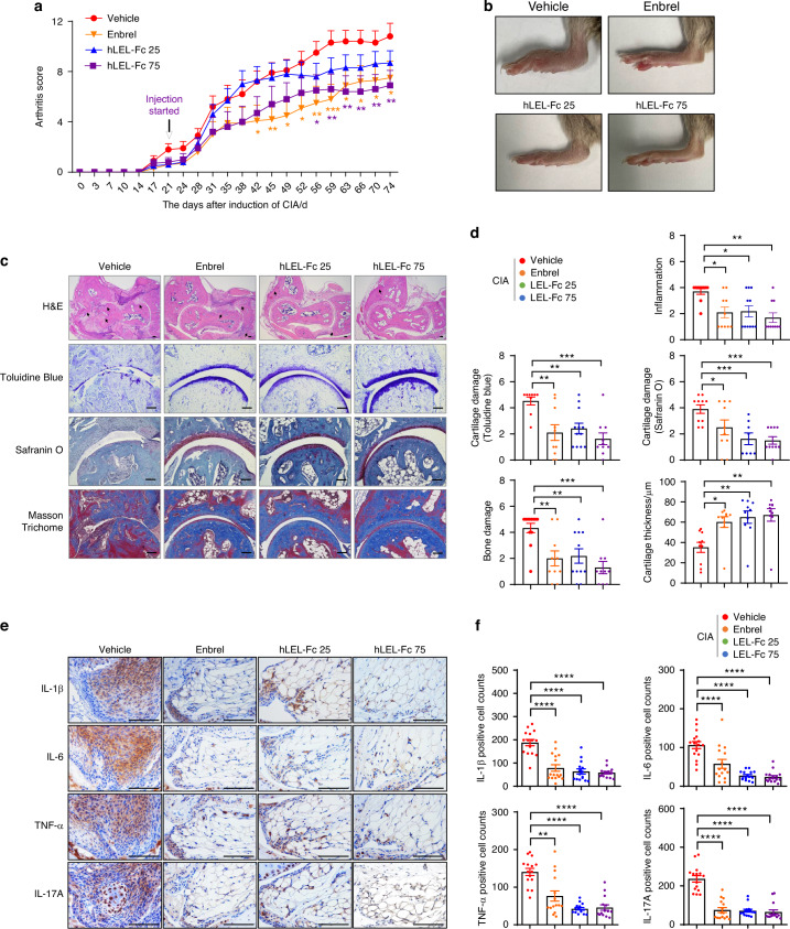
Rheumatoid arthritis (RA) is an autoimmune disease characterized by inflammation and abnormal osteoclast activation, leading to bone destruction. We previously demonstrated that the large extracellular loop (LEL) of Tm4sf19 is important for its function in osteoclast differentiation, and LEL-Fc, a competitive inhibitor of Tm4sf19, effectively suppresses osteoclast multinucleation and prevent bone loss associated with osteoporosis. This study aimed to investigate the role of Tm4sf19 in RA, an inflammatory and abnormal osteoclast disease, using a mouse model of collagen-induced arthritis (CIA). Tm4sf19 expression was observed in macrophages and osteoclasts within the inflamed synovium, and Tm4sf19 expression was increased together with inflammatory genes in the joint bones of CIA-induced mice compared with the sham control group. Inhibition of Tm4sf19 by LEL-Fc demonstrated both preventive and therapeutic effects in a CIA mouse model, reducing the CIA score, swelling, inflammation, cartilage damage, and bone damage. Knockout of Tm4sf19 gene or inhibition of Tm4sf19 activity by LEL-Fc suppressed LPS/IFN-γ-induced TLR4-mediated inflammatory signaling in macrophages. LEL-Fc disrupted not only the interaction between Tm4sf19 and TLR4/MD2, but also the interaction between TLR4 and MD2. μCT analysis showed that LEL-Fc treatment significantly reduced joint bone destruction and bone loss caused by hyperactivated osteoclasts in CIA mice. Taken together, these findings suggest that LEL-Fc may be a potential treatment for RA and RA-induced osteoporosis by simultaneously targeting joint inflammation and bone destruction caused by abnormal osteoclast activation.


Conflict of interest statement

Competing interests: S.J.K. is the founder of Medpacto and serves on its Board of Directors. K.Y., M.W.K., H.J.K., M.J.L., J. B. B., N. P., D.W.K., and S.J.K. are employees of Medpacto. The GILO Foundation has received research support from Medpacto. The other authors declare no competing interests.

Figures

Fig. 1
Fig. 1
The expression of Tm4sf19 is increased in the whole blood of rheumatoid arthritis (RA) patients and the inflamed synovium of mice with collagen-induced arthritis. a Expression of tm4sf19 in the whole blood of RA patients compared to healthy control was analyzed using a public data set (GSE120178). Relative expression of maker genes and tm4sf19 in normal tissue-derived synovial fibroblasts (NDSF), arthritis tissue-derived synovial fibroblasts (ADSF), normal tissue-derived synovial macrophages (NDSM), and arthritis tissue-derived synovial macrophages (ADSM), was analyzed using a public data set (GSE142607) b and in primary culture c. d Representative images of immunofluorescence analysis of Tm4sf19 and F4/80. Bar indicates 50 μm. e Relative expression of marker genes and tm4sf19 in CX3CR1loLy6Chi (R1), CX3CR1loLy6ChiF4/80int (R2’) and CX3CR1hiLy6CintF4/80hi (R3’, arthritis-associated osteoclastogenic macrophages, AtoM) cells, was analyzed using a public data set (GSE117149). f Representative images of co-immunostaining with Tm4sf19 and F4/80 in synovial macrophages and co-localization of Tm4sf19 and TRAP to the articular bones of sham and CIA mice. The bar indicates 50μm. g Relative mRNA expression of tm4sf19 in inflammation condition and during osteoclast differentiation in macrophages. All the quantitative data were presented as mean ± SD, and the significance was calculated by student t-test or one-way ANOVA; *P < 0.05, **P < 0.01, ***P < 0.001, ****P < 0.000 1, ns=no significance
Fig. 2
Fig. 2
mLEL-Fc suppresses inflammation in mice with CIA. a The expression of inflammation markers was analyzed in macrophages in response to LPS/IFN-γ or LPS in BMDM and Raw264.7 cells. The effect of mLEL-Fc on the development of collagen-induced arthritis (CIA) was assessed by arthritis score (b), arthritis incidence (c). d Representative photographs of hind limbs 42 days after CIA-induction. Statistical analysis was performed with data from hIgG1 (n = 12), mLEL-Fc 10 mg/kg (n = 15) and mLEL-Fc 25 mg/kg (n = 15). e The expression of inflammation markers and genes involved in cartilage destruction of joint bones was analyzed by qRT-PCR. f Representative images of H&E, Toluidine Blue, Safranin O and Masson Trichome staining. Black arrows indicate bone damage in H&E. Articular cartilage thickness was measured using Masson Trichome staining. Scale bars represent 200 μm for H&E, 50 μm for Toluidine blue, and 100 μm for Safranin O staining and Masson Trichome staining. g Inflammation, bone damage, cartilage damages and cartilage thickness were analyzed. The significance was calculated by one-way ANOVA; *P < 0.05, **P < 0.01, ***P < 0.001, ****P < 0.000 1, ns=no significance
Fig. 3
Fig. 3
hLEL-Fc showed a therapeutic effect on CIA mouse model. a The effect of hLEL-Fc on CIA was assessed by arthritis score. b Representative images of hind limbs of CIA mice. c Representative photographs of H&E, Toluidine Blue, Safranin O and Masson Trichome staining. Scale bar indicates 100 μm for H&E and Masson Trichome staining, 50 μm for Toluidine blue, and Safranin O staining. Black arrows indicate bone damage in H&E. d Inflammation, bone damage, cartilage damage and cartilage thickness were analyzed from c. e Representative images of immunohistochemistry, examining inflammation makers. Scale bar indicates 100 μm. f Positive staining cells from the immunohistochemistry data e were analyzed. The significance was calculated by one-way ANOVA; *P < 0.05, **P < 0.01, ***P < 0.001, ****P < 0.000 1
Fig. 4
Fig. 4
Tm4sf19 is involved in TLR4-related signaling in inflammation. a The effect of mLEL-Fc on LPS/IFN-γ-induced inflammatory TLR4 downstream signaling pathways was examined by western blotting with the indicated antibody. Relative protein expression was normalized by β-actin and quantified by ImageJ. b Representative immunofluorescence images of the synovium of CIA mice. Scale bar indicates 50 μm. Immunoblot analysis of NF-KB and MAPK signaling in respond to LPS/IFN-γ was performed in macrophages at the indicated time points, showing the effect of mLEL-Fc treatment (c) and deficiency of Tm4sf19 (d). e Immunoprecipitation results of Tm4sf19 and TLR4. f The interaction between TLR4 and WT and deletion mutants of Tm4sf19 was examined. g The interaction between Tm4sf19 and MD2 was confirmed by immunoprecipitation. h Immunoprecipitation results showed the effect of Tm4sf19 on TLR4/MD2 complex formation. The amount of GST-Tm4sf19 was gradually increased. i Immunoprecipitation results of TLR4/MD2 complex from pLenti (control) and Tm4sf19 expressing Raw264.7 cells with or without LPS treatment. j Endogenous interaction of MD2 and TLR4 in WT or Tm4sf19 knock out Raw 264.7 cell lines in the presence of LPS. The effect of hLEL-Fc treatment on the interaction between Tm4sf19 and TLR4 (k) or TLR4 and MD2 (l)
Fig. 5
Fig. 5
Tm4sf19 is involved in M1 macrophage polarization. FACS analysis of spleens from mice with CIA treated with hIgG1 or mLEL-Fc a Surface binding of LEL-Fc on M1 and M2 macrophages. b M1 macrophage polarization. c Representative immunofluorescence images of inflamed synovium in CIA mice with or without hLEL-Fc treatment. Scale bar indicates 50 μm. d Immunoblot analysis of M1 markers was performed after mLEL-Fc treatment. Relative protein expressions were calculated by normalization to β-actin protein expression. e The expression of M1 macrophage markers in response to mLEL-Fc was examined. f Relative mRNA expression of M1 marker genes in BMDM isolated from WT or Tm4sf19 KO mice or Tm4sf19 KO Raw264.7 cell lines. g Immunoblot analysis showed the effect of Tm4sf19 deficiency on M1 macrophage polarization. h qPCR analysis of inflammation marker genes in synovial fibroblast co-culture with M1 differentiated macrophages from WT or KO BMDM. i Proposed model for the inhibitory effect of LEL-Fc on Tm4sf19/TLR4 and TLR4/MD2 complex. Illustration was created in BioRender and modified. All the quantitative data were presented as mean ± SD and the significance was calculated by by student t-test, one-way ANOVA or two-way ANOVA; *P < 0.05, **P < 0.01, ***P < 0.001, ****P < 0.000 1, ns=no significance
Fig. 6
Fig. 6
Treatment with LEL-Fc inhibits the activation of arthritis-associated osteoclastogenic macrophages in inflamed joints. a Representative image showing osteoclast multinucleation in untreated, hIgG1 treated, hLEL-Fc and mLEL-Fc treated osteoclasts in response to an increased amount of M-CSF confirmed by TRAP staining. Scale bar, 200 μm. b Representative images of immunofluorescence of the synovium of CIA mice treated with Vehicle or hLEL-Fc compared to the synovium of sham. Scale bar represents 50 μm. c Relative expression of rankl and osteoclast marker genes in the inflamed joints compared to sham using qRT-PCR. d Bone damages of mice CIA treated with Vehicle, Enbrel or hLEL-Fc was examined. μCT analysis of LEL-Fc treated bones of hind paw and ankle in CIA-induced mice. Representative μCT images (e) and trabecular bone analysis; BMD (bone mineral density), BV/TV (%) (bone volume/tissue volume), Tb.N (Trabucular number) and Tb.Sp (trabecular separation) (f). g TRAP staining analysis of ankle and inflamed synovium of CIA mice. Scale bar indicates 100 μm. h Schematic illustration of working model of the effect of LEL-Fc on arthritis. Illustration was created in BioRender and modified. All quantitative data are presented as mean ± SD, and significance was calculated using one-way ANOVA; *P < 0.05, **P < 0.01, ***P < 0.001, ****P < 0.000 1, ns=no significance

References

    1. Llorente, I., Garcia-Castaneda, N., Valero, C., Gonzalez-Alvaro, I. & Castaneda, S. Osteoporosis in rheumatoid arthritis: dangerous liaisons. Front Med.7, 601618 (2020). - PMC - PubMed
    1. Meirow, Y. et al. Specific inflammatory osteoclast precursors induced during chronic inflammation give rise to highly active osteoclasts associated with inflammatory bone loss. Bone Res.10, 36 (2022). - PMC - PubMed
    1. Niu, Q., Gao, J., Wang, L., Liu, J. & Zhang, L. Regulation of differentiation and generation of osteoclasts in rheumatoid arthritis. Front Immunol.13, 1034050 (2022). - PMC - PubMed
    1. Komatsu, N. & Takayanagi, H. Mechanisms of joint destruction in rheumatoid arthritis - immune cell-fibroblast-bone interactions. Nat. Rev. Rheumatol.18, 415–429 (2022). - PubMed
    1. Lewis, M. J. et al. Molecular portraits of early rheumatoid arthritis identify clinical and treatment response phenotypes. Cell Rep.28, 2455–2470.e2455 (2019). - PMC - PubMed

LinkOut - more resources