. 2025 Mar 24;15(1):10096.
doi: 10.1038/s41598-025-94589-8.

A stable NTN1 fluorescent reporter chicken reveals cell specific molecular signatures during optic fissure closure

Affiliations

A stable NTN1 fluorescent reporter chicken reveals cell specific molecular signatures during optic fissure closure

Brian Ho Ching Chan et al. Sci Rep. .

Abstract

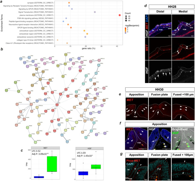
NTN1 is expressed in a wide range of developmental tissues and is essential for normal development. Here we describe the generation of a Netrin-1 reporter chicken line (NTN1-T2A-eGFP) by targeting green fluorescent protein into the NTN1 locus using CRISPR/Cas9 methodology. Our strategy gave 100% transmission of heterozygous (NTN1T2A - eGFP/+) embryos in which GFP localisation faithfully replicated endogenous NTN1 expression in the optic fissure and neural tube floorplate. Furthermore, all NTN1T2A - eGFP/+ embryos and hatched birds appeared phenotypically normal. We applied this resource to a pertinent developmental context - coloboma is a structural eye malformation characterised by failure of epithelial fusion during optic fissure closure (OFC) and NTN1 is specifically expressed in fusion pioneer cells at the edges of the optic fissure. We therefore optimised the isolation of GFP expressing cells from embryonic NTN1T2A - eGFP/+ eyes using spectral fluorescence cell-sorting and applied transcriptomic profiling of pioneer cells, which revealed multiple new OFC markers and novel pathways for developmental tissue fusion and coloboma. This work provides a novel fluorescent NTN1 chicken reporter line with broad experimental utility and is the first to directly molecularly characterise pioneer cells during OFC.


Conflict of interest statement

Declarations. Competing interests: The authors declare no competing interests.

Figures

Fig. 1
Fig. 1
Generation and validation of the NTN1-T2A-GFP chicken line. (a) Expression of endogenous NTN1 in wild type chicken embryo in somite regions (between asterisks), neural tube (arrowheads), and in pioneer cell regions at the fissure edges (arrow, inset), and fluorescent in situ hybridisation for NTN1 mRNA (green, arrowheads. Scale bar, 50 μm. Blue = DAPI; Magenta = Laminin. (b) Schema of genome editing strategy with sgRNA location (red arrow) shown in genome and vector. Abbreviations: EGF, Epidermal growth factor domain; NTR, N-terminal region; PAM, protospacer motif. Blue arrows indicate locations of diagnostic PCR primers. (c) Strategy for generating germ-line gene edited NTN1-T2A-eGFP chicken line via transfection of PGCs and injection of PGC clones into surrogate host embryos. (d) Diagnostic PCRs indicate monoallelic (NTN1GFP/WT) or biallelic (NTN1GFP/GFP) HDR-mediated integration of RTs in clonal PGCs (higher molecular weight band). (e) DNA sequence trace and corresponding amino-acid translation showing successful in-frame HDR in clonally propagated PGCs. Arrow indicates predicted T2A self-cleavage site. (f) RT-PCR for transgene expression in neural tube (NT) and optic fissure (OF) dissected from WT or NTN1NTN1 − T2A−GFP/+ embryos. Crops from separate agarose gels are displayed. (g) Whole NTN1NTN1 − T2A−GFP/+ embryo with GFP expression detected in neural tube (arrowheads), optic fissure (arrows), and somite regions (asterisks). (h) Flat-mount NTN1NTN1 − T2A−GFP/+ HH28/E6 dissected neural tube (Left) opened by cutting dorsally with GFP expression (arrowheads) observed in the floor-plate region, and ventral retina (Right) with GFP detected at the edges of the fissure margin (arrowheads). Full uncropped agarose gels are shown in Supplemental Fig. 1.
Fig. 2
Fig. 2
Isolation and transcriptomic profiling of pioneer cells from NTN1NTN1 − T2A−GFP/+ reveals novel pioneer cell markers with regulated expression during OFC. (a) Schema for optic fissure transcriptomic profiling strategy: manually dissected fissures (red hatched lines) from HH28 NTN1NTN1 − T2A−GFP/+ embryos were dissociated and FACS sorted prior to bulk RNA sequencing. (b) The GFP + ve and GFP-ve sorted cell populations from NTN1NTN1 − T2A−GFP/+ eyes were defined by gating based on using non-fluorescent WT control samples (i.e. no GFP). (c) GFP + ve and GFP-ve sorted NTN1eGFP cells were further separated using spectral flow cytometry to subtract auto fluorescent cells from the GFP + ve population (red arrows). (d) Volcano plot of all DEGs defined by -log10 p-value and Log2 fold change. There were 433 GFP + ve enriched DEGs (LFC2 > 1.0) and 1268 DEGs enriched in GFP-ve (LFC2 < -0.5). GFP + ve DEGs included known pioneer cell markers NTN1 and PAX2, whereas GFP-ve DEGs included periocular mesenchyme markers ALX1 and EMX2. (e) Fluorescence in situ hybridisation confirmed NTN1 and PAX2 mRNA expression was localised to pioneer cells (arrowheads) at HH28. In contrast ALX1 and EMX2 mRNA expression were identified in periocular mesenchyme (arrows). (f) Box plots for novel pioneer cell markers identified in this study, with log2 fold change and adjusted-P values indicated. (g) Colorimetric in situ hybridisation for novel OFM genes at HH28 showed pioneer cell specific expression (arrowheads) in the open fissure at HH28 before fusion. Non pioneer cell is expression indicated by arrows.
Fig. 3
Fig. 3
Pathway analyses profiling reveals HGF/Scatter factor activity during active fusion. (a) Consolidated plots showing gene ratios and counts for significant ontology enrichment terms (adj P < 0.05) associated with GFP + ve enriched DEGs using KEGG Pathway, Reactome Pathway, and GO TERMs. (b) STRING Network analysis of protein-protein interactions within the GFP + ve DEG set indicated MET at the centre of interactions and directly connected to its ligand HGF. (c) Box and whisker plots of absolute gene expression level (transcripts per million, TPM y-axis) for MET and HGF in GFP + ve (green plots) and GFP-ve cells (blue plots). (d) Fluorescence in situ hybridisation analyses at prefusion stage HH28 for MET (filled arrows) and HGF (yellow open arrows) in the distal and medial optic fissure margins. Note non-overlapping expression domains. (e) Fluorescence in situ hybridisation analyses during active fusion (HH30) indicated specific expression of MET (arrowheads) in the neural retina pioneer cell domain. (f) Comparison of MET and HGF expression in the fusing OFM revealed HGF expression (arrows) in cells at the junction of the RPE and pioneer cells, which was immediately adjacent to the MET expressing pioneer cells. (g) Immunofluorescence analysis for phosphorylated MET receptor protein (Phospho-Tyr1234/Tyr1235) identified activity at the folding point of the OFM connecting the RPE border cells to the apical regions of pioneer cells (arrows). Signal was not markedly reduced in the fused seam at 100 μm from the fusion plate in the same eye. Large arrowheads in (e) indicate the midline of the OFM; asterisks represent auto fluorescent red blood cells. Abbreviations: RPE, Retinal pigmented epithelium; POM, periocular mesenchyme; NR, neural retina.

References

    1. Lai Wing Sun, K., Correia, J. P. & Kennedy, T. E. Netrins: Versatile extracellular cues with diverse functions. Development138, 2153–2169. 10.1242/dev.044529 (2011). - PubMed
    1. Chaturvedi, V. & Murray, M. J. Netrins: Evolutionarily conserved regulators of epithelial fusion and closure in development and wound healing. Cells Tissues Organs.10.1159/000513880 (2021). - PubMed
    1. Cirulli, V. & Yebra, M. Netrins: Beyond the brain. Nat. Rev. Mol. Cell. Biol.8, 296–306. 10.1038/nrm2142 (2007). - PubMed
    1. Serafini, T. et al. Netrin-1 Is Required for Commissural Axon Guidance in the Developing Vertebrate Nervous System. Cell (1996). 10.1016/S0092-8674(00)81795-X - PubMed
    1. Bin, J. M. et al. Complete loss of Netrin-1 results in embryonic lethality and severe axon guidance defects without increased neural cell death. Cell. Rep.12, 1099–1106. 10.1016/j.celrep.2015.07.028 (2015). - PubMed

Substances

LinkOut - more resources