. 2025 Mar 24;26(1):112.
doi: 10.1186/s12931-025-03190-0.

Aging shapes infection profiles of influenza A virus and SARS-CoV-2 in human precision-cut lung slices

Affiliations

Aging shapes infection profiles of influenza A virus and SARS-CoV-2 in human precision-cut lung slices

Melanie Brügger et al. Respir Res. .

Abstract

Background: The coronavirus disease 2019 (COVID-19) outbreak revealed the susceptibility of elderly patients to respiratory virus infections, showing cell senescence or subclinical persistent inflammatory profiles and favoring the development of severe pneumonia.

Methods: In our study, we evaluated the potential influence of lung aging on the efficiency of replication of influenza A virus (IAV) and severe acute respiratory syndrome coronavirus 2 (SARS-CoV-2), as well as determining the pro-inflammatory and antiviral responses of the distal lung tissue.

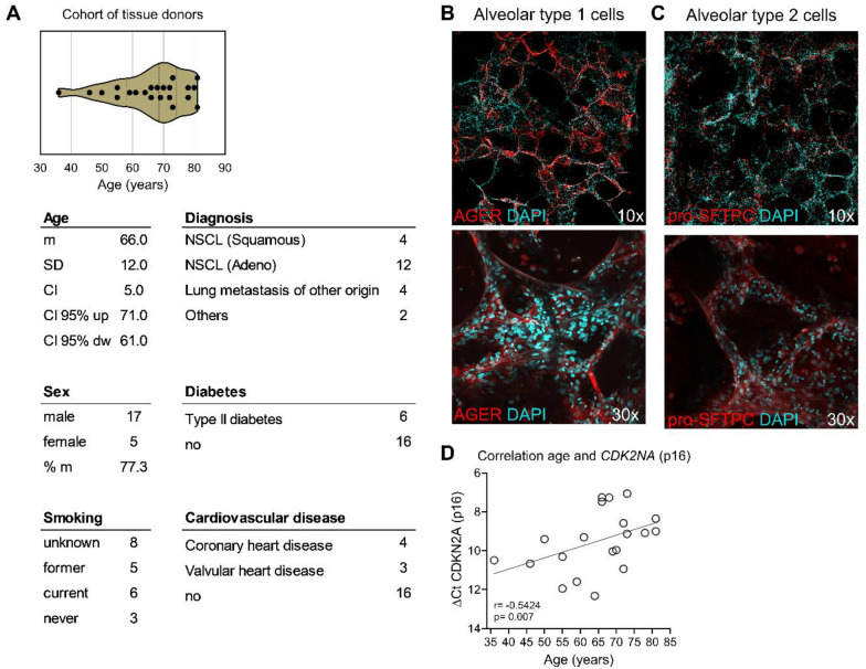
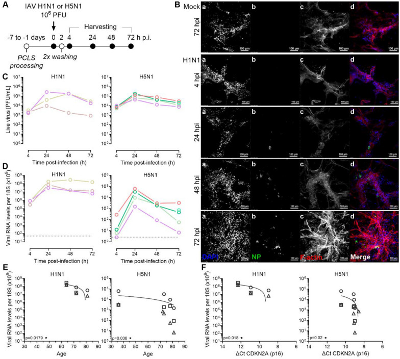
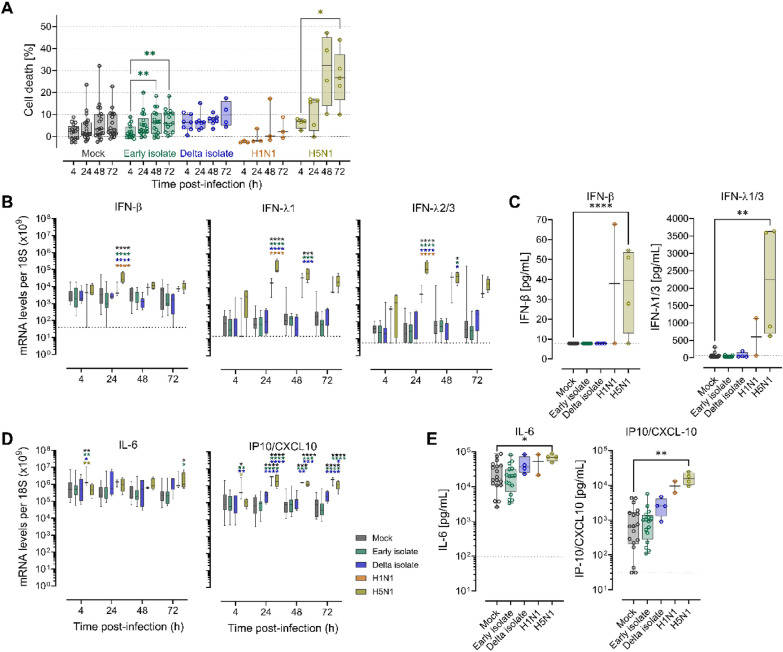
Results: Using precision-cut lung slices (PCLS) from donors of different ages, we found that pandemic H1N1 and avian H5N1 IAV replicated in the lung parenchyma with high efficacy. In contrast to these IAV strains, SARS-CoV-2 Early isolate and Delta variant of concern (VOC) replicated less efficiently in PCLS. Interestingly, both viruses showed reduced replication in PCLS from older compared to younger donors, suggesting that aged lung tissue represents a suboptimal environment for viral replication. Regardless of the age-dependent viral loads, PCLS responded to H5N1 IAV infection by an induction of IL-6 and IP10/CXCL10, both at the mRNA and protein levels, and to H1N1 IAV infection by induction of IP10/CXCL10 mRNA. Finally, while SARS-CoV-2 and H1N1 IAV infection were not causing detectable cell death, H5N1 IAV infection led to more cytotoxicity and induced significant early interferon responses.

Conclusions: In summary, our findings suggest that aged lung tissue might not favor viral dissemination, pointing to a determinant role of dysregulated immune mechanisms in the development of severe disease.

Keywords: Aging; Distal lung; Influenza virus; Precision-cut lung slices; SARS-CoV-2.


Conflict of interest statement

Declarations. Ethical approval and consent to participate: All human tissues were gathered and managed in accordance with the Declaration of Helsinki, and the study was approved by the Swiss Ethics Committee, Bern, Switzerland, approval number KEK-BE_2018-01801. Written informed consent was obtained from all donors before recruitment. Consent for publication: Not applicable. Competing interests: The authors declare no competing interests.

Figures

Fig. 1
Fig. 1
PCLS cultures from aged lungs present a senescent phenotype. A Violin plot representing the age distribution of the individual donors used for the generation of the PCLS cultures. The dashed vertical line indicates the median value, whereas the dotted lines indicate the interquartile ranges. Each symbol represents an individual donor. The table summarizes patient’s characteristics: age (mean, m; standard deviation, SD; confidence interval, CI; down, dw), sex (% male, m), smoking status (n), diagnosis, and other comorbidities, specifically diabetes and cardiovascular disease. B, C Representative confocal microscopy evaluation of PCLS stained for (B) AGER, highly expressed in alveolar type 1 cells, or (C) the alveolar type 2 cell marker pro-SFTPC. Micrographs are projections of 3D captures. Magnification 10X and 30X. AGER and pro-SFTPC, red; DAPI, light blue. D Correlation of deltaCt CDKN2A (p16) (deltaCt = Ct CDKN2A-Ct B2M) and donor age. Association was tested using the one-tailed Spearman rank correlation test, whereas the correlation coefficient (r) is given and a p value lower than 0.05 (*), 0.005 (**), 0.0005 (***), 0.0001 (****) is significant. Each symbol represents an individual donor
Fig. 2
Fig. 2
Human PCLS are productively infected by pandemic and avian IAV strains. A PCLS were infected one to seven days after generation with 106 PFU of IAV H1N1 A/Hamburg/4/2009 or H5N1 A/turkey/Turkey/2005. After 2 h PCLS were washed twice with PBS. Culture medium was changed daily. 4, 24, 48, and 72 h p.i. supernatants and PCLS were harvested for further analysis. B Representative confocal microscopy evaluation of Mock- and H1N1-infected PCLS, 4, 24, 48, and 72 h p.i. a, DAPI; b, IAV nucleoprotein (NP); c, F-actin; d, merge: DAPI, blue; IAV NP, green; F-actin, red. Scale bars, 100 µm. C Supernatants of infected PCLS were analyzed with a PFU assay. Each line represents an individual donor. D Tissue-associated viral RNA loads per 18S rRNA in PCLS over time following IAV infection. Dotted line represents the detection limit. E Correlation of tissue-associated viral RNA and donor age. F Correlation of tissue-associated viral RNA and deltaCt CDKN2A (p16) (deltaCt = Ct CDKN2A-Ct B2M). Association was tested using the Spearman rank correlation test. Each symbol represents an individual donor at 24 h (circle), 48 h (square), or 72 h p.i. (triangle). IAV H1N1, n = 3 and H5N1, n = 4
Fig. 3
Fig. 3
SARS-CoV-2 infection of human PCLS is leading to a low level of replication. A ACE2 and TMPRSS2 mRNA levels per 18S rRNA in PCLS of selected donors. Boxplots indicate the median value (centerline) and interquartile ranges (box edges), with whiskers extending to the lowest and the highest values. (ACE2, n = 15; TMPRSS2, n = 4). Fold difference was calculated by comparing the median ACE2 expression of all the PCLS (n = 15) and WD-NEC (n = 3) donors tested. B PCLS were infected one to seven days after generation with 106 PFU of SARS-CoV-2 (SARS-CoV-2/München1.1/2020/929; Early isolate) and Delta isolate (Delta AY.127, hCoV-19/Switzerland/BE-IFIK-918–4879/2021). After 2 h PCLS were washed twice with PBS. Growth medium was changed daily. 4, 24, 48, and 72 h p.i. supernatants and PCLS were harvested for further analysis. C Histological section of a representative Mock- and SARS-CoV-2 Delta isolate-infected PCLS 48 h p.i., stained for the nucleocapsid (N) protein and counterstained for hematoxylin. D Representative confocal microscopy evaluation of Mock- and SARS-CoV-2 Early isolate-infected PCLS 48 h p.i., stained for DAPI (grey) and SARS-CoV-2 N protein (green). a, Mock; b-e, SARS-CoV-2 Early isolate-infected PCLS. a-c, micrographs are projections of 3D captures. a-e, magnification 10X. E Supernatants of infected PCLS were analyzed with a PFU assay. Early isolate, n = 18; Delta isolate, n = 7. F Extracellular viral RNA in the supernatant of SARS-CoV-2 Early isolate-infected PCLS over time expressed as viral RNA copies per ml (n = 14). G Tissue-associated viral RNA loads per 18S rRNA in PCLS over time following SARS-CoV-2 infection. Dotted line represents the detection limit. Early isolate, n = 15; Delta isolate, n = 7 H Correlation of tissue-associated viral RNA and donor age. I Correlation of tissue-associated viral RNA and deltaCt CDKN2A (p16) (deltaCt = Ct CDKN2A-Ct B2M). Association was tested using the Spearman rank correlation test. Each symbol represents one individual donor at 24 h (circle), 48 h (square), or 72 h p.i. (triangle)
Fig. 4
Fig. 4
IAV is cytotoxic and induces a robust pro-inflammatory response in human PCLS. A Cell death (%), calculated from lactate dehydrogenase (LDH) release in PCLS, exposed to 106 PFU of SARS-CoV-2 Early or Delta isolate, and IAV H1N1 or H5N1 over time. A two-way ANOVA and Tukey’s multiple comparisons test was applied to compare the different groups to Mock (not significant) and the different timepoints of a group to its 4 h p.i. timepoint. Each symbol represents an individual donor. B IFN-β, IFN-λ1, IFN-λ2/3 and (D) IL-6, IP10/CXCL10 mRNA expression levels per 18S rRNA over time. A two-way ANOVA and Tukey’s multiple comparisons test was applied to compare the different groups. C IFN-β and IFN-λ1/3, and E IL-6 and IP10/CXCL10 protein levels in supernatants harvested 48 h p.i. in pg/ml. A Kruskal–Wallis test with Dunn’s multiple comparisons test was applied to compare infection groups to Mock. The dotted line represents the detection limit of the assays. Mock, black; SARS-CoV-2 Early isolate, green; SARS-CoV-2 Delta isolate, blue; IAV H1N1, orange; IAV H5N1, yellow. Boxplots indicate the median value (centerline) and interquartile ranges (box edges), with whiskers extending to the lowest and the highest values

Similar articles

References

    1. Jain S, Self WH, Wunderink RG, Fakhran S, Balk R, Bramley AM, et al. Community-acquired pneumonia requiring hospitalization among U.S. Adults. N Engl J Med. 2015;373(5):415–27. - PMC - PubMed
    1. Kochanek KD, Murphy SL, Xu J, Arias E. Deaths: Final Data for 2017. Natl Viral Stat Rep. 2019. 68(9). - PubMed
    1. Musuuza JS, Watson L, Parmasad V, Putman-Buehler N, Christensen L, Safdar N. Prevalence and outcomes of co-infection and superinfection with SARS-CoV-2 and other pathogens: a systematic review and meta-analysis. PLoS ONE. 2021;16(5): e0251170. - PMC - PubMed
    1. Onder G, Rezza G, Brusaferro S. Case-fatality rate and characteristics of patients dying in relation to COVID-19 in Italy. JAMA. 2020;323(18):1775. - PubMed
    1. Jain S, Kamimoto L, Bramley AM, Schmitz AM, Benoit SR, Louie J, et al. Hospitalized patients with 2009 H1N1 influenza in the United States, April–June 2009. N Engl J Med. 2009;361(20):1935. - PubMed

MeSH terms