. 2025 Mar 24;13(1):82.
doi: 10.1186/s40168-025-02079-w.

Metagenomic signatures of extraintestinal bacterial infection in the febrile term infant gut microbiome

Affiliations

Metagenomic signatures of extraintestinal bacterial infection in the febrile term infant gut microbiome

Anna L DeVeaux et al. Microbiome. .

Abstract

Background: Extraintestinal bacterial infections (EBIs), e.g., urinary tract infection, bacteremia, and meningitis, occur in approximately 10% of febrile infants younger than 60 days. Although many EBI-causing species commonly reside in the infant gut, proof that the digestive system is a pre-infection habitat remains unestablished.

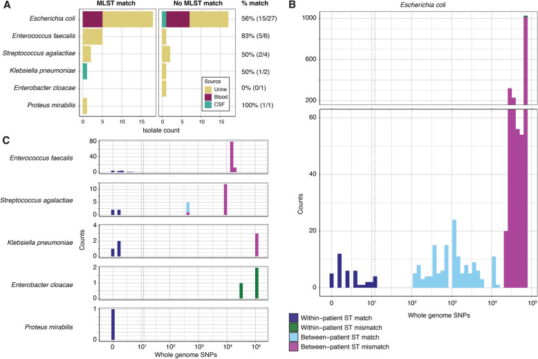
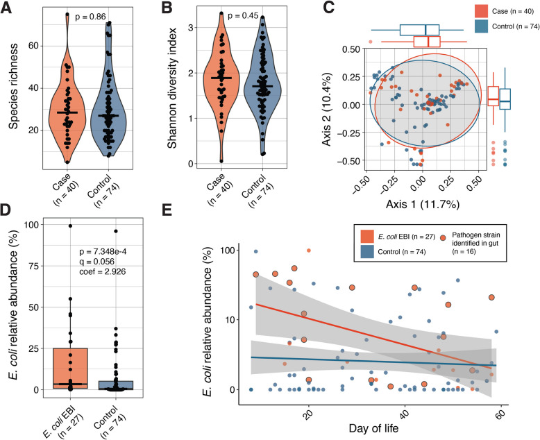
Results: We studied a cohort of febrile term infants < 60 days old who presented to one of thirteen US emergency departments in the Pediatric Emergency Care Applied Research Network from 2016 to 2019. Forty EBI cases and 74 febrile controls matched for age, sex, and race without documented EBIs were selected for analysis. Shotgun sequencing was performed of the gut microbiome and of strains cultured from the gut and extraintestinal site(s) of EBI cases, including blood, urine, and/or cerebrospinal fluid. Using a combination of EBI isolate genomics and fecal metagenomics, we detected an intestinal strain presumptively isogenic to the EBI pathogen (> 99.999% average nucleotide identity) in 63% of infants with EBIs. Although there was no difference in gut microbiome diversity between cases and controls, we observed significantly increased Escherichia coli relative abundance in the gut microbiome of infants with EBIs caused by E. coli. Infants with E. coli infections who were colonized by the putatively isogenic pathogen strain had significantly higher E. coli phylogroup B2 abundance in their gut, and their microbiome was more likely to contain virulence factor loci associated with adherence, exotoxin production, and nutritional/metabolic function.

Conclusions: The intestine plausibly serves as a reservoir for EBI pathogens in a subset of febrile term infants, prompting consideration of new opportunities for surveillance and EBI prevention among colonized, pre-symptomatic infants. Video Abstract.


Conflict of interest statement

Declarations. Ethics approval and consent to participate: This study was approved by the institutional review board at all sites. Secondary analyses conducted here were approved under Washington University HRPO IRB#202106080. Consent for publication: Not applicable. Competing interests: The authors declare no competing interests.

Figures

Fig. 1
Fig. 1
High genomic relatedness between cognate extraintestinal and intestinal isolates. A Bar plot depicting the number of extraintestinal isolates that had a strain of the same species and ST cultured from the gut (MLST match) or not (no MLST match) by species and isolate source. B, C Histograms of whole-genome SNP counts computed through pairwise comparisons within (B) Escherichia coli or (C) other pathogen species. Colors indicate whether the SNPs represent comparisons of samples from the same or different infants and whether the STs matched. The dotted line at 12 SNPs indicates the upper limit observed for within-patient ST matches. CSF cerebrospinal fluid, ST sequence type, SNP single-nucleotide polymorphism
Fig. 2
Fig. 2
Metagenomic read mapping delineates gut pathogen colonization at high abundance in EBI cases. A Genome breadth of coverage (filtered by ≥ 0.5 for visualization) and popANI of pairwise metagenomic read mapping to EBI pathogens. Each dot represents an individual alignment, and colors correspond to whether the metagenome mapped to a genome from the same infant and whether that pathogen had been cultured from the gut (“culture match”). Dot size is scaled to read depth-normalized coverage. Dashed line indicates the minimum popANI (99.999%) that was determined to be specific to within-infant alignments. B Relative abundance of the EBI-causing species compared to normalized coverage for within-infant mappings of metagenomic reads to pathogen genomes for strains determined to be present in the gut via culturing and/or inStrain (as indicated by color). Dashed line indicates the minimum relative abundance (1%) of the pathogen species that enabled metagenomic detection of the EBI-causing strain. Dot size is scaled to popANI. Pearson correlation coefficient (R) and p value are shown
Fig. 3
Fig. 3
Microbiome compositional differences are age- and infection-dependent for infants with E. coli EBI. α-diversity as measured by A species richness and B Shannon diversity from the gut metagenomes of all EBI cases compared to control infants. P values determined through a Wilcoxon rank-sum test. C Principal coordinate analysis (PCoA) plot of Bray–Curtis dissimilarity between samples computed at the species level. PCoA axes have the percent variation explained shown parenthetically. Marginal x- and y-axis boxplots correspond to PCoA axis 1 and 2 values, respectively. D Association between E. coli relative abundance and E. coli EBI status compared to control infants as determined through generalized linear mixed effect models. The BH-adjusted p value (q) and coefficient of effect (coef) are shown. E E. coli relative abundance plotted by day of life for infants with E. coli EBI compared to controls with linear regression lines plotted for each group. The shaded region represents the 95% confidence interval
Fig. 4
Fig. 4
Strain-level signatures of E. coli EBI matches in the gut. A Cladogram of all E. coli reference genomes for which a related strain was identified in at least one E. coli EBI case or E. coli-colonized control. Color strip denotes STs identified in at least three infants (the remainder are grouped as “other”). The bar chart indicates the frequency at which each reference genome was identified for E. coli EBI cases with a matching strain in their gut (match), those without a matching strain identified (non-match), or E. coli-colonized control infants. B Relative abundance of E. coli phylogroup B2 between groups as in A. C Virulence factor (VF) genes encoded by Enterobacteriaceae associated with E. coli EBI matches compared to E. coli-colonized controls as determined through generalized linear mixed effect models. Points are sized by their BH-adjusted p value (q) and colored by their VF functional category

References

    1. DePorre AG, Aronson PL, McCulloh RJ. Facing the ongoing challenge of the febrile young infant. Crit Care. 2017;21:68. - PMC - PubMed
    1. Simonsen KA, Anderson-Berry AL, Delair SF, Davies HD. Early-onset neonatal sepsis. Clin Microbiol Rev. 2014;27:21–47. - PMC - PubMed
    1. Greenhow TL, Hung Y-Y, Herz AM, Losada E, Pantell RH. The changing epidemiology of serious bacterial infections in young infants. Pediatr Infect Dis J. 2014;33:595–9. - PubMed
    1. Burstein B, Anderson G, Yannopoulos A. Prevalence of serious bacterial infections among febrile infants 90 days or younger in a canadian urban pediatric emergency department during the COVID-19 pandemic. Jama Netw Open. 2021;4:e2116919. - PMC - PubMed
    1. Watt K, Waddle E, Jhaveri R. Changing epidemiology of serious bacterial infections in febrile infants without localizing signs. PLoS ONE. 2010;5:e12448. - PMC - PubMed